
Article Figures & Data
Figures
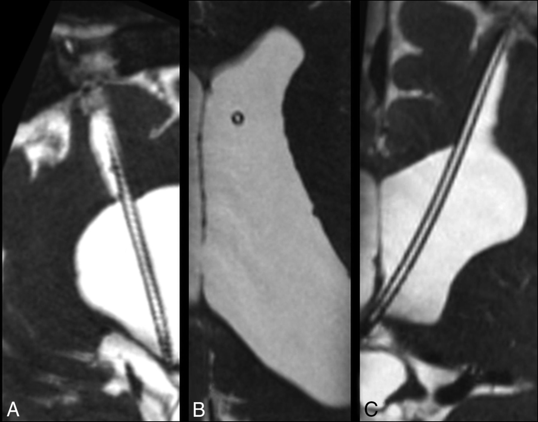
- Fig 1.
3D high-resolution isotropic CISS MR imaging sequence acquired in the sagittal plane demonstrates CSF signal throughout a patent ventriculostomy tube. Images were reconstructed post hoc in the sagittal oblique (A), axial (B), and coronal (C) planes.
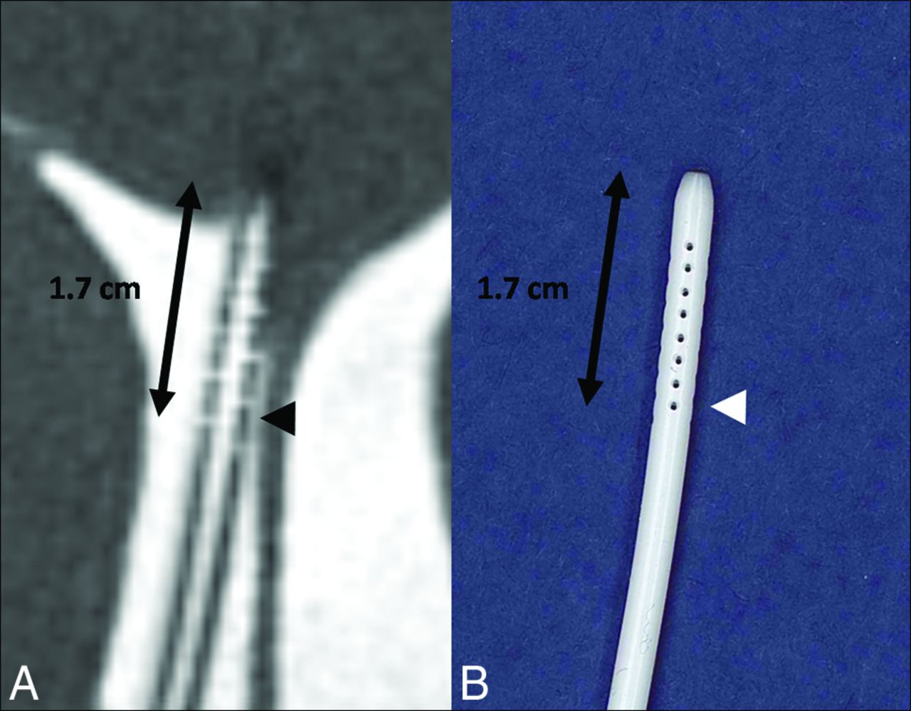
- Fig 2.
A, Isotropic CISS demonstrates a right-parietal-approach catheter with the tip extending just beyond the frontal horn of the right lateral ventricle. The side holes are seen as fluid-filled defects in the proximal aspect of the catheter extending to 1.7 cm from the tip (arrowhead). B, A photograph of a corresponding ventriculostomy catheter sample depicts the same relationship of side holes to the tip of the catheter.
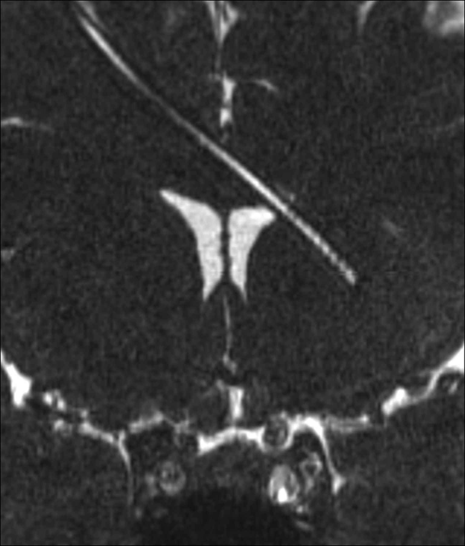
- Fig 3.
Coronal isotropic CISS demonstrates the tip of the ventriculostomy catheter passing beyond the left lateral ventricle. The lumen is fluid-filled.
- Fig 4.
Coronal CISS images from 3 patients demonstrate the position of the catheter tip with precision with respect to the surrounding structures. A, The ventriculostomy catheter abuts the septum pellucidum. B, The ventriculostomy catheter traverses the foramen of Monro to terminate in the third ventricle. C, The ventriculostomy catheter pierces the septum pellucidum, crossing the midline, in a third patient.
- Fig 5.
A, Coronal CISS in a patient with callosal dysgenesis demonstrates the curvilinear path of the ventriculostomy catheter with the tip in the interhemispheric fissure. B, Axial oblique CISS in a patient with previous obstruction at the posterior body of the lateral ventricle demonstrates tubing extending from the right frontal horn (left-pointing arrow) into the temporal horn of the lateral ventricle to terminate in the periventricular region (right-pointing arrow).
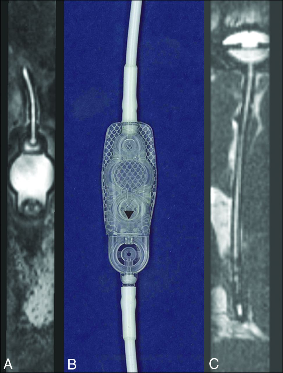
- Fig 6.
The detailed structure of the ventriculostomy tube and shunt reservoir can be evaluated in detail, particularly when the device does not contain metallic components. A, Sagittal oblique CISS through the extracranial component of the shunt device demonstrates the fluid-filled shunt reservoir corresponding to the sample (B) provided by the manufacturer Strata, Medtronic (Minneapolis, Minnesota). In another patient, the intracranial extent of the ventriculostomy tubing is seen extending to the shunt reservoir, of which the internal structures are visible.
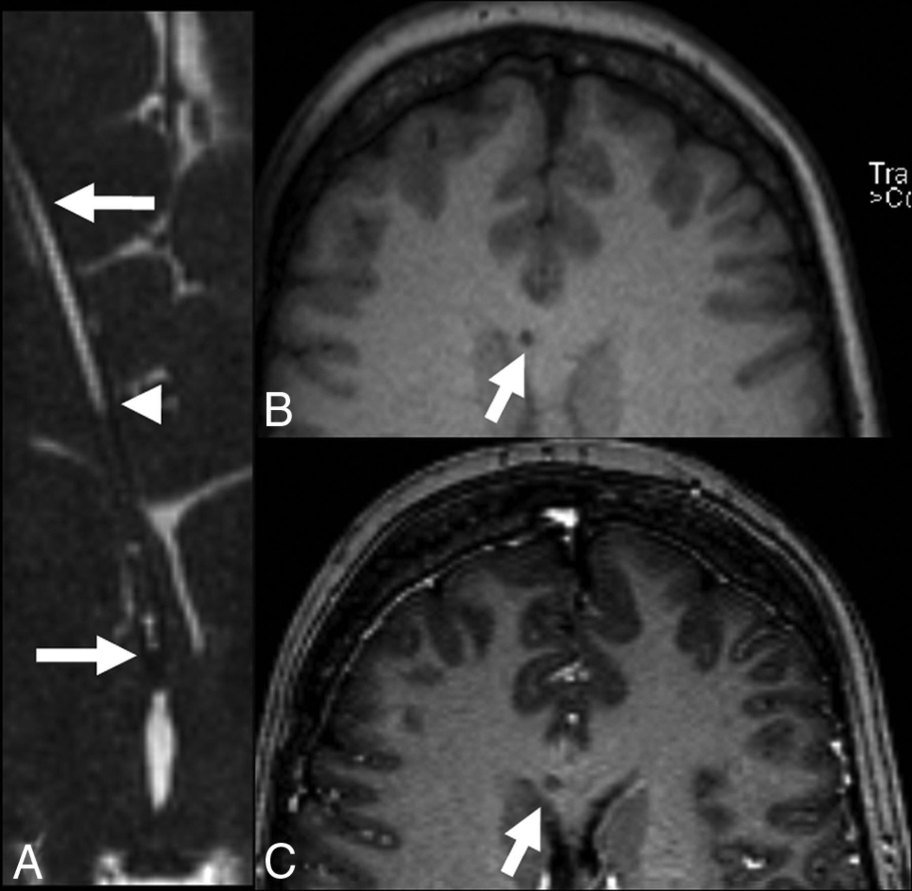
- Fig 7.
Shunt failure. While this case does not feature hydrocephalus, patients may have ventriculoperitoneal shunt placement for a variety of indications, including pseudotumor cerebri, where ventricles are narrowed rather than dilated, as well as cases involving multiple catheters, in which the development of obstructive hydrocephalus is impeded even if 1 of the shunts is partially occluded by debris. A, Axial oblique reconstructed CISS demonstrates the tip of the ventriculostomy tube (right-pointing arrow) extending peripherally through the frontal lobe (left-pointing arrow). CSF is not seen in the proximal catheter (compare with Fig 3) to the point marked with an arrowhead. B, Axial precontrast MPRAGE demonstrates the catheter extending through the corpus callosum. C, Axial postcontrast MPRAGE demonstrates no change in intensity, suggesting debris.
- Fig 8.
The ingrowth of the choroid plexus into the shunt catheter may significantly increase the risk of hemorrhage during shunt removal. A, Precontrast coronal CISS demonstrates a filling defect within the ventriculostomy tube. B, Postcontrast CISS demonstrates an increase in intensity due to enhancement of material filling the midportion of the catheter, suggesting choroid plexus ingrowth. C, Coronal postcontrast MPRAGE demonstrates corresponding hyperintensity, though the relationship to the catheter lumen is not as clear.


Tables
Baseline patient demographic data
Demographic Statistic (No.) (%) Age (mean) (range) (yr) 47.9 (32–74) Sex Male 10 (43.5) Female 13 (56.5) Indication for VP shunting Obstructive hydrocephalus 11 (47.8) Congenital hydrocephalus 7 (30.4) Communicating hydrocephalus 3 (13.0) Pseudotumor cerebri 2 (8.7) Indication for imaging Concern for shunt malfunction 11 (47.8) Alarming neurologic symptoms 6 (26.1) Routine follow-up 6 (26.1) Type or brand of shunt (per chart review) Programmable shunts 8 (34.7) Strataa 6 (26.1) Unspecified 2 (8.7) Nonprogrammable shunts 7 (30.4) Codman 1 (4.3) Raimondi 1 (4.3) Unspecified 5 (21.7) Unspecified/no information found 8 (34.7) Note:—VP indicates ventriculoperitoneal.
↵a Medtronic, Minneapolis, Minnesota.


Jump to section
Related Articles
Cited By...
- No citing articles found.