
Article Figures & Data
Figures
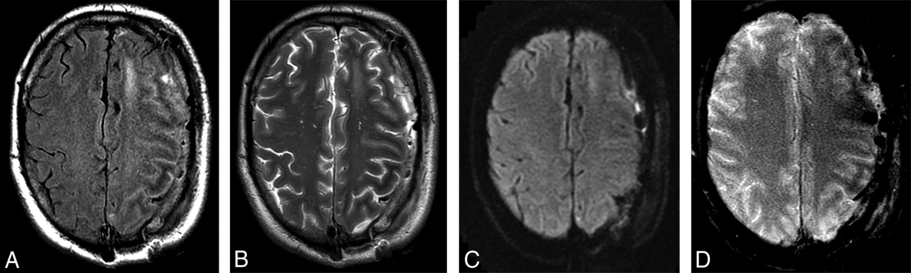
- Fig 1.
Patient admitted to the intensive care unit with persistent seizures 1 day after evacuation of a left-sided subdural hematoma. A, T2 FLAIR, B, FSE T2, C, DWI, and D, GRE-weighted images. Note the decreased T2-weighted signal in the subcortical white matter of the left frontal and parietal lobes on A and B, as well as the lack of corresponding diffusion restriction on C. D, GRE-weighted image shows there is subtle corresponding decreased subcortical white matter signal change.
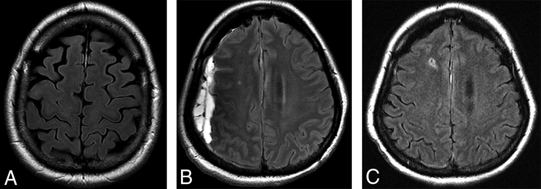
- Fig 2.
A, T2 FLAIR images from a 49-year-old male patient being followed for a left-frontal meningioma with asymptomatic low signal in the subcortical white matter adjacent to the meningioma. Nonspecific failure of suppression of CSF signal in the subjacent sulci was also noted. B, T2 FLAIR images showing low signal in the left occipital lobe in a 46-year-old patient who initially presented with visual seizures. C, T2 FLAIR images from a 48-year-old patient with a left convexity subdural hematoma and seizures. Subcortical low T2 signal is present in the compressed posterior frontal and parietal lobes.
- Fig 3.
A, T2 FLAIR image in the same patient as in Fig 2A shows complete resolution of the white matter changes 2 years after resection of the lesion. B, T2 FLAIR image from a 53-year-old female patient with a spontaneous right-sided acute subdural hematoma. T2 hypointense signal changes are present in the subjacent white matter. C, Follow-up 5 months later shows resolution of both the hematoma and the white matter changes.


Tables
Summary of imaging and clinical findings in patients with abnormally decreased T2 signal
Findings Demographics Patients 29 Sex (male) 19 (65.5%) Age (yr) 49 (range, 18–85) Clinical information Associated seizures 22 (75.9%) Extrinsic compressive lesions 14 (48.3%) Imaging findings Laterality Bilateral 7 (24.1%) Unilateral 22 (75.9%) Multiple lobes involved 21 (72.4%) Frequency of involved lobes Frontal 19 (65.5%) Parietal 17 (58.6%) Temporal 16 (55.2%) Occipital 14 (48.3%) EEG findings Availability 19 (65.5%) Laterality Unilateral 12/12 Cases with MRI T2 changes unilateral Bilateral 7/7 Cases with MRI T2 changes bilateral Follow-up imaging Availability 9 (31%) Changes resolved 6 (66.6%) Associated with compressive lesion 4/6 Nonresolved/gliosis 3 (33.3%) Associated with compressive lesion 1/3


Jump to section
Related Articles
Cited By...
- No citing articles found.