
Article Figures & Data
Figures
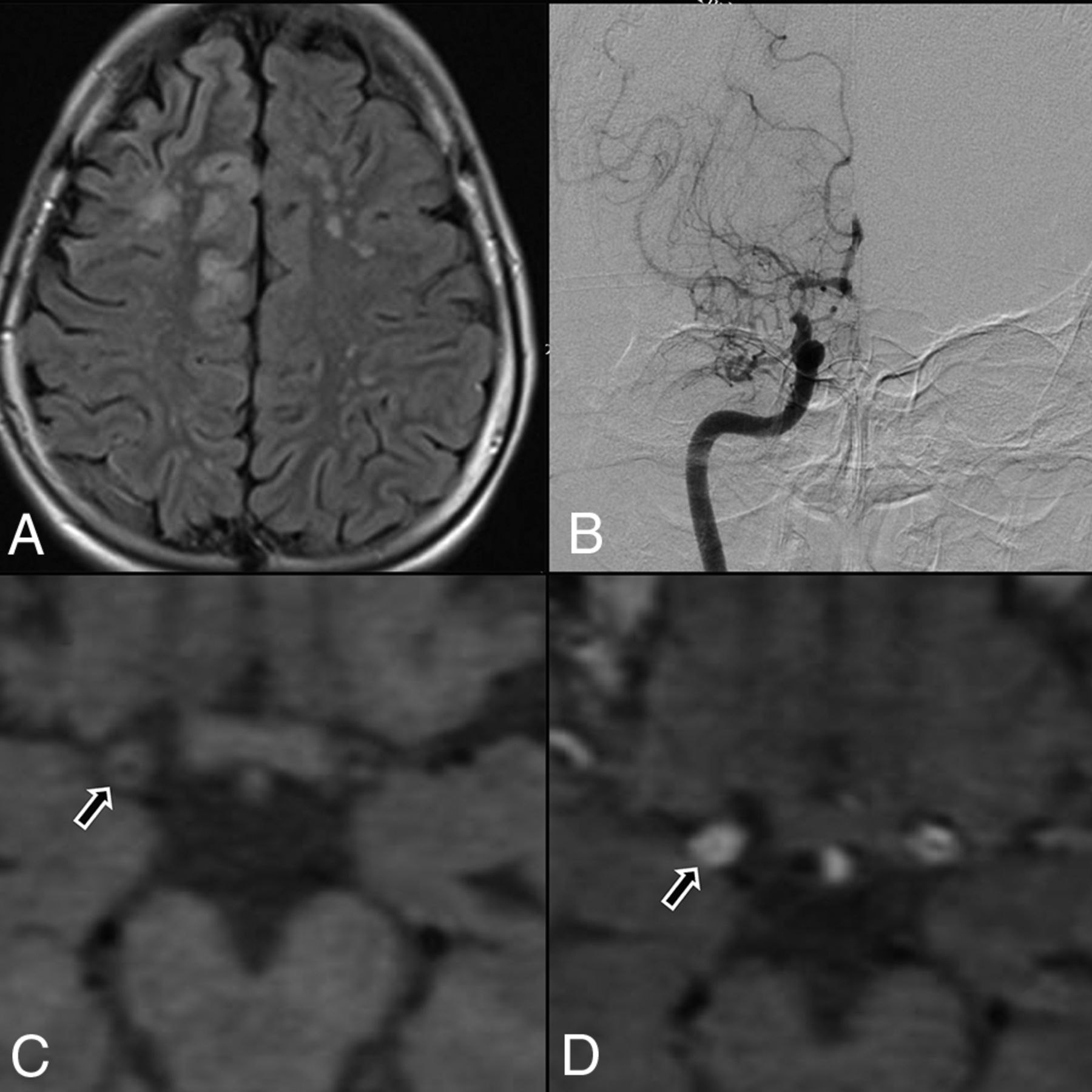
- FIGURE.
A, Flair axial section showing right ACA and MCA territory cortical infarct and multiple discrete MCA-ACA watershed infarct on the left side. B, DSA, showing tight stenosis of the distal ICA, with concomitant narrowing of proximal MCA and ACA, with leptomeningeal collaterals. C, Axial T1 noncontrast, HRVWI, showing circumferential wall thickening (arrow) and narrowing of the lumen of right distal ICA. D, Axial T1 contrast-enhanced, HRVWI, showing circumferential grade II enhancement (arrow) of a right distal ICA (Note:—Enhancement is graded by comparing with pituitary infundibulum).

Tables
Patient Characteristics Results Sex, n (%) Males 17 (58.6) Females 12 (41.6) Age, n (%) <18 y 15 (51.7) ≥18 y 14 (48.3) Age at first eventa, median (IQR), y −12 (6–36) Initial clinical presentation, n (%) Ischemic stroke 10 (34.5) Intracerebral bleed 5 (17.2) TIA 7 (24.2) Syncope 1 (3.4) Seizure 6 (20.7) Recurrent events, n (%) 22 (75.9) Ischemic stroke 10 (34.5) Intracranial bleed (intraventricular hemorrhage) 2 (6.9) Positive family history of MMD, n (%) 2 (6.9) NIHSS on admission, median (IQR) 1 (0–3) mRS on admission, median (IQR) 1 (0–3) Note:–IQR indicates interquartile range; mRS, modified Rankin scale.
↵a The mean ± SD interval between the first symptom and HRVWI was 64.7 ± 80.7 months.
Characteristic n (%) (n = 58 Hemispheres) A. MRI 1. Pattern of infarct Acute Infarcts Chronic Infarcts Watershed 9 (15.5) 15 (25.9) Territorial 3 (5.2) 5 (8.6) Cortical 2 (3.4) 3 (5.2) Isolated subcortical 1 (1.7) 2 (3.4) B. DSA 1. Laterality (n = 29) Bilateral 27 (93.1) Unilateral 2 (6.9) 2. Vessels involved (n = 166 vessels) Narrowed Occluded Distal ICA 19 (11.4) 37 (22.2) Proximal MCA 12 (7.2) 39 (23.5) Proximal ACA 17 (10.2) 25 (15.0) PCA 8 (4.8) 1 (0.6) Basilar artery (29 vessels) 1 (0.6) 0 (0) 3. Suzuki stage Right Left I 2 (7.1) 2 (7.1) II 0 (0) 1 (3.6) III 5 (17.9) 2 (7.1) IV 4 (14.3) 10 (35.7) V 16 (57.1) 13 (46.4) VI 1 (3.6) 0 (0) 4. Collaterals (n = 58 hemispheres) Superficial meningeal Leptomeningeal Anterior 27 (46.6) 24 (41.4) Posterior 21 (36.2) 20 (34.5) Durocortical Anterior 21 (36.2) 18 (31.1) Posterior 3 (5.2) 5 (8.6) Deep parenchymal Subependymal anastomotic Anterior 6 (10.3) 6 (10.3) Posterior 5 (8.6) 5 (8.6) Inner thalamic Anterior 8 (13.8) 6 (10.3) Posterior 8 (13.8) 7 (12.1) HRVWI Characteristics Contrast Enhancement (8 Patients, 10 Lesions) T1 Wall Thickening (9 Patients, 11 Lesions) Pattern, n (%) Concentric 10 (100) 11 (100) Eccentric 0 (0) 0 (0) Vessel involved, n (%) Distal ICA 8 (80) 9 (81.8) Proximal MCA 2 (20) 2 (18.2) Events within 3 mo before and after the imaging, n (%) 7 (87.5) (P = .01) 9 (100) (P ≤ .001) Recent events (DWI positive) within 4 wk of imaging, n (%) 4 (44.4) (P = .20) Grade I contrast enhancement 0 (0) Grade II contrast enhancement 3 (100) (P = .02) Suzuki stage Grade 2 enhancement (4 lesions) Wall thickening (11 lesions) I 1 (25) 1 (9.1) II 1 (25) 2 (18.2) III 0 4 (36.4) IV 2 (50) 4 (36.4) Remodeling index, mean ± SD Right, 0.71 ± 0.13 Left, 0.69 ± 0.13


Jump to section
Related Articles
Cited By...
- Clinical and genetic risk factors and long-term outcomes of MRI vessel wall enhancement in moyamoya disease
- Designing a flow-controlled STA-MCA anastomosis based on the Hagen-Poiseuille law for preventing postoperative hyperperfusion in adult moyamoya disease
- Utilisation of advanced MRI techniques to understand neurovascular complications of PHACE syndrome: a case of arterial stenosis and dissection