Index by author
Rueda-lopes, F.C.
- Pediatric NeuroimagingYou have accessBrain MR Imaging of Patients with Perinatal Chikungunya Virus InfectionD.G. Corrêa, T. A. L. Freddi, H. Werner, F.P.P.L. Lopes, M.E.L. Moreira, F.C.P. de Almeida Di Maio Ferreira, J.M. de Andrade Lopes, F.C. Rueda-Lopes and L.C.H. da CruzAmerican Journal of Neuroradiology January 2020, 41 (1) 174-177; DOI: https://doi.org/10.3174/ajnr.A6339
Rutland, J.W.
- Adult BrainOpen AccessEmerging Use of Ultra-High-Field 7T MRI in the Study of Intracranial Vascularity: State of the Field and Future DirectionsJ.W. Rutland, B.N. Delman, C.M. Gill, C. Zhu, R.K. Shrivastava and P. BalchandaniAmerican Journal of Neuroradiology January 2020, 41 (1) 2-9; DOI: https://doi.org/10.3174/ajnr.A6344
Sahraian, S.
- You have accessRedundant Neurovascular Imaging: Who Is to Blame and What Is the Value?E. Beheshtian, S. Emamzadehfard, S. Sahraian, R. Jalilianhasanpour and D.M. YousemAmerican Journal of Neuroradiology January 2020, 41 (1) 35-39; DOI: https://doi.org/10.3174/ajnr.A6329
Salmeen, A.K.
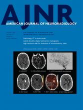
- EDITOR'S CHOICEAdult BrainYou have accessPrediction of Hemorrhage after Successful Recanalization in Patients with Acute Ischemic Stroke: Improved Risk Stratification Using Dual-Energy CT Parenchymal Iodine Concentration Ratio Relative to the Superior Sagittal SinusD. Byrne, J.P. Walsh, H. Schmiedeskamp, F. Settecase, M.K.S. Heran, B. Niu, A.K. Salmeen, B. Rohr, T.S. Field, N. Murray and A. RohrAmerican Journal of Neuroradiology January 2020, 41 (1) 64-70; DOI: https://doi.org/10.3174/ajnr.A6345
The authors evaluated whether, in acute ischemic stroke, iodine concentration within contrast-stained parenchyma compared with an internal reference in the superior sagittal sinus on dual-energy CT could predict subsequent intracerebral hemorrhage in 71 patients. Forty-three of 71 patients had parenchymal hyperdensity on initial dual-energy CT. The median relative iodine concentration compared with the superior sagittal sinus was significantly higher in those with subsequent intracerebral hemorrhage (137.9% versus 109.2%). They conclude that in dual-energy CT performed within 1 hour following thrombectomy that the relative iodine concentration within contrast-stained brain parenchyma compared with that in the superior sagittal sinus was a more reliable predictor of ICH compared with the absolute maximum iodine concentration.
Santhanam, P.
- Adult BrainOpen AccessStructural and Volumetric Brain MRI Findings in Mild Traumatic Brain InjuryJ.B. Patel, S.H. Wilson, T.R. Oakes, P. Santhanam and L.K. WeaverAmerican Journal of Neuroradiology January 2020, 41 (1) 92-99; DOI: https://doi.org/10.3174/ajnr.A6346
Schmidbauer, V.
- FunctionalYou have accessLesion-Specific Language Network Alterations in Temporal Lobe EpilepsyO. Foesleitner, K.-H. Nenning, L. Bartha-Doering, C. Baumgartner, E. Pataraia, D. Moser, M. Schwarz, V. Schmidbauer, J.A. Hainfellner, T. Czech, C. Dorfer, G. Langs, D. Prayer, S. Bonelli and G. KasprianAmerican Journal of Neuroradiology January 2020, 41 (1) 147-154; DOI: https://doi.org/10.3174/ajnr.A6350
Schmiedeskamp, H.
- EDITOR'S CHOICEAdult BrainYou have accessPrediction of Hemorrhage after Successful Recanalization in Patients with Acute Ischemic Stroke: Improved Risk Stratification Using Dual-Energy CT Parenchymal Iodine Concentration Ratio Relative to the Superior Sagittal SinusD. Byrne, J.P. Walsh, H. Schmiedeskamp, F. Settecase, M.K.S. Heran, B. Niu, A.K. Salmeen, B. Rohr, T.S. Field, N. Murray and A. RohrAmerican Journal of Neuroradiology January 2020, 41 (1) 64-70; DOI: https://doi.org/10.3174/ajnr.A6345
The authors evaluated whether, in acute ischemic stroke, iodine concentration within contrast-stained parenchyma compared with an internal reference in the superior sagittal sinus on dual-energy CT could predict subsequent intracerebral hemorrhage in 71 patients. Forty-three of 71 patients had parenchymal hyperdensity on initial dual-energy CT. The median relative iodine concentration compared with the superior sagittal sinus was significantly higher in those with subsequent intracerebral hemorrhage (137.9% versus 109.2%). They conclude that in dual-energy CT performed within 1 hour following thrombectomy that the relative iodine concentration within contrast-stained brain parenchyma compared with that in the superior sagittal sinus was a more reliable predictor of ICH compared with the absolute maximum iodine concentration.
Schwarz, M.
- FunctionalYou have accessLesion-Specific Language Network Alterations in Temporal Lobe EpilepsyO. Foesleitner, K.-H. Nenning, L. Bartha-Doering, C. Baumgartner, E. Pataraia, D. Moser, M. Schwarz, V. Schmidbauer, J.A. Hainfellner, T. Czech, C. Dorfer, G. Langs, D. Prayer, S. Bonelli and G. KasprianAmerican Journal of Neuroradiology January 2020, 41 (1) 147-154; DOI: https://doi.org/10.3174/ajnr.A6350
Senthilvelan, S.
- FELLOWS' JOURNAL CLUBAdult BrainOpen AccessVessel Wall Thickening and Enhancement in High-Resolution Intracranial Vessel Wall Imaging: A Predictor of Future Ischemic Events in Moyamoya DiseaseA. Kathuveetil, P.N. Sylaja, S. Senthilvelan, K. Chandrasekharan, M. Banerjee and B. Jayanand SudhirAmerican Journal of Neuroradiology January 2020, 41 (1) 100-105; DOI: https://doi.org/10.3174/ajnr.A6360
Twenty-nine patients with Moyamoya disease were enrolled in this study. The median age at symptom onset was 12 years. A total of 166 steno-occlusive lesions were detected by high-resolution intracranial vessel wall imaging. Eleven lesions with concentric wall thickening (6.6%) were noted in 9 patients. Ten concentric contrast-enhancing lesions were observed in 8 patients, of which 4 lesions in 3 patients showed grade II enhancement. The presence of contrast enhancement and wall thickening showed a statistically significant association with ischemic events within 3 months before and after the vessel wall imaging. The presence of wall thickening and enhancement may predict future ischemic events in patients with MMD.
Settecase, F.
- EDITOR'S CHOICEAdult BrainYou have accessPrediction of Hemorrhage after Successful Recanalization in Patients with Acute Ischemic Stroke: Improved Risk Stratification Using Dual-Energy CT Parenchymal Iodine Concentration Ratio Relative to the Superior Sagittal SinusD. Byrne, J.P. Walsh, H. Schmiedeskamp, F. Settecase, M.K.S. Heran, B. Niu, A.K. Salmeen, B. Rohr, T.S. Field, N. Murray and A. RohrAmerican Journal of Neuroradiology January 2020, 41 (1) 64-70; DOI: https://doi.org/10.3174/ajnr.A6345
The authors evaluated whether, in acute ischemic stroke, iodine concentration within contrast-stained parenchyma compared with an internal reference in the superior sagittal sinus on dual-energy CT could predict subsequent intracerebral hemorrhage in 71 patients. Forty-three of 71 patients had parenchymal hyperdensity on initial dual-energy CT. The median relative iodine concentration compared with the superior sagittal sinus was significantly higher in those with subsequent intracerebral hemorrhage (137.9% versus 109.2%). They conclude that in dual-energy CT performed within 1 hour following thrombectomy that the relative iodine concentration within contrast-stained brain parenchyma compared with that in the superior sagittal sinus was a more reliable predictor of ICH compared with the absolute maximum iodine concentration.
In this issue