
Article Figures & Data
Figures
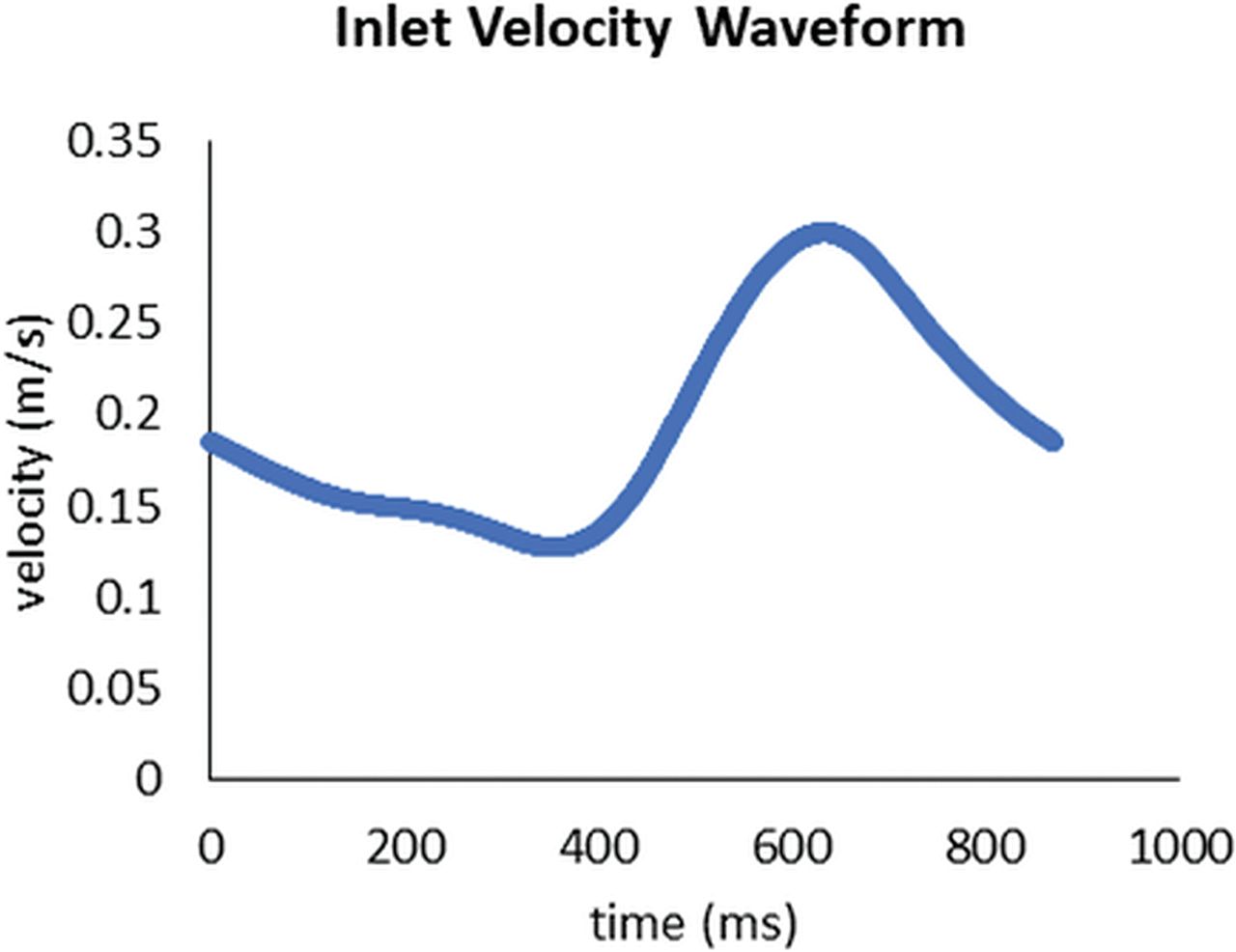
- Fig 1.
Flow waveform at the inflow representing the inflow conditions used for CFD analysis. A homogeneous velocity profile across the complete inflow cross-section with the velocities given by the inflow curve was applied as inflow boundary conditions. The outflow boundary conditions were defined as zero pressure.
- Fig 2.
Images exemplarily show measurements of the aneurysmal ostium (A and B) and the aneurysm itself (C and D) for calculation of the ostium size, maximum diameter, and aneurysmal volume.
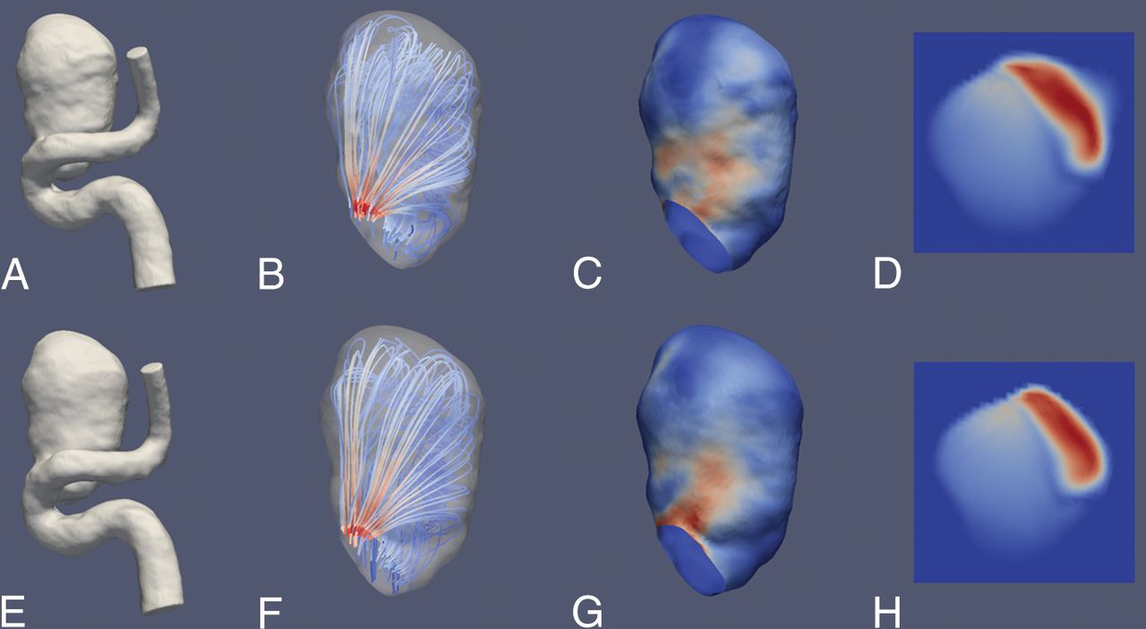
- Fig 3.
Illustrative case 1. CFD simulations show a saccular aneurysm of the ICA. Upper row (A–D) and lower row (E–H) show reconstruction results derived from a 3D-DSA and a 4D-DSA dataset, respectively. A and E, 3D view of the aneurysm using a volume-rendering technique and demonstrating comparable aneurysmal morphology. B and F, A color-coded visualization of intra-aneurysmal flow using streamlines (red and blue indicate high- and low-velocity magnitudes). The peak velocities show a laminar distribution along the longitudinal axis. C and G, In concordance, note maximum systolic wall shear stress (red and blue indicate high and low WSS) in the corresponding area. D and H, The aneurysmal ostium, the associated flow conditions, and the corresponding areas with high-velocity magnitude (red and blue indicate high- and low-velocity magnitude). Overall, the congruence of hemodynamic characteristics between 3D- and 4D-DSA shows geometric accordance between both techniques.
- Fig 4.
Illustrative case 2. CFD simulations show a large saccular aneurysm of the carotid T. Upper row (A–D) and lower row (E–H) show reconstruction results derived from a 3D-DSA and a 4D-DSA dataset, respectively. A and E, 3D view of the aneurysm using a volume-rendering technique and demonstrating comparable aneurysmal morphology. B and F, Color-coded visualization of intra-aneurysmal flow using streamlines (red and blue indicate high- and low-velocity magnitudes). The peak velocities show a predominant basal distribution. C and G, In concordance, note maximum systolic wall shear stress (red and blue indicate high and low WSS) in the corresponding area. D and H, Aneurysmal ostium, the associated flow conditions, and the match of areas with high-velocity magnitude (red and blue indicate high- and low-velocity magnitude). Overall, the congruence of hemodynamic characteristics between 3D- and 4D-DSA shows geometric accordance between the techniques.


Tables
Parameter 3D-DSA (Median, IQR) 4D-DSA (Median, IQR) r Max. diameter (mm) 11 (9–20) 11 (9–20) 0.988 (P < .001) Max. volume (mm3) 356 (161–1196) 381 (159–1065) 0.976 (P < .001) Ostium size1 (mm) 5 (4–7) 5 (4–7) 0.976 (P < .001) Ostium size2 (mm) 5 (4–6) 5 (4–6) 0.857 (P = .002) Note:—Max. indicates maximum; IQR, interquartile range.
Parameter 3D-DSA (Mean) 4D-DSA (Mean) r Intra-aneurysmal pressuresystole (Pa) 502.8 ± 242.24 492.2 ± 300.07 0.903 (P < .001) Intra-aneurysmal pressurediastole (Pa) 189.3 ± 99.13 181.4 ± 114.4 0.842 (P = .002) Flow velocitysystole (mm/s) 80.8 ± 50.94 74.89 ± 48.02 0.930 (P < .001) Flow velocitydiastole (mm/s) 31.61 ± 22.2 29.9 ± 21.45 0.952 (P < .001) AWSSsystole (Pa) 0.933 ± 1.02 0.835 ± 0.853 0.936 (P < .001) AWSSdiastole (Pa) 0.26 ± 0.28 0.24 ± 0.25 0.918 (P < .001) MWSS systole (Pa) 5.95 ± 1.83 5.63 ± 1.64 0.927 (P < .001) MWSS diastole (Pa) 1.84 ± 0.73 1.78 ± 0.69 0.879 (P = .001) IZ (mm2) 12.4 ± 14.2 8.9 ± 6.81 0.957 (P < .001) LSZ (mm2) 254.6 ± 203.55 263.9 ± 199.08 1 (P < .01)


