Advertisement

Main menu

  • Home
  • Content
    • Current Issue
    • Accepted Manuscripts
    • Article Preview
    • Past Issue Archive
    • Video Articles
    • AJNR Case Collection
    • Case of the Week Archive
    • Case of the Month Archive
    • Classic Case Archive
  • Special Collections
    • AJNR Awards
    • Low-Field MRI
    • Alzheimer Disease
    • ASNR Foundation Special Collection
    • Photon-Counting CT
    • View All
  • Multimedia
    • AJNR Podcasts
    • AJNR SCANtastic
    • Trainee Corner
    • MRI Safety Corner
    • Imaging Protocols
  • For Authors
    • Submit a Manuscript
    • Submit a Video Article
    • Submit an eLetter to the Editor/Response
    • Manuscript Submission Guidelines
    • Statistical Tips
    • Fast Publishing of Accepted Manuscripts
    • Graphical Abstract Preparation
    • Imaging Protocol Submission
    • Author Policies
  • About Us
    • About AJNR
    • Editorial Board
    • Editorial Board Alumni
  • More
    • Become a Reviewer/Academy of Reviewers
    • Subscribers
    • Permissions
    • Alerts
    • Feedback
    • Advertisers
    • ASNR Home

User menu

  • Alerts
  • Log in

Search

  • Advanced search
American Journal of Neuroradiology

American Journal of Neuroradiology

ASHNR American Society of Functional Neuroradiology ASHNR American Society of Pediatric Neuroradiology ASSR
  • Alerts
  • Log in

Advanced Search

  • Home
  • Content
    • Current Issue
    • Accepted Manuscripts
    • Article Preview
    • Past Issue Archive
    • Video Articles
    • AJNR Case Collection
    • Case of the Week Archive
    • Case of the Month Archive
    • Classic Case Archive
  • Special Collections
    • AJNR Awards
    • Low-Field MRI
    • Alzheimer Disease
    • ASNR Foundation Special Collection
    • Photon-Counting CT
    • View All
  • Multimedia
    • AJNR Podcasts
    • AJNR SCANtastic
    • Trainee Corner
    • MRI Safety Corner
    • Imaging Protocols
  • For Authors
    • Submit a Manuscript
    • Submit a Video Article
    • Submit an eLetter to the Editor/Response
    • Manuscript Submission Guidelines
    • Statistical Tips
    • Fast Publishing of Accepted Manuscripts
    • Graphical Abstract Preparation
    • Imaging Protocol Submission
    • Author Policies
  • About Us
    • About AJNR
    • Editorial Board
    • Editorial Board Alumni
  • More
    • Become a Reviewer/Academy of Reviewers
    • Subscribers
    • Permissions
    • Alerts
    • Feedback
    • Advertisers
    • ASNR Home
  • Follow AJNR on Twitter
  • Visit AJNR on Facebook
  • Follow AJNR on Instagram
  • Join AJNR on LinkedIn
  • RSS Feeds

AJNR Awards, New Junior Editors, and more. Read the latest AJNR updates

Research ArticleAdult Brain
Open Access

Defining Ischemic Core in Acute Ischemic Stroke Using CT Perfusion: A Multiparametric Bayesian-Based Model

K. Nael, E. Tadayon, D. Wheelwright, A. Metry, J.T. Fifi, S. Tuhrim, R.A. De Leacy, A.H. Doshi, H.L. Chang and J. Mocco
American Journal of Neuroradiology September 2019, 40 (9) 1491-1497; DOI: https://doi.org/10.3174/ajnr.A6170
K. Nael
aFrom the Department of Radiology (K.N., E.T., A.M., A.H.D.), Neuroimaging Advanced and Exploratory Lab
  • Find this author on Google Scholar
  • Find this author on PubMed
  • Search for this author on this site
  • ORCID record for K. Nael
E. Tadayon
aFrom the Department of Radiology (K.N., E.T., A.M., A.H.D.), Neuroimaging Advanced and Exploratory Lab
  • Find this author on Google Scholar
  • Find this author on PubMed
  • Search for this author on this site
  • ORCID record for E. Tadayon
D. Wheelwright
bDepartments of Neurology (D.W., J.F., S.T.)
  • Find this author on Google Scholar
  • Find this author on PubMed
  • Search for this author on this site
  • ORCID record for D. Wheelwright
A. Metry
aFrom the Department of Radiology (K.N., E.T., A.M., A.H.D.), Neuroimaging Advanced and Exploratory Lab
  • Find this author on Google Scholar
  • Find this author on PubMed
  • Search for this author on this site
  • ORCID record for A. Metry
J.T. Fifi
bDepartments of Neurology (D.W., J.F., S.T.)
cNeurosurgery (J.F., R.A.D.L., J.M.)
  • Find this author on Google Scholar
  • Find this author on PubMed
  • Search for this author on this site
  • ORCID record for J.T. Fifi
S. Tuhrim
bDepartments of Neurology (D.W., J.F., S.T.)
  • Find this author on Google Scholar
  • Find this author on PubMed
  • Search for this author on this site
  • ORCID record for S. Tuhrim
R.A. De Leacy
cNeurosurgery (J.F., R.A.D.L., J.M.)
  • Find this author on Google Scholar
  • Find this author on PubMed
  • Search for this author on this site
  • ORCID record for R.A. De Leacy
A.H. Doshi
aFrom the Department of Radiology (K.N., E.T., A.M., A.H.D.), Neuroimaging Advanced and Exploratory Lab
  • Find this author on Google Scholar
  • Find this author on PubMed
  • Search for this author on this site
  • ORCID record for A.H. Doshi
H.L. Chang
dPopulation Health Science and Policy (H.C.), Icahn School of Medicine at Mount Sinai, New York, New York.
  • Find this author on Google Scholar
  • Find this author on PubMed
  • Search for this author on this site
  • ORCID record for H.L. Chang
J. Mocco
cNeurosurgery (J.F., R.A.D.L., J.M.)
  • Find this author on Google Scholar
  • Find this author on PubMed
  • Search for this author on this site
  • ORCID record for J. Mocco
  • Article
  • Figures & Data
  • Info & Metrics
  • Responses
  • References
  • PDF
Loading

Article Figures & Data

Figures

  • Tables
  • Fig 1.
    • Download figure
    • Open in new tab
    • Download powerpoint
    Fig 1.

    Multiparametric voxel-based model for infarction.

  • Fig 2.
    • Download figure
    • Open in new tab
    • Download powerpoint
    Fig 2.

    Stacked bar graph visualization of errors in ischemic core volume estimation compared with final infarction volume by Bayesian-logit and oSVD postprocessing. The error values are noticeably greater with oSVD (black bars) compared with Bayesian-logit (gray bars).

  • Fig 3.
    • Download figure
    • Open in new tab
    • Download powerpoint
    Fig 3.

    A 74-year-old woman who presented with left M1 occlusion and a baseline NIHSS score of 26. She underwent successful mechanical thrombectomy (TICI 2c). The time from CTP to recanalization was 78 minutes, and the time from CTP to MR imaging was 19 hours. The 4 CTP maps included in our final model are shown. Note that the estimated ischemic core derived from our multiparametric Bayesian-logit model provides more accurate estimation of final infarction on MR imaging in comparison with what is estimated from oSVD-CBF <30% (current clinical practice).
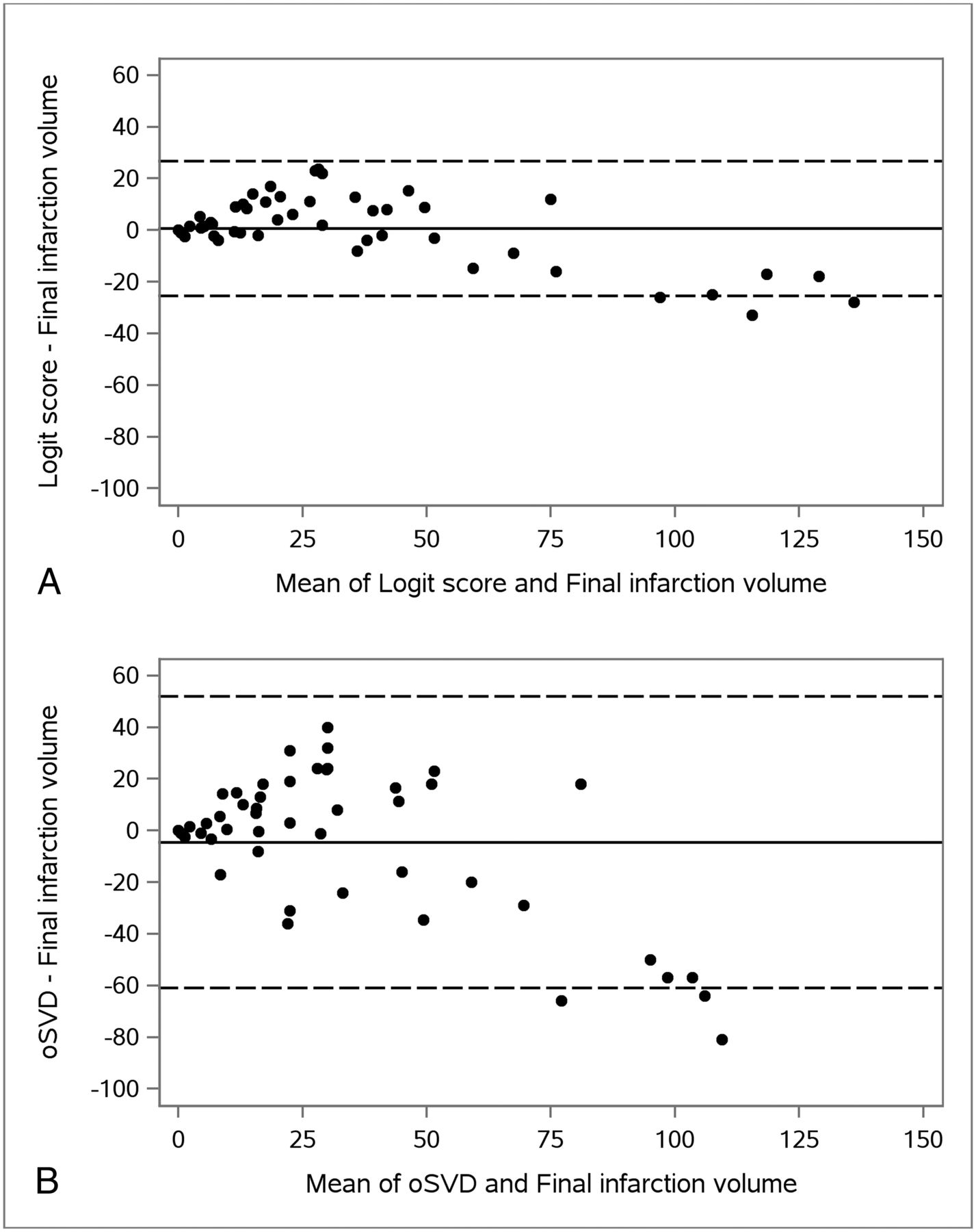
  • Fig 4.
    • Download figure
    • Open in new tab
    • Download powerpoint
    Fig 4.

    Bland-Altman plots of calculated final infarct volume and estimated ischemic core volume using the Bayesian-based logit score (A) and oSVD-rCBF <30% (B). Solid lines represent the mean differences. Dashed lines indicate 2 SDs above and below the mean differences. The limits of agreement were −25 to 27 for the Bayesian-based logit score and −61 to 52 for oSVD-CBF <30%.

Tables

  • Figures
    • View popup
    Table 1:

    Voxel-based univariate analysis of infarction and noninfarction voxelsa

    VariableInfarctionNoninfarctionP Valueb
    TTP32.00 (1.95)24.30 (1.19)<.001
    rCBF24.78 (2.81)48.43 (6.49)<.001
    rCBV2.81 (0.46)4.09 (0.56).02
    ATD3.58 (0.18)1.49 (0.16)<.001
    MTT6.58 (0.58)5.33 (0.15).01
    rCBFdiff−25.60 (10.93)3.20 (1.20).02
    rCBVdiff−1.15 (0.52)0.06 (0.06).03
    TTPdiff8.39 (0.41)0.25 (0.17)<.001
    ATDdiff2.15 (0.17)−0.16 (0.06)<.001
    MTTdiff2.19 (0.49)−0.12 (0.06)<.001
    • Note:—rCBV indicates relative CBV.

    • ↵a The values are presented as mean (SD). The SDs reported are the Huber-White (robust) standard errors. All units are in seconds, except for rCBF and rCBV, which are unitless.

    • ↵b P values are based on a linear regression model with the presence of infarct as the independent variable and imaging parameters as the outcome.

    • View popup
    Table 2:

    Optimal threshold, sensitivity, specificity, and accuracy for TTP, rCBF, ATDdiff, MTTdiff, and the final model in identifying infarcted voxels

    VariableThresholdSensitivitySpecificityAccuracyAUC
    TTP28.82 seconds65.3%77.9%76.5%0.76
    rCBF22.1060.0%72.9%71.5%0.73
    ATDdiff0.87 seconds68.1%80.2%78.9%0.80
    MTTdiff1.38 seconds56.2%74.5%72.5%0.69
    Final modela0.109b74.2%80.0%79.4%0.84
    • ↵a The final model consisted of TTP, rCBF, ATDdiff, and MTTdiff as the independent variables and the presence of infarct as the outcome.

    • ↵b Equation of the final model: logit(P) = −3.9170 + 0.0601 × TTP − 0.0095 × rCBF + 0.4629 × ATDdiff + 0.0989 × MTTdiff where logit(P) = estimated log odds of infarction for a given voxel. If logit(P) is greater than the optimal threshold of 0.109, the voxel is classified as infarct.

PreviousNext
Back to top

In this issue

American Journal of Neuroradiology: 40 (9)
American Journal of Neuroradiology
Vol. 40, Issue 9
1 Sep 2019
  • Table of Contents
  • Index by author
  • Complete Issue (PDF)
Advertisement
Print
Download PDF
Email Article

Thank you for your interest in spreading the word on American Journal of Neuroradiology.

NOTE: We only request your email address so that the person you are recommending the page to knows that you wanted them to see it, and that it is not junk mail. We do not capture any email address.

Enter multiple addresses on separate lines or separate them with commas.
Defining Ischemic Core in Acute Ischemic Stroke Using CT Perfusion: A Multiparametric Bayesian-Based Model
(Your Name) has sent you a message from American Journal of Neuroradiology
(Your Name) thought you would like to see the American Journal of Neuroradiology web site.
CAPTCHA
This question is for testing whether or not you are a human visitor and to prevent automated spam submissions.
Cite this article
K. Nael, E. Tadayon, D. Wheelwright, A. Metry, J.T. Fifi, S. Tuhrim, R.A. De Leacy, A.H. Doshi, H.L. Chang, J. Mocco
Defining Ischemic Core in Acute Ischemic Stroke Using CT Perfusion: A Multiparametric Bayesian-Based Model
American Journal of Neuroradiology Sep 2019, 40 (9) 1491-1497; DOI: 10.3174/ajnr.A6170

Citation Manager Formats

  • BibTeX
  • Bookends
  • EasyBib
  • EndNote (tagged)
  • EndNote 8 (xml)
  • Medlars
  • Mendeley
  • Papers
  • RefWorks Tagged
  • Ref Manager
  • RIS
  • Zotero
0 Responses
Respond to this article
Share
Bookmark this article
Defining Ischemic Core in Acute Ischemic Stroke Using CT Perfusion: A Multiparametric Bayesian-Based Model
K. Nael, E. Tadayon, D. Wheelwright, A. Metry, J.T. Fifi, S. Tuhrim, R.A. De Leacy, A.H. Doshi, H.L. Chang, J. Mocco
American Journal of Neuroradiology Sep 2019, 40 (9) 1491-1497; DOI: 10.3174/ajnr.A6170
del.icio.us logo Twitter logo Facebook logo Mendeley logo
  • Tweet Widget
  • Facebook Like
  • Google Plus One
Purchase

Jump to section

  • Article
    • Abstract
    • ABBREVIATIONS:
    • Materials and Methods
    • Results
    • Discussion
    • Conclusions
    • Footnotes
    • REFERENCES
  • Figures & Data
  • Info & Metrics
  • Responses
  • References
  • PDF

Related Articles

  • No related articles found.
  • PubMed
  • Google Scholar

Cited By...

  • Development and validation of a SCORing systEm for pre-thrombectomy diagnosis of IntraCranial Atherosclerotic Disease (Score-ICAD)
  • Development and validation of a SCORing systEm for pre-thrombectomy diagnosis of IntraCranial Atherosclerotic Disease (Score-ICAD)
  • Crossref
  • Google Scholar

This article has not yet been cited by articles in journals that are participating in Crossref Cited-by Linking.

More in this TOC Section

  • Diagnostic Neuroradiology of Monoclonal Antibodies
  • NCCT vs. MRI for Brain Atrophy in Acute Stroke
  • Clinical Outcomes After Chiari I Decompression
Show more Adult Brain

Similar Articles

Advertisement

Indexed Content

  • Current Issue
  • Accepted Manuscripts
  • Article Preview
  • Past Issues
  • Editorials
  • Editor's Choice
  • Fellows' Journal Club
  • Letters to the Editor
  • Video Articles

Cases

  • Case Collection
  • Archive - Case of the Week
  • Archive - Case of the Month
  • Archive - Classic Case

More from AJNR

  • Trainee Corner
  • Imaging Protocols
  • MRI Safety Corner
  • Book Reviews

Multimedia

  • AJNR Podcasts
  • AJNR Scantastics

Resources

  • Turnaround Time
  • Submit a Manuscript
  • Submit a Video Article
  • Submit an eLetter to the Editor/Response
  • Manuscript Submission Guidelines
  • Statistical Tips
  • Fast Publishing of Accepted Manuscripts
  • Graphical Abstract Preparation
  • Imaging Protocol Submission
  • Evidence-Based Medicine Level Guide
  • Publishing Checklists
  • Author Policies
  • Become a Reviewer/Academy of Reviewers
  • News and Updates

About Us

  • About AJNR
  • Editorial Board
  • Editorial Board Alumni
  • Alerts
  • Permissions
  • Not an AJNR Subscriber? Join Now
  • Advertise with Us
  • Librarian Resources
  • Feedback
  • Terms and Conditions
  • AJNR Editorial Board Alumni

American Society of Neuroradiology

  • Not an ASNR Member? Join Now

© 2025 by the American Society of Neuroradiology All rights, including for text and data mining, AI training, and similar technologies, are reserved.
Print ISSN: 0195-6108 Online ISSN: 1936-959X

Powered by HighWire