
Article Figures & Data
Figures
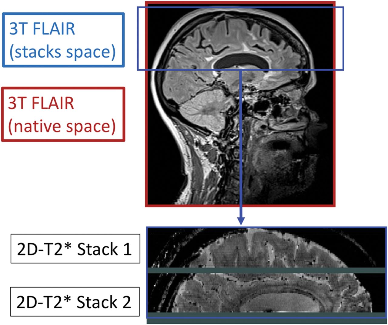
- Fig 1.
Multicontrast protocol volumetric area. Due to the sensitivity of 7T T2*-weighted acquisitions to magnetic susceptibility inhomogeneities present at the level of the posterior fossa and inferior temporal lobes, coverage of this interleaved, multislice acquisition is limited to the superior temporal lobes and above. Representation of the common volume of the brain is considered when comparing CL types detected on 3T and 7T.
- Fig 2.
Cortical lesion classification. 7T T2* MR imaging cortical lesion classification (Bø et al 2003).5
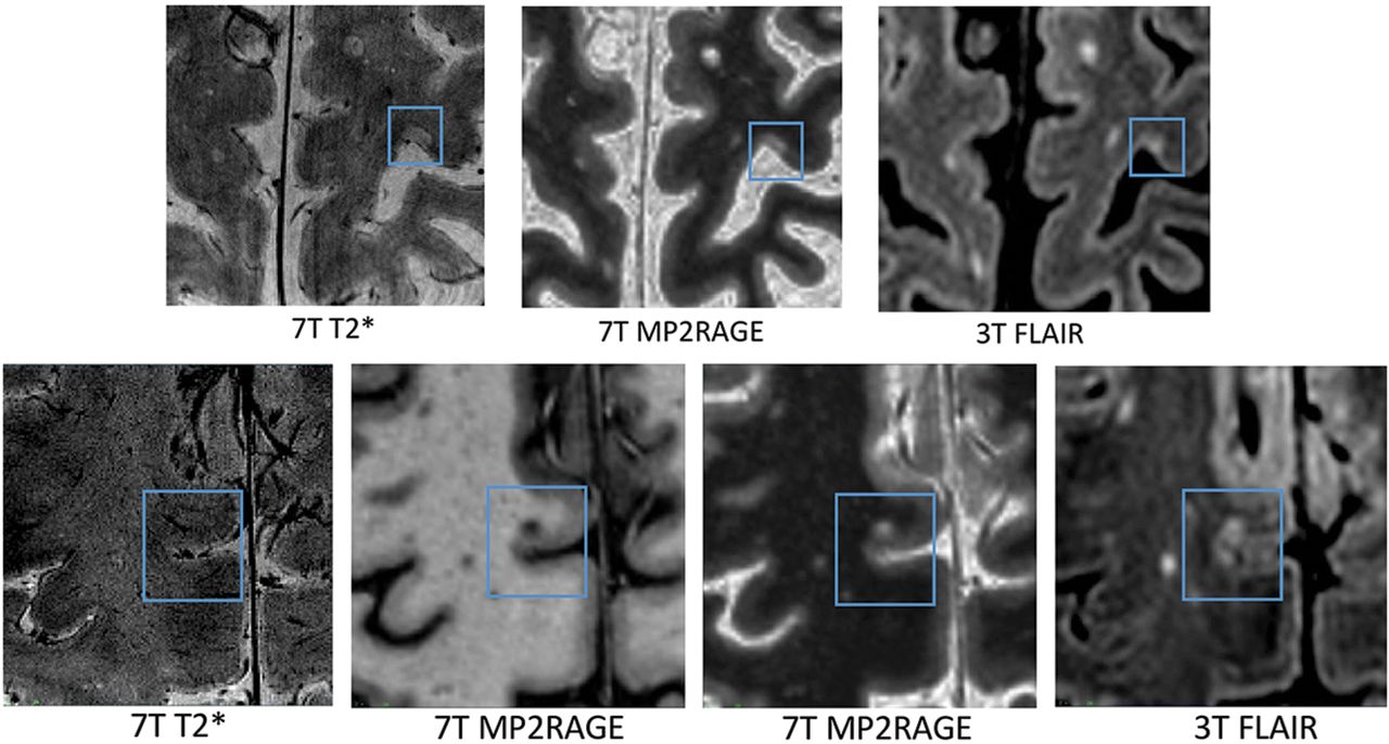
- Fig 3.
Example of CL lesions detected by 3T and 7T protocols. Example of 3T–7T common SP lesions (upper row) and 7T-negative LC lesions (lower row).
- Fig 4.
CL count correlations. Leukocortical and subpial lesion count correlations across scanners: 3T LC lesion counts and 7T LC lesion counts; 3T SP lesion counts and 7T SP lesion counts.


Tables
Patients with RRMS Patients with SPMS Significant Differences (P Value) Male/female 3:7 3:7 EDSS score Median, 1.5 Median, 4.5 .001a Range, 1–3 Range, 3–6.5 Age at Onset (yr) Mean, 34 Mean, 30 .15b SD, 8.2 SD, 6.5 Disease duration (yr) Mean, 12 Mean, 18 .04b SD, 7.8 SD, 6.8 White matter lesion volume (cm3) Median, 9.3 Median, 12.9 .18a Range, 0.8–44.3 Range, 3.7–50.4 3T T1WI 3T PDw/T2WI 3T FLAIR 3T MT On/Off 7T T1WI 7T T2* Sequence 3D-MP2RAGE 2D-dual echo TSE 3D-TSE 3D-GRE 3D-MP2RAGE 2D-GRE Orientation Sagittal Axial Sagittal Axial Sagittal Axial TR (ms) 5000 2350 6000 36 6000 1000 TE (ms) 2.98 22 356 3.86 2.7 22 87 TI (ms) 700 NA 2200 NA 800 NA 2500 2700 Flip angle 4° 120° 180° 10° 4° 55° 5° 5° Slices 176 120 176 192 224 60 (2 Stacks of 30) Voxel size (mm) 1 × 1 × 1 1 × 1 × 1.5 1 × 1 × 1 1 × 1 × 1 0.7 × 0.7 × 0.7 0.3 × 0.3 × 1 Scan time (min:sec) 8:22 5:26 8:44 8:10 10:14 8:26 (per stack) Note:—PDw indicates proton density-weighted; MT, magnetization transfer; GRE, gradient recalled-echo; NA, not applicable.


Jump to section
Related Articles
Cited By...
- Spinal Cord Versus Brain Imaging Biomarkers of Multiple Sclerosis Trajectory Combining 7T and 3T MRI
- Fluid and White Matter Suppression Contrasts MRI Improves Deep Learning Detection of Multiple Sclerosis Cortical Lesions
- Cortical lesions uniquely predict motor disability accrual and form rarely in the absence of new white matter lesions in multiple sclerosis
- Evaluation of the Statistical Detection of Change Algorithm for Screening Patients with MS with New Lesion Activity on Longitudinal Brain MRI
- Evaluation of the Statistical Detection of Change Algorithm for Screening Patients with MS with New Lesion Activity on Longitudinal Brain MRI
- Imaging cortical multiple sclerosis lesions with ultra-high field MRI
- Pathologic correlates of the magnetization transfer ratio in multiple sclerosis
- Conversion of Diffusely Abnormal White Matter to Focal Lesions is Linked to Progression in Secondary Progressive Multiple Sclerosis