
Article Figures & Data
Figures
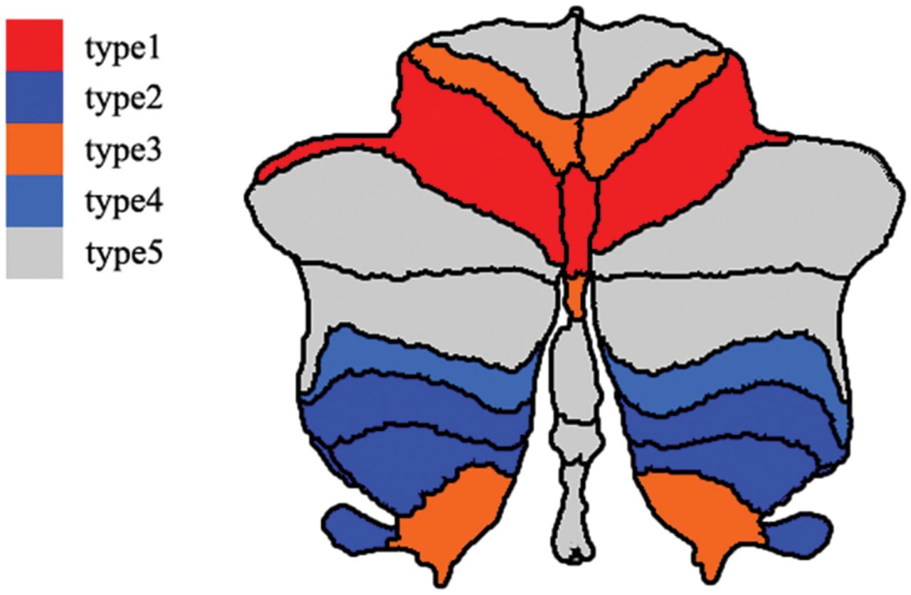
- FIGURE.
Spatial distribution of the occult cerebellar cortical damage in neuromyelitis optica spectrum disorder. Different colors represent the different types of normal-appearing cerebellar damage. Red denotes cerebellar subregions with occult damage related only to spinal lesions (type 1). Dark blue represents cerebellar subregions with occult damage related only to antibodies against aquaporin-4 antibody (type 2). Orange indicates cerebellar subregions with mixed occult damage mainly related to spinal lesions (type 3). Light blue suggests cerebellar subregions with mixed occult damage mainly related to the aquaporin-4 antibody (type 4). Gray denotes cerebellar subregions without occult damage (type 5).

Tables
Types of Occult Cerebellar Damage SCI vs HC AQP4-Ab (+) vs HC Without Controlling for AQP4-Ab Status Controlling for AQP4-Ab Status Without Controlling for LSCL Controlling for LSCL Type 1: only related to spinal lesions Significant or not Significant Nonsignificant Nonsignificant Type 2: only related to AQP4-Ab Nonsignificant Nonsignificant Significant or not Significant Type 3: mainly related to spinal lesions Significant Nonsignificant Nonsignificant Nonsignificant Type 4: mainly related to AQP4-Ab Nonsignificant Nonsignificant Significant Nonsignificant Type 5: no cerebellar damage Nonsignificant Nonsignificant Nonsignificant Nonsignificant Note:—+ indicates seropositive.
SCI SCN AQP4-Ab (+) AQP4-Ab (−) HC P1 P2 P3 P4 P5 P6 No. 17 19 27 9 20 NA NA NA NA NA NA Age (yr) 41.0 (20.5) 41.0 (15.0) 41.0 (12.0) 41.0 (28.0) 42.5 (7.5) .297b .428b .827b .336b .390a .565b Sex (M/F) 3:14 4:15 2:25 5:4 7:13 .383c .522c .797c .026c,d .422c .006c,d Education (yr) 9.0 (8.0) 12.0 (3.0) 12.0 (6.0) 12.0 (3.5) 9.0 (6.3) .517b .095b .076b .570b .594b .667b LSCL (VS) 3.0 (5.0) 0.0 (0.0) 2.0 (5.0) 0.0 (2.5) 0.0 (0.0) NA NA NA NA NA .218b AQP4-Ab (+/−) 15/2 14/5 27/0 0/9 0/0 NA NA .271c NA NA NA Note:—VS indicates vertebral segments; +, seropositive; −, seronegative; NA, not applicable.
↵a P1: comparison between the SCI and HC groups; P2: comparison between the SCN and HC groups; P3: comparison between the SCI and SCN groups; P4: comparison between the AQP4-Ab-seropositive and HC groups; P5: comparison between the AQP4-Ab-seronegative and HC groups; P6: comparison between the AQP4-Ab-seropositive and -seronegative groups. Variables are presented as median (interquartile range).
↵b The P value was obtained with the Mann-Whitney U test.
↵c The P value was obtained with the χ2 test.
↵d Significant.


