
Article Figures & Data
Figures
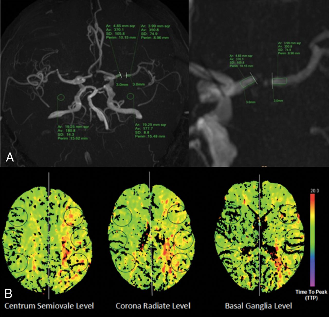
- Fig 1.
Illustration of fractional flow measurement and cerebral perfusion assessment. A, FF measurement: measurement of signal intensities proximal and distal to a stenosis located at the left MCA on a 3D-MIP image of TOF-MRA. B, Cerebral perfusion assessment: 6 circular ROIs were symmetrically drawn in the bilateral MCA territories at the centrum semiovale, corona radiata, and basal ganglia levels.
- Fig 2.
Color-coded perfusion parameter maps show the degree of cerebral hypoperfusion. A1–4, Normal perfusion. B1–4, Hypoperfuison on the stressed autoregulation compensated stage (ischemic penumbra without core infarction). C1–4, Hypoperfuison on the SAG-D stage (ischemic penumbra with core infarction). Short arrows show ischemic penumbra regions (increased TTP and MTT, normal CBF and CBV). The long arrows show core infarction regions (increased TTP and MTT, decreased CBF and CBV).
- Fig 3.
Receiver operating characteristic (ROC) curve of fractional flow to differentiate normal perfusion and hypoperfusion.
- Fig 4.
Correlations between fractional flow and the degree of cerebral hypoperfusion.


Tables
With Normal Perfusion on Brain CTP (n = 29) With Hypoperfusion on Brain CTP (n = 62) P Value Age (mean) (yr) 58.8 ± 9.2 61.5 ± 10.9 .237 Male sex (No.) (%) 16 (17.6%) 47 (51.6%) .261 Current smoker (No.) (%) 4 (4.4%) 30 (33.0%) .001 Hypertension history (No.) (%) 13 (14.3%) 38 (41.8%) .073 Diabetes mellitus history (No.) (%) 5 (5.5%) 19 (20.9%) .204 Blood pressure (mean) (mm Hg) SBP 139.6 ± 25.9 140.7 ± 24.1 .874 DBP 79.3 ± 14.3 83.1 ± 13.9 .283 Fasting blood glucose (mean) (mmol/L) 5.5 ± 1.1 6.4 ± 2.5 .069 Cholesterol (mean) (mmol/L) Total cholesterol 4.1 ± 0.8 4.0 ± 1.2 .814 HDL 1.1 ± 0.3 1.1 ± 0.4 .988 LDL 2.2 ± 0.7 2.2 ± 1.0 .954 Triglycerides 1.7 ± 0.9 1.5 ± 0.9 .494 Admitting diagnosis (No.) (%) Transient ischemic attack 10 (11.0%) 11 (12.1%) .916 Acute ischemic stroke 8 (8.8%) 26 (28.6%) .086 Chronic ischemic stroke 11 (12.1%) 25 (27.5%) .541 Time from onset to CT/MRI (mean) (day) 29.1 ± 19.6 24.0 ± 18.9 .408 Median (interquartile range 25%–75%) 24 (15.5∼46) 22 (11∼30) .536 Anatomic severity of lesion on CTA (No.) (%) Severe 10 (11.0%) 49 (53.8%) .004 Moderate 19 (20.9%) 13 (14.3%) .674 Stenosis length (mean) (mm) 4.7 ± 2.5 4.9 ± 2.7 .795 Collateral scoring on CTA 4.5 ± 0.4 3.7 ± 1.5 .002 Fractional flow on MRA 0.9 ± 0.2 0.7 ± 0.2 .000 Note:—HDL indicates high density lipoprotein; LDL, low density lipoprotein; SBP, systolic blood pressure; DBP, diastolic blood pressure.
- Table 2:
Association of participant characteristics with cerebral hypoperfusion in univariable and multivariable logistic regression models
Univariable Model Multivariable Model P Value OR (95% CI) P Value OR (95% CI) Age (per 1-yr increase) .542 0.98 (0.90–1.05) Male vs female .254 0.38 (0.07–2.01) Current smoker (Y vs N) .043 8.06 (1.06–60.71) .017 7.20 (1.43–36.40) Hypertension history (Y vs N) .072 3.99 (0.89–18.06) Diabetes mellitus history (Y vs N) .847 1.18 (0.21–6.51) SBP (per 1-mm Hg increase) .141 0.96 (0.91–1.01) DBP (per 1-mm Hg increase) .615 1.02 (0.94–1.16) Fasting blood glucose (per 1-mmol/L increase) .081 1.75 (0.93–3.14) HDL (per 1-mmol/L increase) .427 3.19 (0.14–75.54) LDL (per 1-mmol/L increase) .299 3.37 (0.19–60.23) Triglycerides (per 1-mmol/L increase) .180 0.53 (0.23–1.41) Total cholesterol (per 1-mmol/L increase) .300 0.17 (0.07–4.72) Time from onset to CT/MRI (per 1-day lapse) .451 0.98 (0.95–1.03) Anatomic severity of ICAS on CTA (severe vs moderate) .274 0.59 (0.48–12.86) Stenosis length (per 1-mm increase) .262 0.54 (0.45–13.06) Collateral score on CTA (per 1-score increase) .049 0.28 (0.06–1.22) .041 0.26 (0.07–0.97) Fractional flow on MRA (≤0.90 vs >0.90) .046 7.28 (1.03–51.35) .027 3.68 (1.63–11.62) Note:—Y indicates yes; N, no; HDL, high density lipoprotein; LDL, low density lipoprotein; SBP, systolic blood pressure; DBP, diastolic blood pressure.
Groups Collateral Score Current Smoking All 0∼3 (n = 26; 28.6%) 4∼5 (n = 65; 71.4%) Yes (n = 34; 37.4%) No (n = 57; 62.6%) (n = 91; 100%) r P r P r P r P r P rTTP −0.485 .001 −0.250 .004 −0.509 .018 −0.502 .017 −0.517 .000 rMTT −0.414 .001 −0.361 .002 −0.454 .019 −0.507 .011 −0.569 .000 rCBF −0.103 .739 0.079 .592 0.270 .046 0.203 .167 0.213 .043 rCBV 0.033 .914 0.004 .977 −0.172 .400 0.128 .384 0.155 .143 Note:—rCBV indicates relative CBV; rCBF, relative CBF.


