
Article Figures & Data
Figures
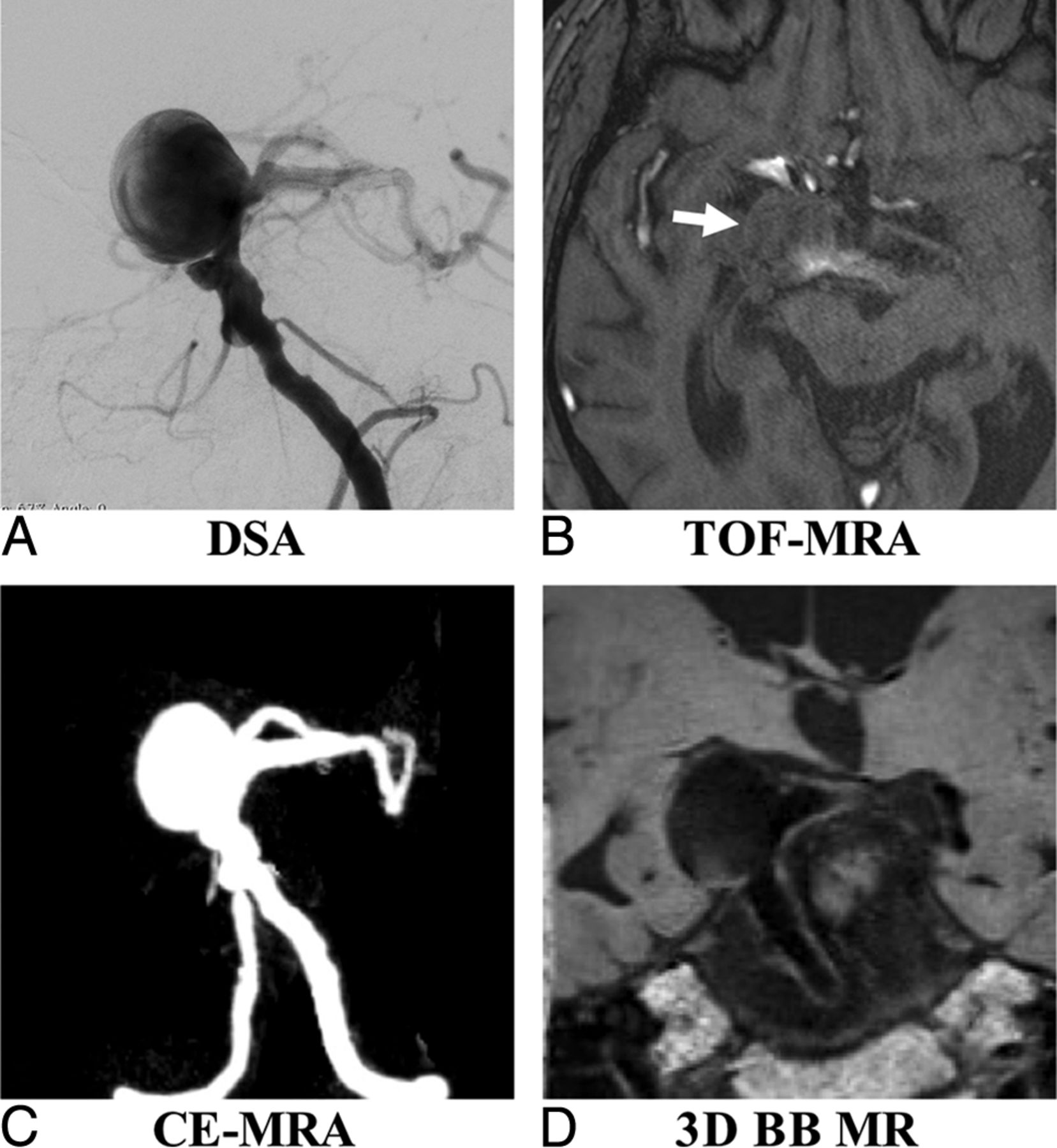
- Fig 1.
A 61-year-old woman with a right posterior communicating artery aneurysm on DSA (A), TOF-MRA (B), CE-MRA (C), and 3D-BB MR imaging (D). Aneurysm height (red line), width (yellow line), and neck (white line) measurements are demonstrated in A.
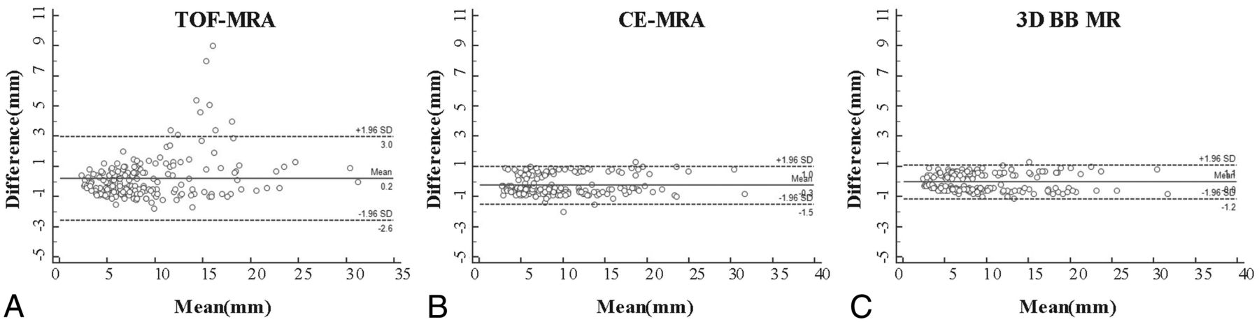
- Fig 2.
Bland-Altman plots of aneurysm measurements of TOF-MRA (A), CE-MRA (B), and 3D-BB MR imaging (C) versus 3DRA as the reference standard. Solid horizontal lines define the reference standard, and upper and lower dashed lines define the LOA.
- Fig 3.
A 53-year-old man with multiple basilar artery aneurysms on DSA (A). The aneurysm shows isointensity on TOF-MRA (B) because of the slow flow (arrow). The aneurysm sac and neck were integrally visualized on CE-MRA (C) and 3D-BB MR imaging (D).
- Fig 4.
A 63-year-old woman with a right middle cerebral artery aneurysm on DSA (A). 3D-BB SPACE (D) can clearly visualize the sac and intraluminal thrombus of the aneurysm, which is superior to DSA (A), TOF-MRA (B), and CE-MRA (C).


Tables
- Table 1:
Comparison of aneurysm height, width, and neck measurements among MR and DSA imaging modalitiesa
TOF-MRA CE-MRA 3D-BB MRI 3DRA P Value Height (mm) 7.7 (5.5–11.1) 8.0 (6.0–12.8) 7.8 (5.4–12.6) 7.7 (5.5–12.6) .918 Width (mm) 7.5 (5.8–11.5) 8.4 (5.8–14.0) 7.8 (5.5–13.9) 8.1 (5.9–13.3) .957 Neck (mm) 6.8 (5.3–10.4) 6.7 (5.2–10.6) 7.0 (4.8–10.2) 6.9 (4.6–9.8) .774 ↵a Data are median (interquartile range).
Mean (mm) SD CV (100%) Bias LOA ICC Height TOF-MRA 9.21 1.50 15.43 −0.55 (−3.49–2.39) 0.96 (0.93–0.98) CE-MRA 9.90 0.62 6.32 0.14 (−1.08–1.36) 0.98 (0.97–0.99) 3D-BB MRI 9.73 0.59 6.04 −0.03 (−1.19–1.13) 0.99 (0.99–0.99) Width TOF-MRA 9.53 1.56 15.78 −0.33 (−2.73–3.39) 0.96 (0.94–0.97) CE-MRA 10.12 0.61 6.18 0.26 (−0.94–1.46) 0.99 (0.98–0.99) 3D-BB MRI 9.96 0.58 5.87 0.10 (−1.04–1.24) 0.99 (0.99–0.99) Neck TOF-MRA 8.00 0.98 12.73 0.31 (−1.61–2.23) 0.96 (0.93–0.98) CE-MRA 8.08 0.68 8.81 0.39 (−0.94–1.72) 0.97 (0.95–0.99) 3D-BB MRI 7.71 0.54 7.04 0.02 (−1.04–1.08) 0.98 (0.98–0.99) Overall TOF-MRA 8.91 1.41 15.54 −0.19 (−2.95–2.57) 0.96 (0.95–0.97) CE-MRA 9.37 0.64 7.03 0.26 (−0.99–1.51) 0.98 (0.98–0.99) 3D-BB MRI 9.14 0.57 6.26 0.03 (−1.08–1.15) 0.99 (0.99–0.99) Aneurysm Growth TOF-MRA (CV = 15.54%) CE-MRA (CV = 7.03%) SPACE (CV = 6.26%) 5% 406 84 66 10% 102 21 17 20% 26 6 5 CV (100%) Bias LOA ICC Intrareader agreement Height TOF-MRA 7.02 0.19 (−0.87–1.25) 0.99 (0.98–0.99) CE-MRA 6.71 0.14 (−1.09–1.37) 0.99 (0.98–0.99) 3D-BB MRI 6.37 −0.08 (−1.18–1.02) 0.99 (0.98–0.99) 3DRA 6.87 0.14 (−1.04–1.32) 0.99 (0.98–0.99) Width TOF-MRA 6.94 0.06 (−0.99–1.12) 0.99 (0.98–0.99) CE-MRA 6.45 0.10 (−1.13–1.33) 0.99 (0.98–0.99) 3D-BB MRI 6.31 0.18 (−0.92–1.28) 0.99 (0.98–0.99) 3DRA 6.25 0.11 (−1.00–1.23) 0.99 (0.98–0.99) Neck TOF-MRA 7.29 0.18 (−1.04–1.39) 0.98 (0.98–0.99) CE-MRA 7.05 0.13 (−0.93–1.19) 0.99 (0.98–0.99) 3D-BB MRI 6.72 0.14 (−0.90–1.18) 0.99 (0.98–0.99) 3DRA 6.98 −0.08 (−1.17–1.02) 0.99 (0.98–0.99) Overall TOF-MRA 7.08 0.14 (−0.78–1.06) 0.99 (0.98–0.99) CE-MRA 6.78 0.12 (−1.06–1.30) 0.99 (0.98–0.99) 3D-BB MRI 6.51 0.08 (−0.96–1.12) 0.99 (0.98–0.99) 3DRA 6.75 0.06 (−1.04–1.16) 0.99 (0.98–0.99) Interreader agreement Height TOF-MRA 8.12 0.02 (−1.45–1.49) 0.98 (0.98–0.99) CE-MRA 8.19 0.23 (−1.33–1.80) 0.98 (0.98–0.99) 3D-BB MRI 8.03 0.03 (−1.50–1.56) 0.98 (0.98–0.99) 3DRA 7.87 −0.07 (−1.58–1.44) 0.98 (0.98–0.99) Width TOF-MRA 8.03 −0.03 (−1.50–1.44) 0.98 (0.98–0.99) CE-MRA 7.55 0.17 (−1.30–1.64) 0.99 (0.98–0.99) 3D-BB MRI 8.37 0.03 (−1.61–1.67) 0.98 (0.98–0.99) 3DRA 7.15 −0.13 (−1.54–1.28) 0.99 (0.98–0.99) Neck TOF-MRA 8.96 0.18 (−0.74–1.10) 0.97 (0.95–0.98) CE-MRA 8.81 0.07 −0.81–1.07) 0.98 (0.97–0.99) 3D-BB MRI 8.95 0.14 (−0.72–1.00) 0.98 (0.97–0.99) 3DRA 8.82 0.10 (−0.82–0.98) 0.98 (0.97–0.99) Overall TOF-MRA 8.44 0.06 (−0.78–1.06) 0.98 (0.98–0.99) CE-MRA 8.16 0.16 (−0.74–0.98) 0.98 (0.98–0.99) 3D-BB MRI 8.43 0.07 (−0.63–0.93) 0.98 (0.98–0.99) 3DRA 8.15 −0.03 (−0.72–1.00) 0.98 (0.98–0.99)


Jump to section
Related Articles
Cited By...
- No citing articles found.