Advertisement

Main menu

  • Home
  • Content
    • Current Issue
    • Accepted Manuscripts
    • Article Preview
    • Past Issue Archive
    • Video Articles
    • AJNR Case Collection
    • Case of the Week Archive
    • Case of the Month Archive
    • Classic Case Archive
  • Special Collections
    • AJNR Awards
    • Low-Field MRI
    • Alzheimer Disease
    • ASNR Foundation Special Collection
    • Photon-Counting CT
    • View All
  • Multimedia
    • AJNR Podcasts
    • AJNR SCANtastic
    • Trainee Corner
    • MRI Safety Corner
    • Imaging Protocols
  • For Authors
    • Submit a Manuscript
    • Submit a Video Article
    • Submit an eLetter to the Editor/Response
    • Manuscript Submission Guidelines
    • Statistical Tips
    • Fast Publishing of Accepted Manuscripts
    • Graphical Abstract Preparation
    • Imaging Protocol Submission
    • Author Policies
  • About Us
    • About AJNR
    • Editorial Board
    • Editorial Board Alumni
  • More
    • Become a Reviewer/Academy of Reviewers
    • Subscribers
    • Permissions
    • Alerts
    • Feedback
    • Advertisers
    • ASNR Home

User menu

  • Alerts
  • Log in

Search

  • Advanced search
American Journal of Neuroradiology

American Journal of Neuroradiology

ASHNR American Society of Functional Neuroradiology ASHNR American Society of Pediatric Neuroradiology ASSR
  • Alerts
  • Log in

Advanced Search

  • Home
  • Content
    • Current Issue
    • Accepted Manuscripts
    • Article Preview
    • Past Issue Archive
    • Video Articles
    • AJNR Case Collection
    • Case of the Week Archive
    • Case of the Month Archive
    • Classic Case Archive
  • Special Collections
    • AJNR Awards
    • Low-Field MRI
    • Alzheimer Disease
    • ASNR Foundation Special Collection
    • Photon-Counting CT
    • View All
  • Multimedia
    • AJNR Podcasts
    • AJNR SCANtastic
    • Trainee Corner
    • MRI Safety Corner
    • Imaging Protocols
  • For Authors
    • Submit a Manuscript
    • Submit a Video Article
    • Submit an eLetter to the Editor/Response
    • Manuscript Submission Guidelines
    • Statistical Tips
    • Fast Publishing of Accepted Manuscripts
    • Graphical Abstract Preparation
    • Imaging Protocol Submission
    • Author Policies
  • About Us
    • About AJNR
    • Editorial Board
    • Editorial Board Alumni
  • More
    • Become a Reviewer/Academy of Reviewers
    • Subscribers
    • Permissions
    • Alerts
    • Feedback
    • Advertisers
    • ASNR Home
  • Follow AJNR on Twitter
  • Visit AJNR on Facebook
  • Follow AJNR on Instagram
  • Join AJNR on LinkedIn
  • RSS Feeds

AJNR Awards, New Junior Editors, and more. Read the latest AJNR updates

Research ArticlePediatrics

Pediatric Atypical Teratoid/Rhabdoid Tumors of the Brain: Identification of Metabolic Subgroups Using In Vivo 1H-MR Spectroscopy

B. Tamrazi, S. Venneti, A. Margol, D. Hawes, S.Y. Cen, M. Nelson, A. Judkins, J. Biegel and S. Blüml
American Journal of Neuroradiology May 2019, 40 (5) 872-877; DOI: https://doi.org/10.3174/ajnr.A6024
B. Tamrazi
aFrom the Departments of Radiology (B.T., M.N., S.B.)
  • Find this author on Google Scholar
  • Find this author on PubMed
  • Search for this author on this site
  • ORCID record for B. Tamrazi
S. Venneti
dDepartment of Pathology (S.V.), University of Michigan, Ann Arbor, Michigan
  • Find this author on Google Scholar
  • Find this author on PubMed
  • Search for this author on this site
  • ORCID record for S. Venneti
A. Margol
bPediatrics (A.M.) and Division of Hematology Oncology
  • Find this author on Google Scholar
  • Find this author on PubMed
  • Search for this author on this site
  • ORCID record for A. Margol
D. Hawes
cPathology (D.H., A.J., J.B.), Children's Hospital Los Angeles, Los Angeles, California
  • Find this author on Google Scholar
  • Find this author on PubMed
  • Search for this author on this site
  • ORCID record for D. Hawes
S.Y. Cen
eDepartment of Radiology and Neurology (S.Y.C.), University of Southern California, Los Angeles, California
  • Find this author on Google Scholar
  • Find this author on PubMed
  • Search for this author on this site
  • ORCID record for S.Y. Cen
M. Nelson
aFrom the Departments of Radiology (B.T., M.N., S.B.)
  • Find this author on Google Scholar
  • Find this author on PubMed
  • Search for this author on this site
  • ORCID record for M. Nelson
A. Judkins
cPathology (D.H., A.J., J.B.), Children's Hospital Los Angeles, Los Angeles, California
  • Find this author on Google Scholar
  • Find this author on PubMed
  • Search for this author on this site
  • ORCID record for A. Judkins
J. Biegel
cPathology (D.H., A.J., J.B.), Children's Hospital Los Angeles, Los Angeles, California
  • Find this author on Google Scholar
  • Find this author on PubMed
  • Search for this author on this site
  • ORCID record for J. Biegel
S. Blüml
aFrom the Departments of Radiology (B.T., M.N., S.B.)
fRudi Schulte Research Institute (S.B.), Santa Barbara, California.
  • Find this author on Google Scholar
  • Find this author on PubMed
  • Search for this author on this site
  • ORCID record for S. Blüml
  • Article
  • Figures & Data
  • Info & Metrics
  • Responses
  • References
  • PDF
Loading

Article Figures & Data

Figures

  • Tables
  • Fig 1.
    • Download figure
    • Open in new tab
    • Download powerpoint
    Fig 1.

    T2-weighted MR imaging and MR spectra of supratentorial AT/RTs with ASCL1 expression (upper row), a posterior fossa AT/RT with ASCL1 expression (middle row), and posterior fossa AT/RT without ASCL1 expression (lower row). Cr, Cho, and mI are readily detectable in the spectra of supratentorial and posterior fossa ASCL1-expressing AT/RTs, whereas only Cho is clearly detectable in the ASCL1-nonexpressing posterior fossa AT/RT. All spectra show prominent signal from lipids and lactate (Lac). Shown are the unprocessed data (thin black lines) with the superimposed fit (thick gray lines) used for quantification. The “box” seen in the images represents the voxel placement.
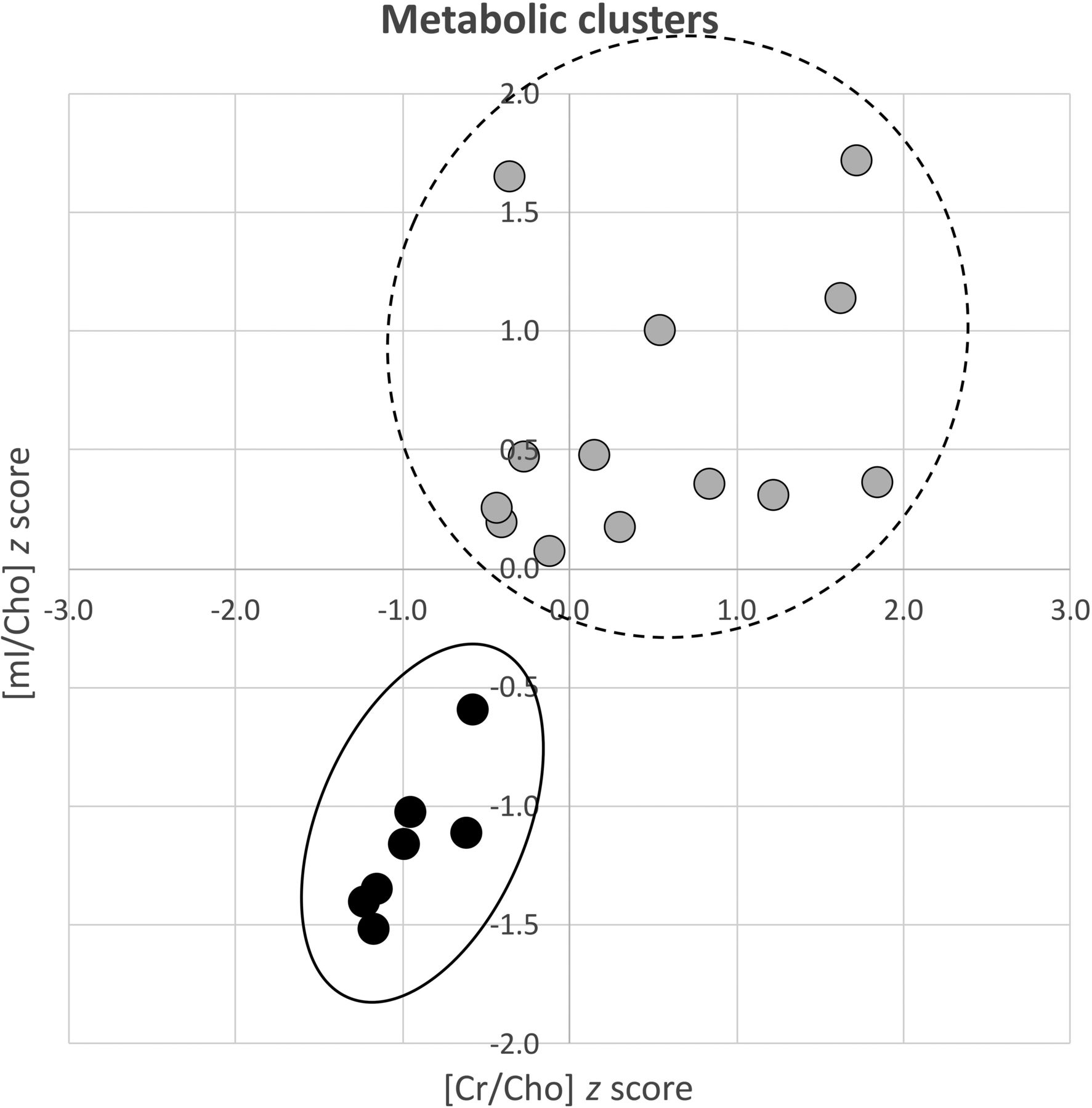
  • Fig 2.
    • Download figure
    • Open in new tab
    • Download powerpoint
    Fig 2.

    K-means cluster analysis graph demonstrating the metabolic clusters based on Cr/Cho and mI/Cho ratios. Ratios were transformed to z scores for better illustration. Findings suggest that a subgroup of AT/RTs (solid line) are homogeneously characterized by very low Cr and mI levels (relative to Cho) and are well-separated from other AT/RTs. It is, however, unclear whether the remaining larger cluster of AT/RTs (dashed line) forms a single group or multiple separate groups as suggested by the molecular data.

Tables

  • Figures
    • View popup
    Table 1:

    Patient demographics and clinical and biologic lesion characterization

    Age (yr)SexOverall Survival (mo)LocationLMASCL1CKBGermline Mutation
    0.15F0.5aPosterior fossa0Negative5.05
    0.32F17.0Posterior fossa0Positive6.31Yes
    0.39M8.2bPosterior fossa1Positive
    0.64M7.0Parietal lobe0Negative5.10
    0.67M0.5aPosterior fossa1Positive
    0.68M5.1Intraventricular0ND
    0.92F0.5aPosterior fossa1PositiveYes
    0.95M71.9bPosterior fossa0ND
    1.09M13.0Posterior fossa0Negative5.31
    1.14M7.0Temporal lobe0Positive6.05Yes
    1.15M59.0Posterior fossa0Negative6.31
    1.42F9.0Posterior fossa1ND
    1.54M32.0Temporal lobe0Positive6.53Yes
    1.55M63.2bPosterior fossa0Negative
    1.73M13.0Pineal0Positive
    1.84F18.0Frontotemporal0Negative5.21
    1.96FNATemporal lobe1ND
    2.86M18.0Frontotemporal1Positive6.61
    7.43F40.0Frontotemporal0Positive6.42
    13.72M40.0Intraventricular0Positive5.71
    • Note:—NA indicates patient lost to follow up; LM, leptomeningeal disease present (1) versus absent (0) at presentation; ND, not determined.

    • ↵a Patients who died from surgical/other complications with no MR imaging or clinical evidence of progressive tumor.

    • ↵b Patients who were still alive at the time of the completion of this study.

    • View popup
    Table 2:

    Features of AT/RTs with expression of ASCL1a

    ASCL1 Pos.ASCL1 Neg.P Value
    Age (mean) (yr)3.4 ± 4.51.8 ± 0.8NS
    Sex (M/F)6:35:2NS
    Location PF vs not PF3 vs 64 vs 3NS
    Crb3.4 ± 1.11.8 ± 0.8<.05
    Chob3.2 ± 0.83.8 ± 2.1NS
    mIb9.0 ± 1.54.7 ± 3.6<.05
    Lacb5.0 ± 3.92.6 ± 2.6NS
    Lipidsb44 ± 2080 ± 30NSc
    CKB (mean)6.3 ± 0.35.4 ± 0.5<0.05
    Months of follow-up (mean)22 ± 1432 ± 27NS
    • Note:—NS indicates not significant; Pos., positive; Neg., negative; PF, posterior fossa.

    • ↵a Tissue from 16 of 20 AT/RTs was available for testing of ASCL1 status.

    • ↵b Means in IU.

    • ↵c Approaching significance with P = .07.

PreviousNext
Back to top

In this issue

American Journal of Neuroradiology: 40 (5)
American Journal of Neuroradiology
Vol. 40, Issue 5
1 May 2019
  • Table of Contents
  • Index by author
  • Complete Issue (PDF)
Advertisement
Print
Download PDF
Email Article

Thank you for your interest in spreading the word on American Journal of Neuroradiology.

NOTE: We only request your email address so that the person you are recommending the page to knows that you wanted them to see it, and that it is not junk mail. We do not capture any email address.

Enter multiple addresses on separate lines or separate them with commas.
Pediatric Atypical Teratoid/Rhabdoid Tumors of the Brain: Identification of Metabolic Subgroups Using In Vivo 1H-MR Spectroscopy
(Your Name) has sent you a message from American Journal of Neuroradiology
(Your Name) thought you would like to see the American Journal of Neuroradiology web site.
CAPTCHA
This question is for testing whether or not you are a human visitor and to prevent automated spam submissions.
Cite this article
B. Tamrazi, S. Venneti, A. Margol, D. Hawes, S.Y. Cen, M. Nelson, A. Judkins, J. Biegel, S. Blüml
Pediatric Atypical Teratoid/Rhabdoid Tumors of the Brain: Identification of Metabolic Subgroups Using In Vivo 1H-MR Spectroscopy
American Journal of Neuroradiology May 2019, 40 (5) 872-877; DOI: 10.3174/ajnr.A6024

Citation Manager Formats

  • BibTeX
  • Bookends
  • EasyBib
  • EndNote (tagged)
  • EndNote 8 (xml)
  • Medlars
  • Mendeley
  • Papers
  • RefWorks Tagged
  • Ref Manager
  • RIS
  • Zotero
0 Responses
Respond to this article
Share
Bookmark this article
Pediatric Atypical Teratoid/Rhabdoid Tumors of the Brain: Identification of Metabolic Subgroups Using In Vivo 1H-MR Spectroscopy
B. Tamrazi, S. Venneti, A. Margol, D. Hawes, S.Y. Cen, M. Nelson, A. Judkins, J. Biegel, S. Blüml
American Journal of Neuroradiology May 2019, 40 (5) 872-877; DOI: 10.3174/ajnr.A6024
del.icio.us logo Twitter logo Facebook logo Mendeley logo
  • Tweet Widget
  • Facebook Like
  • Google Plus One
Purchase

Jump to section

  • Article
    • Abstract
    • ABBREVIATIONS:
    • Materials and Methods
    • Results
    • Discussion
    • Conclusions
    • Footnotes
    • REFERENCES
  • Figures & Data
  • Info & Metrics
  • Responses
  • References
  • PDF

Related Articles

  • No related articles found.
  • PubMed
  • Google Scholar

Cited By...

  • Identifying Reproducible Transcription Regulator Coexpression Patterns with Single Cell Transcriptomics
  • Crossref
  • Google Scholar

This article has not yet been cited by articles in journals that are participating in Crossref Cited-by Linking.

More in this TOC Section

  • Early Ultrasonic Monitoring of Brain Growth and Later Neurodevelopmental Outcome in Very Preterm Infants
  • Diagnostic Value of Sylvian Fissure Hyperechogenicity in Fetal SAH
  • Feasibility and Added Value of Fetal DTI Tractography in the Evaluation of an Isolated Short Corpus Callosum: Preliminary Results
Show more Pediatrics

Similar Articles

Advertisement

Indexed Content

  • Current Issue
  • Accepted Manuscripts
  • Article Preview
  • Past Issues
  • Editorials
  • Editor's Choice
  • Fellows' Journal Club
  • Letters to the Editor
  • Video Articles

Cases

  • Case Collection
  • Archive - Case of the Week
  • Archive - Case of the Month
  • Archive - Classic Case

More from AJNR

  • Trainee Corner
  • Imaging Protocols
  • MRI Safety Corner
  • Book Reviews

Multimedia

  • AJNR Podcasts
  • AJNR Scantastics

Resources

  • Turnaround Time
  • Submit a Manuscript
  • Submit a Video Article
  • Submit an eLetter to the Editor/Response
  • Manuscript Submission Guidelines
  • Statistical Tips
  • Fast Publishing of Accepted Manuscripts
  • Graphical Abstract Preparation
  • Imaging Protocol Submission
  • Evidence-Based Medicine Level Guide
  • Publishing Checklists
  • Author Policies
  • Become a Reviewer/Academy of Reviewers
  • News and Updates

About Us

  • About AJNR
  • Editorial Board
  • Editorial Board Alumni
  • Alerts
  • Permissions
  • Not an AJNR Subscriber? Join Now
  • Advertise with Us
  • Librarian Resources
  • Feedback
  • Terms and Conditions
  • AJNR Editorial Board Alumni

American Society of Neuroradiology

  • Not an ASNR Member? Join Now

© 2025 by the American Society of Neuroradiology All rights, including for text and data mining, AI training, and similar technologies, are reserved.
Print ISSN: 0195-6108 Online ISSN: 1936-959X

Powered by HighWire