Advertisement

Main menu

  • Home
  • Content
    • Current Issue
    • Accepted Manuscripts
    • Article Preview
    • Past Issue Archive
    • Video Articles
    • AJNR Case Collection
    • Case of the Week Archive
    • Case of the Month Archive
    • Classic Case Archive
  • Special Collections
    • AJNR Awards
    • Low-Field MRI
    • Alzheimer Disease
    • ASNR Foundation Special Collection
    • Photon-Counting CT
    • View All
  • Multimedia
    • AJNR Podcasts
    • AJNR SCANtastic
    • Trainee Corner
    • MRI Safety Corner
    • Imaging Protocols
  • For Authors
    • Submit a Manuscript
    • Submit a Video Article
    • Submit an eLetter to the Editor/Response
    • Manuscript Submission Guidelines
    • Statistical Tips
    • Fast Publishing of Accepted Manuscripts
    • Graphical Abstract Preparation
    • Imaging Protocol Submission
    • Author Policies
  • About Us
    • About AJNR
    • Editorial Board
    • Editorial Board Alumni
  • More
    • Become a Reviewer/Academy of Reviewers
    • Subscribers
    • Permissions
    • Alerts
    • Feedback
    • Advertisers
    • ASNR Home

User menu

  • Alerts
  • Log in

Search

  • Advanced search
American Journal of Neuroradiology

American Journal of Neuroradiology

ASHNR American Society of Functional Neuroradiology ASHNR American Society of Pediatric Neuroradiology ASSR
  • Alerts
  • Log in

Advanced Search

  • Home
  • Content
    • Current Issue
    • Accepted Manuscripts
    • Article Preview
    • Past Issue Archive
    • Video Articles
    • AJNR Case Collection
    • Case of the Week Archive
    • Case of the Month Archive
    • Classic Case Archive
  • Special Collections
    • AJNR Awards
    • Low-Field MRI
    • Alzheimer Disease
    • ASNR Foundation Special Collection
    • Photon-Counting CT
    • View All
  • Multimedia
    • AJNR Podcasts
    • AJNR SCANtastic
    • Trainee Corner
    • MRI Safety Corner
    • Imaging Protocols
  • For Authors
    • Submit a Manuscript
    • Submit a Video Article
    • Submit an eLetter to the Editor/Response
    • Manuscript Submission Guidelines
    • Statistical Tips
    • Fast Publishing of Accepted Manuscripts
    • Graphical Abstract Preparation
    • Imaging Protocol Submission
    • Author Policies
  • About Us
    • About AJNR
    • Editorial Board
    • Editorial Board Alumni
  • More
    • Become a Reviewer/Academy of Reviewers
    • Subscribers
    • Permissions
    • Alerts
    • Feedback
    • Advertisers
    • ASNR Home
  • Follow AJNR on Twitter
  • Visit AJNR on Facebook
  • Follow AJNR on Instagram
  • Join AJNR on LinkedIn
  • RSS Feeds

AJNR Awards, New Junior Editors, and more. Read the latest AJNR updates

Research ArticleHead & Neck

The Black Turbinate Sign, A Potential Diagnostic Pitfall: Evaluation of the Normal Enhancement Patterns of the Nasal Turbinates

Q. Han and E.J. Escott
American Journal of Neuroradiology May 2019, 40 (5) 855-861; DOI: https://doi.org/10.3174/ajnr.A6037
Q. Han
aFrom the Departments of Radiology (Q.H.)
  • Find this author on Google Scholar
  • Find this author on PubMed
  • Search for this author on this site
  • ORCID record for Q. Han
E.J. Escott
bRadiology and Otolaryngology-Head and Neck Surgery (E.J.E.), University of Kentucky, Lexington, Kentucky.
  • Find this author on Google Scholar
  • Find this author on PubMed
  • Search for this author on this site
  • ORCID record for E.J. Escott
  • Article
  • Figures & Data
  • Supplemental
  • Info & Metrics
  • Responses
  • References
  • PDF
Loading

Article Figures & Data

Figures

  • Tables
  • Fig 1.
    • Download figure
    • Open in new tab
    • Download powerpoint
    Fig 1.

    A 27-year-old woman with relapsed pre-B-cell acute lymphoblastic leukemia was admitted for chemotherapy. She had an MR imaging examination due to headache. Axial (A) and coronal (B) T1-weighted postcontrast images with fat saturation showed nonenhancement centered in the left middle turbinate (long arrows) with irregular and invasive borders extending into the left inferior turbinate (short arrow). The patient had tissue-proved invasive fungal rhinosinusitis and went through a series of 12 aggressive surgical debridements and eventually died.
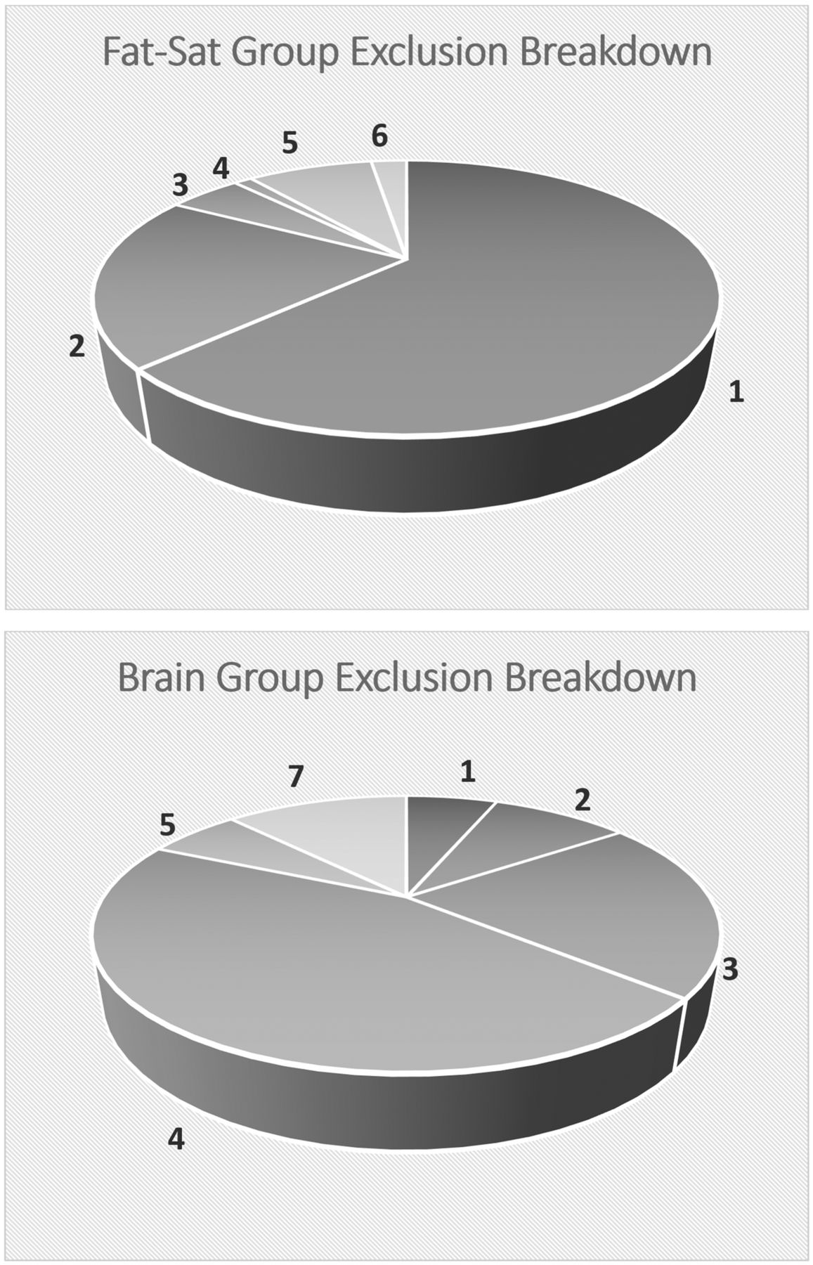
  • Fig 2.
    • Download figure
    • Open in new tab
    • Download powerpoint
    Fig 2.

    Breakdown of excluded cases: 1) insufficient inclusion of nasal turbinates, 2) significant postoperative or posttreatment changes involving the nasal cavity, 3) inappropriate scan sequence, 4) specific protocols, 5) significant motion or other artifacts, 6) significant nasal mass, and 7) scans from outside or satellite hospitals.

  • Fig 3.
    • Download figure
    • Open in new tab
    • Download powerpoint
    Fig 3.

    A 24-year-old immunocompetent woman with a history of a gestational trophoblastic neoplasm who had a brain MR imaging for a metastatic work-up. Coronal (A) and sagittal (B) postcontrast T1-weighted images show nonenhancement of the posterior aspect of the left inferior turbinate (long arrows) with thin rim enhancement (short arrows). The posterior aspect of the inferior turbinate is a common location of a black turbinate in immunocompetent patients.

  • Fig 4.
    • Download figure
    • Open in new tab
    • Download powerpoint
    Fig 4.

    A 14-year-old immunocompetent adolescent girl with right-sided blurred vision, who had orbit and brain MR imaging for a multiple sclerosis work-up. The postcontrast images for the orbit MR imaging were obtained before those for the brain MR imaging. Axial (A) and coronal (B) postcontrast T1-weighted images with fat saturation show nonenhancement most pronounced in the posterior aspects of both inferior turbinates (long arrows) with thin rim enhancement (short arrows) and scattered nonenhancing foci in the right middle turbinate (arrowhead). The inferior turbinate, especially the posterior portion, is a common location of a BT in immunocompetent patients. The sagittal postcontrast image from the brain MR imaging (C), which is the last postcontrast sequence available, shows that the BT of the right interior turbinate is almost completely resolved and has transitioned into mildly scattered nonenhancing foci (curved arrow).

  • Fig 5.
    • Download figure
    • Open in new tab
    • Download powerpoint
    Fig 5.

    Axial postcontrast T1-weighted images with fat saturation from 3 different patients. A, A 43-year-old woman shows extensive nonenhancing “dots” in both inferior turbinates, left more than right, with relative sparing of the anterior aspects, which is a typical pattern of scattered nonenhancing foci. B, A 12-year-old girl shows the more homogeneous and intense enhancement of the nasal turbinates, which is typical of pediatric patients. C, A 36-year-old woman shows relatively homogeneous enhancement, but it is less intense than that in the pediatric patient, and there are a few small nonenhancing dots (arrows).

Tables

  • Figures
    • View popup
    Table 1:

    Timing guideline of postcontrast sequences for 3T scannersa

    Skyra adult protocols
        OrbitAxial T1 FSCoronal T1 FSCor MPRAGE (O)
            Timing04:1004:4204:14
        FaceCoronal T1 FSAxial T1 FSSagittal T1 FS
            Timing03:5804:3105:03
        NeckSagittal T1 FSCoronal T1 FSAxial T1 FS
            Timing06:4304:3908:02
        Skull baseAxial FLAIRAxial T1 FSCoronal T1 FSSagittal FLAIR FS
            Timing01:5406:5806:4202:54
        BrainAxial T1 FLAIRCoronal T1 FLAIRSagittal T1 FLAIR
            Timing01:5402:5402:54
    Verio adult protocols
        OrbitAxial T1 FSCoronal T1 FS
            Timing03:1401:51
        FaceCoronal T1 FSAxial T1 FSSagittal T1 FS
            Timing04:0004:0005:04
        NeckAxial T1 FSCoronal T1 FSAxial T1 FS
            Timing03:5604:0405:03
        Skull baseAxial T1 FSCoronal T1 FSAxial T1Sagittal T1 FS
            Timing05:1305:1301:4105:07
        BrainAxial T1Coronal T1Sagittal T1 FLAIRCor MPRAGE (O)
            Timing01:2101:4501:3904:18
    • Note:—Cor indicates coronal; FS, fat saturation; O, optional sequence; VIBE, volume interpolated gradient echo.

    • ↵a Skyra and Verio scanners (Siemens, Erlangen, Germany). All timing is in minutes and seconds.

    • View popup
    Table 2:

    Grading criteria for imaging findingsa

    CategoryGrades
    123
    Size symmetryMarkedly asymmetricMildly-to-moderately asymmetricSymmetric
    Degree of enhancementLess than extraocular muscleSimilar to extraocular muscleGreater than extraocular muscle
    T2 signal intensitySimilar to extraocular muscleSimilar to gray matterSimilar to vitreous
    • ↵a Grading criteria for the symmetry of the size of the nasal turbinates, their overall degree of enhancement, and their signal intensity on the T2-weighted images. Scores range from 1 to 3, with scores of 1.5 or 2.5 representing the intermediate grade between 2 levels.

    • View popup
    Table 3:

    Imaging findings by groupa

    Grades
    11.522.53
    FS group (16 cases)
        Size symmetry106
        Enhancement level12121
        T2 signal intensity817
    Brain group (29 cases)
        Size symmetry38117
        Enhancement level524
        T2 signal intensity10172
    • ↵a Grading criteria for the symmetry of the size of the nasal turbinates, their overall degree of enhancement, and their signal intensity on the T2-weighted images. Scores range from 1 to 3, with scores of 1.5 or 2.5 representing the intermediate grade between 2 levels.

    • View popup
    Table 4:

    BT site of involvement and temporal characteristicsa

    GroupsSuperiorMiddleInferiorTemporal-ImprovingbTemporal-Transientc
    FS616511
    Brain8281316
    • ↵a The location of the black turbinates and the change in enhancement during multiple series are shown. Because a patient can demonstrate BT in >1 turbinate (ie, both the middle and inferior turbinates can show BT simultaneously), the total number of turbinates seen with BT is larger than the number of cases in each group.

    • ↵b BT persists across all sequences but improves gradually.

    • ↵c BT resolves on later sequences.

    • View popup
    Table 5:

    Relationship of turbinate size to BT, presence of BT at 2 time points, and nasal cyclinga

    GroupsCases with BTCases with Size AsymmetryBT Seen in Smaller TurbinatesCases with Prior ExamBT Seen on Prior ExamCases with Nasal Cycling
    FS16106940
    Brain2912413103
    • ↵a The occurrence of BT relative to turbinate size shows that BT did not primarily occur in the smaller turbinate when size asymmetry was present. Patients who had a BT on 1 scan often did not always have a BT on a prior exam. Nasal cycling indicates a change in nasal turbinate symmetry between the index and prior study. This occurred relatively infrequently.

    • View popup
    Table 6:

    Demographic summary of the recruited patientsa

    TotalFemaleMaleP (M vs F)Pediatric PatientsAdult PatientsP (Adult vs Pediatric)
    FS group7540 (53%)35 (47%).74b16 (21%) (6 mo to 14 yr)59 (79%) (range, 18–81 yr).74b
    Brain group7541 (55%)34 (45%).62b17 (23%) (2 mo to 17 yr)58 (77%) (range, 24–86 yr)1.00b
    P FS vs Brain1.00c1.00c
    • Note:—P indicates P value; M, male; F, female.

    • ↵a All P values were calculated using the Fisher exact test.

    • ↵b To compare the sex and age composition of each group with the general population, assuming even sex distribution and approximately 24% pediatric population.

    • ↵c To compare the sex and age composition between the FS and brain groups.

    • View popup
    Table 7:

    Demographic summary of the patients with black turbinatesa

    TotalFemaleMaleP (M vs F)Pediatric PatientsAdult PatientsP (Adult vs Pediatric)
    FS group1611 (69%)5 (31%).28b2 (12%) (11 and 14 yr)14 (88%) (range, 31–74 yr).73b
    Brain group2917 (59%)12 (41%).83b1 (3%) (10 yr)28 (97%) (range, 24–79 yr).02b,c
    P FS vs brain.54d.28d
    • ↵a All P values were calculated using the Fisher exact test.

    • ↵b To compare the sex and age compositions of each BT group with the general population, assuming even sex distribution and approximately 24% pediatric population.

    • c Significant.

    • ↵d To compare the sex and age composition between the FS and brain BT groups.

    • View popup
    Table 8:

    Imaging features of benign BT in immunocompetent patients compared with those of invasive fungal rhinosinusitis in immunocompromised patients

    Incidental BT in Immunocompetent PatientsPatients with IFRS
    Enhancement patternNonenhancing centrally with smooth, thin peripheral enhancement, likely normal mucosal enhancementInfiltrative nonenhancement extending into adjacent structures, without smooth, thin enhancing margin
    T2 characteristicIntermediate to hyperintense, but not hypointenseVariable: hypointense, intermediate, or hyperintense
    Internal featureFaint septa or confluent dotsConfluent nonenhancement
    Most common location for BTPosterior and midportions of inferior turbinateMiddle turbinate
    NE confined to turbinate?YesNo
    Temporal featureIncreased enhancement with timePersistent nonenhancement
PreviousNext
Back to top

In this issue

American Journal of Neuroradiology: 40 (5)
American Journal of Neuroradiology
Vol. 40, Issue 5
1 May 2019
  • Table of Contents
  • Index by author
  • Complete Issue (PDF)
Advertisement
Print
Download PDF
Email Article

Thank you for your interest in spreading the word on American Journal of Neuroradiology.

NOTE: We only request your email address so that the person you are recommending the page to knows that you wanted them to see it, and that it is not junk mail. We do not capture any email address.

Enter multiple addresses on separate lines or separate them with commas.
The Black Turbinate Sign, A Potential Diagnostic Pitfall: Evaluation of the Normal Enhancement Patterns of the Nasal Turbinates
(Your Name) has sent you a message from American Journal of Neuroradiology
(Your Name) thought you would like to see the American Journal of Neuroradiology web site.
CAPTCHA
This question is for testing whether or not you are a human visitor and to prevent automated spam submissions.
Cite this article
Q. Han, E.J. Escott
The Black Turbinate Sign, A Potential Diagnostic Pitfall: Evaluation of the Normal Enhancement Patterns of the Nasal Turbinates
American Journal of Neuroradiology May 2019, 40 (5) 855-861; DOI: 10.3174/ajnr.A6037

Citation Manager Formats

  • BibTeX
  • Bookends
  • EasyBib
  • EndNote (tagged)
  • EndNote 8 (xml)
  • Medlars
  • Mendeley
  • Papers
  • RefWorks Tagged
  • Ref Manager
  • RIS
  • Zotero
0 Responses
Respond to this article
Share
Bookmark this article
The Black Turbinate Sign, A Potential Diagnostic Pitfall: Evaluation of the Normal Enhancement Patterns of the Nasal Turbinates
Q. Han, E.J. Escott
American Journal of Neuroradiology May 2019, 40 (5) 855-861; DOI: 10.3174/ajnr.A6037
del.icio.us logo Twitter logo Facebook logo Mendeley logo
  • Tweet Widget
  • Facebook Like
  • Google Plus One
Purchase

Jump to section

  • Article
    • Abstract
    • ABBREVIATIONS:
    • Materials and Methods
    • Results
    • Discussion
    • Conclusions
    • Footnotes
    • REFERENCES
  • Figures & Data
  • Supplemental
  • Info & Metrics
  • Responses
  • References
  • PDF

Related Articles

  • PubMed
  • Google Scholar

Cited By...

  • No citing articles found.
  • Crossref
  • Google Scholar

This article has not yet been cited by articles in journals that are participating in Crossref Cited-by Linking.

More in this TOC Section

  • Revisiting CT Signs of Unilateral Vocal Fold Paralysis: A Single, Blinded Study
  • COVID-19–Associated Bifacial Weakness with Paresthesia Subtype of Guillain-Barré Syndrome
  • MRI Findings of Immune Checkpoint Inhibitor–Induced Hypophysitis: Possible Association with Fibrosis
Show more Head & Neck

Similar Articles

Advertisement

Indexed Content

  • Current Issue
  • Accepted Manuscripts
  • Article Preview
  • Past Issues
  • Editorials
  • Editor's Choice
  • Fellows' Journal Club
  • Letters to the Editor
  • Video Articles

Cases

  • Case Collection
  • Archive - Case of the Week
  • Archive - Case of the Month
  • Archive - Classic Case

More from AJNR

  • Trainee Corner
  • Imaging Protocols
  • MRI Safety Corner
  • Book Reviews

Multimedia

  • AJNR Podcasts
  • AJNR Scantastics

Resources

  • Turnaround Time
  • Submit a Manuscript
  • Submit a Video Article
  • Submit an eLetter to the Editor/Response
  • Manuscript Submission Guidelines
  • Statistical Tips
  • Fast Publishing of Accepted Manuscripts
  • Graphical Abstract Preparation
  • Imaging Protocol Submission
  • Evidence-Based Medicine Level Guide
  • Publishing Checklists
  • Author Policies
  • Become a Reviewer/Academy of Reviewers
  • News and Updates

About Us

  • About AJNR
  • Editorial Board
  • Editorial Board Alumni
  • Alerts
  • Permissions
  • Not an AJNR Subscriber? Join Now
  • Advertise with Us
  • Librarian Resources
  • Feedback
  • Terms and Conditions
  • AJNR Editorial Board Alumni

American Society of Neuroradiology

  • Not an ASNR Member? Join Now

© 2025 by the American Society of Neuroradiology All rights, including for text and data mining, AI training, and similar technologies, are reserved.
Print ISSN: 0195-6108 Online ISSN: 1936-959X

Powered by HighWire