
Article Figures & Data
Figures
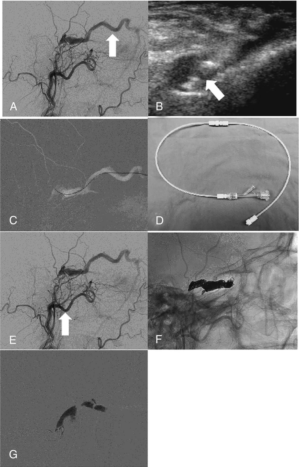
- Fig 1.
dCCF of the right cavernous sinus in a 56-year-old woman. A, Lateral-view angiogram from the right external carotid artery, obtained before embolization, shows a dCCF. The inferior petrosal sinus is thrombosed at the level of its inferior portion. There is predominant venous drainage toward the SOV (arrow). B, Direct percutaneous sonographically guided SOV puncture with a 22- or 24-ga angiocatheter cannula (arrow) using a high-frequency 15- to 7-MHz hockey stick sonography probe. C, Lateral-view roadmap, obtained after injecting the right external carotid artery, shows how a Transend 0.014-inch guidewire is advanced to the posterior aspect of the cavernous sinus. This allows subsequent exchange of a 2F inner for a 4F outer piece of a micropuncture set over a V-18 guidewire. D, Connecting system of two 30-cm connecting tubes and a Tuohy Borst hemostatic valve, which is connected to the micropuncture cannula. E, Lateral-view angiogram from the right external carotid artery shows the location in the posterior part of the cavernous sinus where a PX Slim microcatheter is placed (arrow). F, Lateral subtracted-view angiogram from the right external carotid artery shows how the coils are placed from posterior to anterior. G, Lateral blank subtraction roadmap demonstrating Gelfoam embolization of SOV from downstream to upstream while withdrawing the micropuncture cannula.
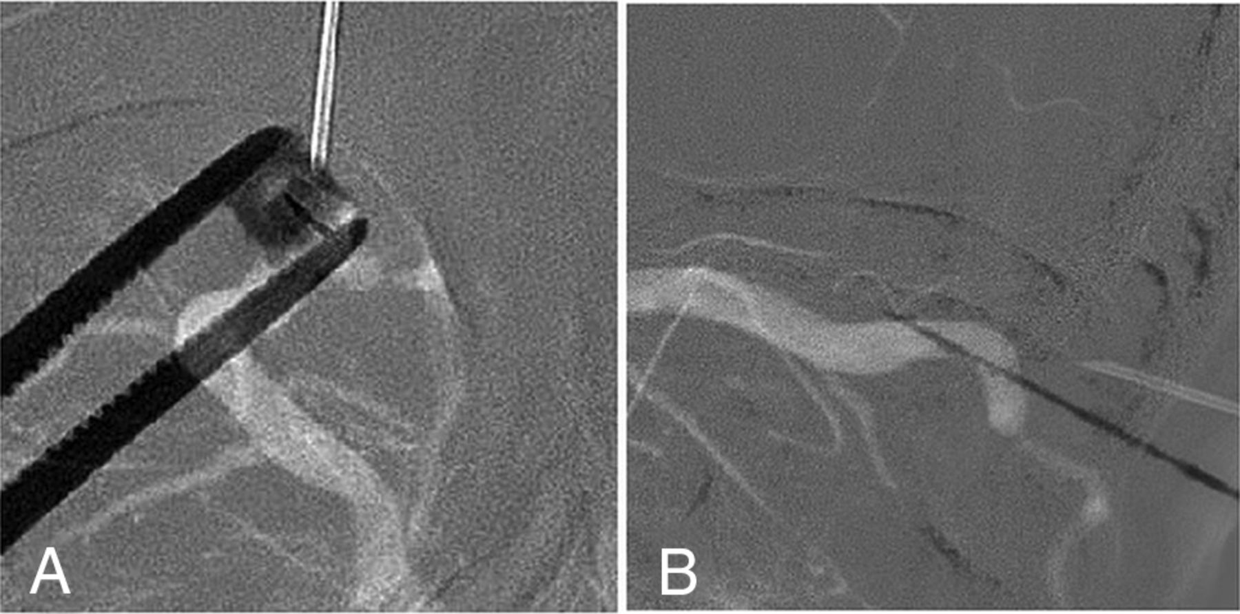
- Fig 2.
dCCF of the right cavernous sinus in a 66-year-old man. Anteroposterior (A) and lateral (B) view roadmap, obtained after injecting the right external carotid artery, shows how the needle is directed toward the SOV.


Tables
Twenty patients who underwent endovascular treatment for dCCF with transvenous embolization after direct imaging-guided percutaneous SOV puncturea
Age (yr)/Sex Side IPS Outflow Imaging Puncture Hematoma 67/F Left None US Preseptal None 66/M Right None US Preseptal None 59/M Left None Biplane roadmap Postseptal Preseptal 18/F Right None US Preseptal Preseptal 59/F Left None US Postseptal None 47/F Bilateral None US Preseptal Preseptal 98/F Right None US Preseptal None 77/F Left None US Preseptal None 71/F Left None US Preseptal None 70/M Right None US Preseptal None 84/F Left Minimal US Preseptal None 69/F Right None Biplane roadmap Preseptal Preseptal 66/M Right None US Postseptal Retrobulbar 66/F Bilateral Minimal US Preseptal None 75/M Right None US Preseptal None 27/F Right None Biplane roadmap Postseptal Retrobulbar 67/F Bilateral Minimal Biplane roadmap Preseptal None 76/F Right None US Preseptal None 56/F Right None US Preseptal None 73/F Left None US Preseptal None Note:—US indicates ultrasound.
↵a All patients achieved an angiographic cure.


Jump to section
Related Articles
Cited By...
- No citing articles found.