Advertisement

Main menu

  • Home
  • Content
    • Current Issue
    • Accepted Manuscripts
    • Article Preview
    • Past Issue Archive
    • Video Articles
    • AJNR Case Collection
    • Case of the Week Archive
    • Case of the Month Archive
    • Classic Case Archive
  • Special Collections
    • AJNR Awards
    • Low-Field MRI
    • Alzheimer Disease
    • ASNR Foundation Special Collection
    • Photon-Counting CT
    • View All
  • Multimedia
    • AJNR Podcasts
    • AJNR SCANtastic
    • Trainee Corner
    • MRI Safety Corner
    • Imaging Protocols
  • For Authors
    • Submit a Manuscript
    • Submit a Video Article
    • Submit an eLetter to the Editor/Response
    • Manuscript Submission Guidelines
    • Statistical Tips
    • Fast Publishing of Accepted Manuscripts
    • Graphical Abstract Preparation
    • Imaging Protocol Submission
    • Author Policies
  • About Us
    • About AJNR
    • Editorial Board
    • Editorial Board Alumni
  • More
    • Become a Reviewer/Academy of Reviewers
    • Subscribers
    • Permissions
    • Alerts
    • Feedback
    • Advertisers
    • ASNR Home

User menu

  • Alerts
  • Log in

Search

  • Advanced search
American Journal of Neuroradiology

American Journal of Neuroradiology

ASHNR American Society of Functional Neuroradiology ASHNR American Society of Pediatric Neuroradiology ASSR
  • Alerts
  • Log in

Advanced Search

  • Home
  • Content
    • Current Issue
    • Accepted Manuscripts
    • Article Preview
    • Past Issue Archive
    • Video Articles
    • AJNR Case Collection
    • Case of the Week Archive
    • Case of the Month Archive
    • Classic Case Archive
  • Special Collections
    • AJNR Awards
    • Low-Field MRI
    • Alzheimer Disease
    • ASNR Foundation Special Collection
    • Photon-Counting CT
    • View All
  • Multimedia
    • AJNR Podcasts
    • AJNR SCANtastic
    • Trainee Corner
    • MRI Safety Corner
    • Imaging Protocols
  • For Authors
    • Submit a Manuscript
    • Submit a Video Article
    • Submit an eLetter to the Editor/Response
    • Manuscript Submission Guidelines
    • Statistical Tips
    • Fast Publishing of Accepted Manuscripts
    • Graphical Abstract Preparation
    • Imaging Protocol Submission
    • Author Policies
  • About Us
    • About AJNR
    • Editorial Board
    • Editorial Board Alumni
  • More
    • Become a Reviewer/Academy of Reviewers
    • Subscribers
    • Permissions
    • Alerts
    • Feedback
    • Advertisers
    • ASNR Home
  • Follow AJNR on Twitter
  • Visit AJNR on Facebook
  • Follow AJNR on Instagram
  • Join AJNR on LinkedIn
  • RSS Feeds

AJNR Awards, New Junior Editors, and more. Read the latest AJNR updates

Research ArticleAdult Brain
Open Access

Leptomeningeal Contrast Enhancement Is Related to Focal Cortical Thinning in Relapsing-Remitting Multiple Sclerosis: A Cross-Sectional MRI Study

N. Bergsland, D. Ramasamy, E. Tavazzi, D. Hojnacki, B. Weinstock-Guttman and R. Zivadinov
American Journal of Neuroradiology April 2019, 40 (4) 620-625; DOI: https://doi.org/10.3174/ajnr.A6011
N. Bergsland
aFrom the Buffalo Neuroimaging Analysis Center (N.B., D.R., E.T., R.Z.)
  • Find this author on Google Scholar
  • Find this author on PubMed
  • Search for this author on this site
  • ORCID record for N. Bergsland
D. Ramasamy
aFrom the Buffalo Neuroimaging Analysis Center (N.B., D.R., E.T., R.Z.)
  • Find this author on Google Scholar
  • Find this author on PubMed
  • Search for this author on this site
  • ORCID record for D. Ramasamy
E. Tavazzi
aFrom the Buffalo Neuroimaging Analysis Center (N.B., D.R., E.T., R.Z.)
  • Find this author on Google Scholar
  • Find this author on PubMed
  • Search for this author on this site
  • ORCID record for E. Tavazzi
D. Hojnacki
bJacobs Comprehensive MS Treatment and Research Center (D.H., B.W.-G.), Department of Neurology
  • Find this author on Google Scholar
  • Find this author on PubMed
  • Search for this author on this site
  • ORCID record for D. Hojnacki
B. Weinstock-Guttman
bJacobs Comprehensive MS Treatment and Research Center (D.H., B.W.-G.), Department of Neurology
  • Find this author on Google Scholar
  • Find this author on PubMed
  • Search for this author on this site
  • ORCID record for B. Weinstock-Guttman
R. Zivadinov
aFrom the Buffalo Neuroimaging Analysis Center (N.B., D.R., E.T., R.Z.)
cJacobs School of Medicine and Biomedical Sciences, Center for Biomedical Imaging at Clinical Translational Science Institute (R.Z.), University at Buffalo, State University of New York, Buffalo, New York.
  • Find this author on Google Scholar
  • Find this author on PubMed
  • Search for this author on this site
  • ORCID record for R. Zivadinov
  • Article
  • Figures & Data
  • Info & Metrics
  • Responses
  • References
  • PDF
Loading

Article Figures & Data

Figures

  • Tables
  • Fig 1.
    • Download figure
    • Open in new tab
    • Download powerpoint
    Fig 1.

    A representative case depicting the T2WI FLAIR precontrast, T2WI FLAIR postcontrast, and T2WI FLAIR pre-/postcontrast subtraction images. The FreeSurfer-derived white and pial surfaces are shown in yellow and red, respectively. The white arrow shows a focus of leptomeningeal contrast enhancement.
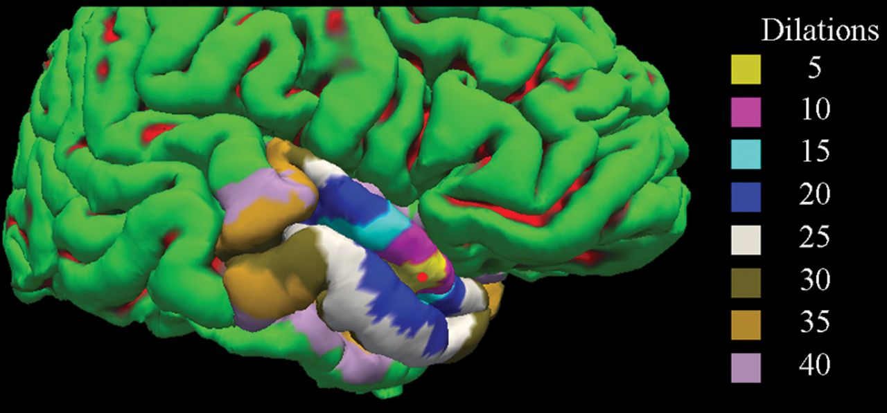
  • Fig 2.
    • Download figure
    • Open in new tab
    • Download powerpoint
    Fig 2.

    A representative cortical reconstruction from FreeSurfer is shown with dilated ROIs overlaid on the cortical surface. Gyri are shown in green, while sulci are shown in red. The red circle corresponds to the position of the mapped focus of leptomeningeal contrast enhancement. Different-sized ROIs are shown in varying colors.

Tables

  • Figures
    • View popup
    Table 1:

    Demographic, clinical, and MR imaging characteristics of the cohort

    RRMS (n = 43)SPMS (n = 15)P Valuea
    Women (No.) (%)38 (88.4)11 (73.3).658
    Age (mean) (SD) (yr)52.8 (12.1)60.5 (6.7).023b
    Disease duration (mean) (SD) (yr)15.2 (8.9)21.9 (8.3).014b
    EDSS (median) (IQR)3.0 (2.0–3.5)6.0 (3.6–6.5).003b
    Age at onset (mean) (SD) (yr)37.6 (12.0)38.7 (8.5).428
    Disease-modifying therapy (No.) (%)
        Interferon β-1a I.M.14 (32.6)2 (13.3).111
        Interferon β-1a S.C.3 (9.3)0 (0)
        Glatiramer acetate8 (18.6)2 (13.3)
        Natalizumab4 (9.3)1 (6.7)
        Dimethyl fumarate2 (4.7)0 (0)
        Fingolimod3 (7.0)0 (0)
        Teriflunomide1 (2.3)3 (20.0)
        Rituximab1 (2.3)0 (0)
        Ocrelizumab0 (0)1 (6.7)
        IVIG1 (2.3)1 (6.7)
        No therapy6 (14.0)5 (33.3)
    No. of LMCE foci1 (n = 33)1 (n = 8).292
    2 (n = 8)2 (n = 4)
    3 (n = 2)3 (n = 3)
    LMCE shape (No.).637
        Nodular3616
        Linear75
        Plate-like124
    T2 lesion volume (mean) (SD) (mL)16.0 (16.2)15.8 (11.4).965
    Gd lesion volume (median) (IQR) (mL)0 (0–0)0 (0–0).097
    • Note:—EDSS indicates Expanded Disability Status Scale; I.M., intramuscular; S.C., subcutaneous; IVIG, intravenous immunoglobulin; Gd, gadolinium.

    • ↵a P value represents differences between the patients with RRMS and SPMS. The differences between the groups were analyzed using the Fisher exact, Student t, Mann-Whitney U, or χ2 test.

    • ↵b P values < .05.

    • View popup
    Table 2:

    Cortical thickness measurements in the ipsilateral region surrounding focal areas of leptomeningeal contrast enhancement and in the contralateral regiona

    Entire Cohort (80 LMCE Foci)Patients with RRMS (55 LMCE Foci)Patients with SPMS (25 LMCE Foci)
    DilationsIpsilateralContralateralPct. Diff.P/FDR-PIpsilateralContralateralPct. Diff.P/FDR-PIpsilateralContralateralPct. Diff.P/FDR-P
    52.043 (.515)2.149 (.556)−5.06.07/.1872.031 (.534)2.212 (.567)−8.53.005/.04b2.070 (.480)2.000 (.510)3.44.562/.823
    102.063 (.437)2.160 (.476)−4.59.016/.1282.099 (.455)2.211 (.501)−5.20.011/.044b1.979 (.387)2.039 (.396)−2.99.493/.823
    152.088 (.411)2.153 (.413)−3.07.039/.1562.141 (.414)2.208 (.432)−3.08.055/.1471.964 (.383)2.023 (.339)−2.96.384/.823
    202.114 (.286)2.136 (.385)−1.04.409/.5542.167 (.394)2.196 (.395)−1.33.365/.4171.989 (.342)2.000 (.325)−.55.895/.895
    252.116 (.362)2.131 (.368)−.71.485/.5542.167 (.376)2.194 (.377)−1.24.306/.4081.996 (.301)1.983 (.306)0.65.749/.856
    302.113 (.348)2.131 (.354)−.85.342/.5542.162 (.363)2.195 (.361)−1.51.137/.2741.997 (.284)1.980 (.291)0.85.617/.823
    352.115 (.338)2.127 (.342)−.57.483/.5542.165 (.353)2.190 (.347)−1.15.191/.3061.998 (.273)1.977 (.284)1.06.509/.823
    402.117 (.327)2.120 (.332)−.14.882/.8822.168 (.341)2.180 (.336)−.55.529/.5291.998 (.261)1.979 (.282)0.95.485/.823
    • Note:—Pct. Diff. indicates percentage difference; FDR, false discovery rate.

    • Ipsilateral and contralateral columns represent mean (standard deviation) cortical thickness measures in millimeters.

    • ↵a Paired t tests comparing the ipsilateral and contralateral regions were used to calculate P values. The Benjamini-Hochberg procedure was used to control the false discovery rate.

    • ↵b Corrected P values < .05.

PreviousNext
Back to top

In this issue

American Journal of Neuroradiology: 40 (4)
American Journal of Neuroradiology
Vol. 40, Issue 4
1 Apr 2019
  • Table of Contents
  • Index by author
  • Complete Issue (PDF)
Advertisement
Print
Download PDF
Email Article

Thank you for your interest in spreading the word on American Journal of Neuroradiology.

NOTE: We only request your email address so that the person you are recommending the page to knows that you wanted them to see it, and that it is not junk mail. We do not capture any email address.

Enter multiple addresses on separate lines or separate them with commas.
Leptomeningeal Contrast Enhancement Is Related to Focal Cortical Thinning in Relapsing-Remitting Multiple Sclerosis: A Cross-Sectional MRI Study
(Your Name) has sent you a message from American Journal of Neuroradiology
(Your Name) thought you would like to see the American Journal of Neuroradiology web site.
CAPTCHA
This question is for testing whether or not you are a human visitor and to prevent automated spam submissions.
Cite this article
N. Bergsland, D. Ramasamy, E. Tavazzi, D. Hojnacki, B. Weinstock-Guttman, R. Zivadinov
Leptomeningeal Contrast Enhancement Is Related to Focal Cortical Thinning in Relapsing-Remitting Multiple Sclerosis: A Cross-Sectional MRI Study
American Journal of Neuroradiology Apr 2019, 40 (4) 620-625; DOI: 10.3174/ajnr.A6011

Citation Manager Formats

  • BibTeX
  • Bookends
  • EasyBib
  • EndNote (tagged)
  • EndNote 8 (xml)
  • Medlars
  • Mendeley
  • Papers
  • RefWorks Tagged
  • Ref Manager
  • RIS
  • Zotero
0 Responses
Respond to this article
Share
Bookmark this article
Leptomeningeal Contrast Enhancement Is Related to Focal Cortical Thinning in Relapsing-Remitting Multiple Sclerosis: A Cross-Sectional MRI Study
N. Bergsland, D. Ramasamy, E. Tavazzi, D. Hojnacki, B. Weinstock-Guttman, R. Zivadinov
American Journal of Neuroradiology Apr 2019, 40 (4) 620-625; DOI: 10.3174/ajnr.A6011
del.icio.us logo Twitter logo Facebook logo Mendeley logo
  • Tweet Widget
  • Facebook Like
  • Google Plus One
Purchase

Jump to section

  • Article
    • Abstract
    • ABBREVIATIONS:
    • Materials and Methods
    • Results
    • Discussion
    • Conclusions
    • Footnotes
    • REFERENCES
  • Figures & Data
  • Info & Metrics
  • Responses
  • References
  • PDF

Related Articles

  • No related articles found.
  • PubMed
  • Google Scholar

Cited By...

  • Alternative Venous Pathways: A Potential Key Imaging Feature for Early Diagnosis of Sturge-Weber Syndrome Type 1
  • Leptomeningeal Enhancement in Multiple Sclerosis and Other Neurological Diseases: A Systematic Review and Meta-Analysis
  • Imaging meningeal inflammation in CNS autoimmunity identifies a therapeutic role for BTK inhibition
  • Crossref
  • Google Scholar

This article has not yet been cited by articles in journals that are participating in Crossref Cited-by Linking.

More in this TOC Section

  • Diagnostic Neuroradiology of Monoclonal Antibodies
  • NCCT vs. MRI for Brain Atrophy in Acute Stroke
  • Clinical Outcomes After Chiari I Decompression
Show more Adult Brain

Similar Articles

Advertisement

Indexed Content

  • Current Issue
  • Accepted Manuscripts
  • Article Preview
  • Past Issues
  • Editorials
  • Editor's Choice
  • Fellows' Journal Club
  • Letters to the Editor
  • Video Articles

Cases

  • Case Collection
  • Archive - Case of the Week
  • Archive - Case of the Month
  • Archive - Classic Case

More from AJNR

  • Trainee Corner
  • Imaging Protocols
  • MRI Safety Corner
  • Book Reviews

Multimedia

  • AJNR Podcasts
  • AJNR Scantastics

Resources

  • Turnaround Time
  • Submit a Manuscript
  • Submit a Video Article
  • Submit an eLetter to the Editor/Response
  • Manuscript Submission Guidelines
  • Statistical Tips
  • Fast Publishing of Accepted Manuscripts
  • Graphical Abstract Preparation
  • Imaging Protocol Submission
  • Evidence-Based Medicine Level Guide
  • Publishing Checklists
  • Author Policies
  • Become a Reviewer/Academy of Reviewers
  • News and Updates

About Us

  • About AJNR
  • Editorial Board
  • Editorial Board Alumni
  • Alerts
  • Permissions
  • Not an AJNR Subscriber? Join Now
  • Advertise with Us
  • Librarian Resources
  • Feedback
  • Terms and Conditions
  • AJNR Editorial Board Alumni

American Society of Neuroradiology

  • Not an ASNR Member? Join Now

© 2025 by the American Society of Neuroradiology All rights, including for text and data mining, AI training, and similar technologies, are reserved.
Print ISSN: 0195-6108 Online ISSN: 1936-959X

Powered by HighWire