
Article Figures & Data
Figures
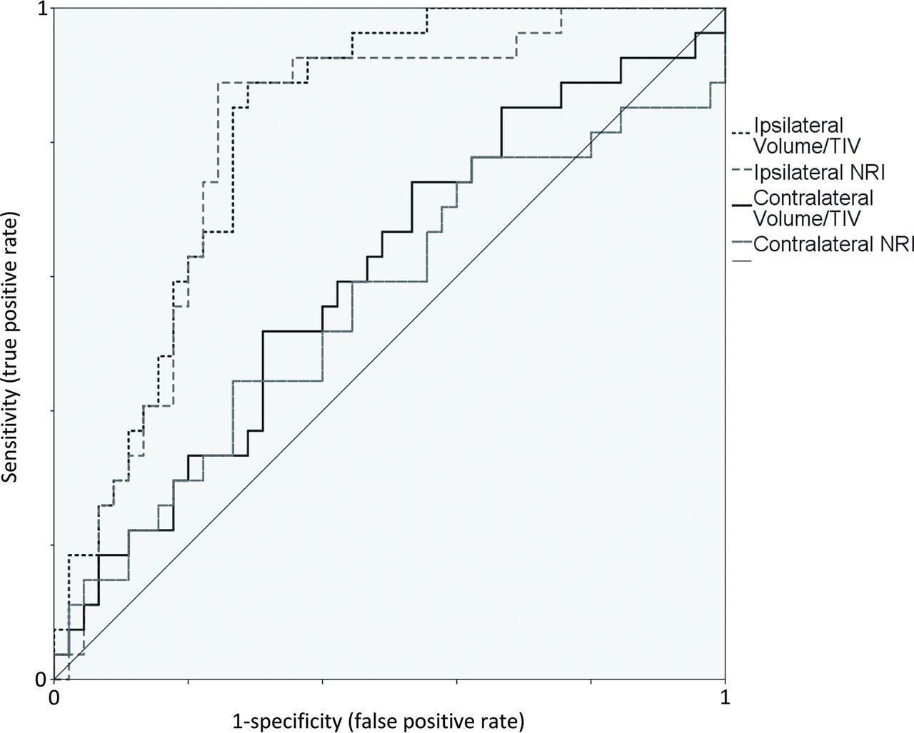
- Fig 1.
Receiver operating characteristic curve for mesial temporal sclerosis. The volume of the hippocampi corrected for total intracranial volume and the Neuroreader Index, a nonparametric comparative statistic derived from age- and sex-adjusted normative data, were calculated and evaluated for their ability to classify patients with pathology-proved mesial temporal sclerosis. Receiver operating characteristic curves from ipsilateral and contralateral hippocampal volume measures relative to the subsequent side of surgery are presented. Note that contralateral volumes appear skewed away from the 45° diagonal.
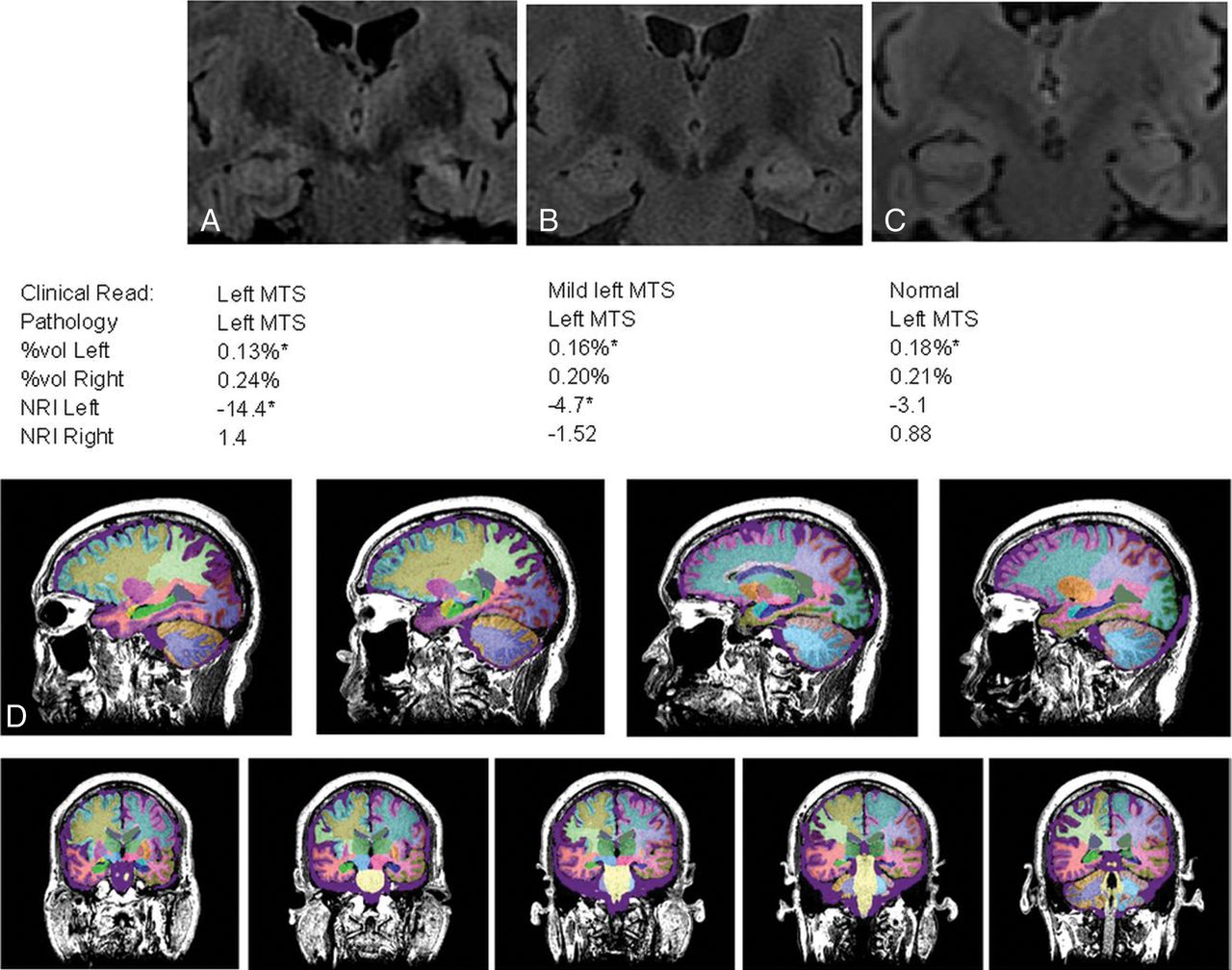
- Fig 2.
Volumetric analysis. A–C, Three patients with left mesial temporal sclerosis of varying conspicuity on MR imaging. Clinical reads and volumetric measures of the abnormal left and normal right hippocampal formations are presented below the images. D, Representative registration and segmentation. Hippocampal volumes are denoted by bright green (right) and dark blue (left) in a patient with left mesial temporal sclerosis.


Tables
MTS (n = 26) Normal or Other Findings (n = 45) Age at operation (mean) (SD) (yr) 43.7 (12.9) 42.0 (13.1) Female sex (No.) (%) 14 (54%) 18 (40%) Left temporal lobectomy (No.) (%) 14 (54%) 23 (51.1%) Normal pathologic findings (No.) (%) 0 (0%) 23 (51.1%) Other pathology (No.) (%) Low-grade glioma 0 (0%) 7 (15.5%) Dysplasia 1 (3.8%) 3 (6.7%) Cavernous malformation 0 (0%) 3 (6.7%) Gliosis or old infarct 0 (0%) 4 (8.9%) Volumetric T1 acquisition (No.) (%) Axial only 6 (23%) 8 (17.8%) Coronal only 6 (23%) 10 (22.2%) Both 14 (54%) 27 (60%) - Table 2:
Comparison of Neuroreader measures with radiologist interpretation for detection of MTS
SENS SPEC ACCUR AUC P P, McNemara %Vol <0.193 89% 71% 77% 0.818 <0.001 <0.001 NRI ≤−3.807 89% 76% 81% 0.800 <0.001 <0.001 Radiologist 50% 87% 73% NA NA NA Note:—SENS indicates sensitivity; SPEC, specificity; ACCUR, accuracy; NA, nonapplicable; AUC, area under the curve.
↵a The McNemar nonparametric test demonstrates significant differences using the provided thresholds compared with the interpretation of a radiologist for detection of mesial temporal sclerosis.


