Advertisement

Main menu

  • Home
  • Content
    • Current Issue
    • Accepted Manuscripts
    • Article Preview
    • Past Issue Archive
    • Video Articles
    • AJNR Case Collection
    • Case of the Week Archive
    • Case of the Month Archive
    • Classic Case Archive
  • Special Collections
    • AJNR Awards
    • Low-Field MRI
    • Alzheimer Disease
    • ASNR Foundation Special Collection
    • Photon-Counting CT
    • View All
  • Multimedia
    • AJNR Podcasts
    • AJNR SCANtastic
    • Trainee Corner
    • MRI Safety Corner
    • Imaging Protocols
  • For Authors
    • Submit a Manuscript
    • Submit a Video Article
    • Submit an eLetter to the Editor/Response
    • Manuscript Submission Guidelines
    • Statistical Tips
    • Fast Publishing of Accepted Manuscripts
    • Graphical Abstract Preparation
    • Imaging Protocol Submission
    • Author Policies
  • About Us
    • About AJNR
    • Editorial Board
    • Editorial Board Alumni
  • More
    • Become a Reviewer/Academy of Reviewers
    • Subscribers
    • Permissions
    • Alerts
    • Feedback
    • Advertisers
    • ASNR Home

User menu

  • Alerts
  • Log in

Search

  • Advanced search
American Journal of Neuroradiology

American Journal of Neuroradiology

ASHNR American Society of Functional Neuroradiology ASHNR American Society of Pediatric Neuroradiology ASSR
  • Alerts
  • Log in

Advanced Search

  • Home
  • Content
    • Current Issue
    • Accepted Manuscripts
    • Article Preview
    • Past Issue Archive
    • Video Articles
    • AJNR Case Collection
    • Case of the Week Archive
    • Case of the Month Archive
    • Classic Case Archive
  • Special Collections
    • AJNR Awards
    • Low-Field MRI
    • Alzheimer Disease
    • ASNR Foundation Special Collection
    • Photon-Counting CT
    • View All
  • Multimedia
    • AJNR Podcasts
    • AJNR SCANtastic
    • Trainee Corner
    • MRI Safety Corner
    • Imaging Protocols
  • For Authors
    • Submit a Manuscript
    • Submit a Video Article
    • Submit an eLetter to the Editor/Response
    • Manuscript Submission Guidelines
    • Statistical Tips
    • Fast Publishing of Accepted Manuscripts
    • Graphical Abstract Preparation
    • Imaging Protocol Submission
    • Author Policies
  • About Us
    • About AJNR
    • Editorial Board
    • Editorial Board Alumni
  • More
    • Become a Reviewer/Academy of Reviewers
    • Subscribers
    • Permissions
    • Alerts
    • Feedback
    • Advertisers
    • ASNR Home
  • Follow AJNR on Twitter
  • Visit AJNR on Facebook
  • Follow AJNR on Instagram
  • Join AJNR on LinkedIn
  • RSS Feeds

AJNR Awards, New Junior Editors, and more. Read the latest AJNR updates

Research ArticleSpine Imaging and Spine Image-Guided Interventions

Unintended Consequences: Review of New Artifacts Introduced by Iterative Reconstruction CT Metal Artifact Reduction in Spine Imaging

D.R. Wayer, N.Y. Kim, B.J. Otto, A.M. Grayev and A.D. Kuner
American Journal of Neuroradiology November 2019, 40 (11) 1973-1975; DOI: https://doi.org/10.3174/ajnr.A6238
D.R. Wayer
aFrom the Department of Radiology (D.R.W., N.Y.K., B.J.O., A.M.G., A.D.K.), University of Wisconsin, Madison, Wisconsin
  • Find this author on Google Scholar
  • Find this author on PubMed
  • Search for this author on this site
  • ORCID record for D.R. Wayer
N.Y. Kim
aFrom the Department of Radiology (D.R.W., N.Y.K., B.J.O., A.M.G., A.D.K.), University of Wisconsin, Madison, Wisconsin
  • Find this author on Google Scholar
  • Find this author on PubMed
  • Search for this author on this site
  • ORCID record for N.Y. Kim
B.J. Otto
aFrom the Department of Radiology (D.R.W., N.Y.K., B.J.O., A.M.G., A.D.K.), University of Wisconsin, Madison, Wisconsin
  • Find this author on Google Scholar
  • Find this author on PubMed
  • Search for this author on this site
  • ORCID record for B.J. Otto
A.M. Grayev
aFrom the Department of Radiology (D.R.W., N.Y.K., B.J.O., A.M.G., A.D.K.), University of Wisconsin, Madison, Wisconsin
  • Find this author on Google Scholar
  • Find this author on PubMed
  • Search for this author on this site
  • ORCID record for A.M. Grayev
A.D. Kuner
aFrom the Department of Radiology (D.R.W., N.Y.K., B.J.O., A.M.G., A.D.K.), University of Wisconsin, Madison, Wisconsin
bthe University of Wisconsin School of Medicine and Public Health (A.D.K.), Madison, Wisconsin
  • Find this author on Google Scholar
  • Find this author on PubMed
  • Search for this author on this site
  • ORCID record for A.D. Kuner
  • Article
  • Figures & Data
  • Info & Metrics
  • Responses
  • References
  • PDF
Loading

Abstract

SUMMARY: Metal hardware serves as a common artifact source in spine CT imaging in the form of beam-hardening, photon starvation, and streaking. Postprocessing metal artifact reduction techniques have been developed to decrease these artifacts, which has been proved to improve visualization of soft-tissue structures and increase diagnostic confidence. However, metal artifact reduction reconstruction introduces its own novel artifacts that can mimic pathology.

ABBREVIATION:

MAR
metal artifact reduction

Metal artifacts are an obstacle to obtaining high-fidelity CT images in postoperative spine imaging, which is increasingly problematic as the number of instrumented spinal fusions grows. There were approximately 463,200 spinal fusions in 2014 in the United States,1 a 12% increase compared with 20112 and nearly triple the amount from 1998.3 Unfortunately, up to one-third of patients experience no improvement or worsening of symptoms following surgical intervention4 and require appropriate spine imaging.4⇓⇓–7

The main artifacts introduced by metallic spinal implants include beam-hardening, photon starvation, and streaking,8 which diminish overall image quality and impair the identification of pathology.9,10 Metal artifact reduction (MAR; GE Healthcare, Milwaukee, Wisconsin) postprocessing techniques have been developed11 to recover image quality and detail in affected areas and to diminish the artifacts themselves.12 Unfortunately, MAR introduces different artifacts that can mimic pathology, which radiologists need to recognize.12,13 The purpose of this article was to review MAR-related artifacts seen on GE Healthcare scanners. While only 1 manufacturer is included in this article, many of the concepts are applicable to different vendor products that use similar techniques.

MAR Technique and Its Benefits

MAR is an automated postprocessing, projection-based technique developed by GE Healthcare that addresses metal artifacts in 3 stages. First, metal artifacts are identified in the source tomogram projection using a density threshold. Second, the data lost from metal artifacts are reconstructed into an inpainted projection with corrected data, which is generated by estimation from artifact-free areas. Finally, the corrected projection is formed by combining the inpainted projection and the original projection, which reveal details of structures obscured by artifacts.14,15

MAR reconstruction has been shown to significantly improve visualization of obscured soft-tissue structures and diagnostic confidence compared with the standard weighted filtered back-projection reconstruction method.12 Paraspinal soft tissues adjacent to the fusion hardware are obscured on traditional images, while the MAR image allows adequate visualization of the underlying structures (Fig 1). Despite the clear benefits of applying MAR to decrease metal artifacts, MAR introduces its own unique artifacts.

Fig 1.
  • Download figure
  • Open in new tab
  • Download powerpoint
Fig 1.

Axial lumbar CT images of a 15-year-old boy with spinal muscular atrophy type II who underwent posterior spinal fusion of T2–L5 due to severe scoliosis. A, MAR images demonstrating improved visualizations of soft tissues surrounding the hardware. B, Uncorrected images show artifacts covering soft tissues surrounding the hardware.

MAR-Related Artifacts and Limitations

MAR-induced artifacts have been described in the hip and elbow,15 but there are limited descriptions of these artifacts in the spine12,16 where MAR artifacts frequently mimic pathology. The primary artifacts include the following: perihardware lucency, pedicle screw lucency, factitious subarachnoid material on myelography, and misrepresentation of intraosseous cement.

Perihardware Lucency.

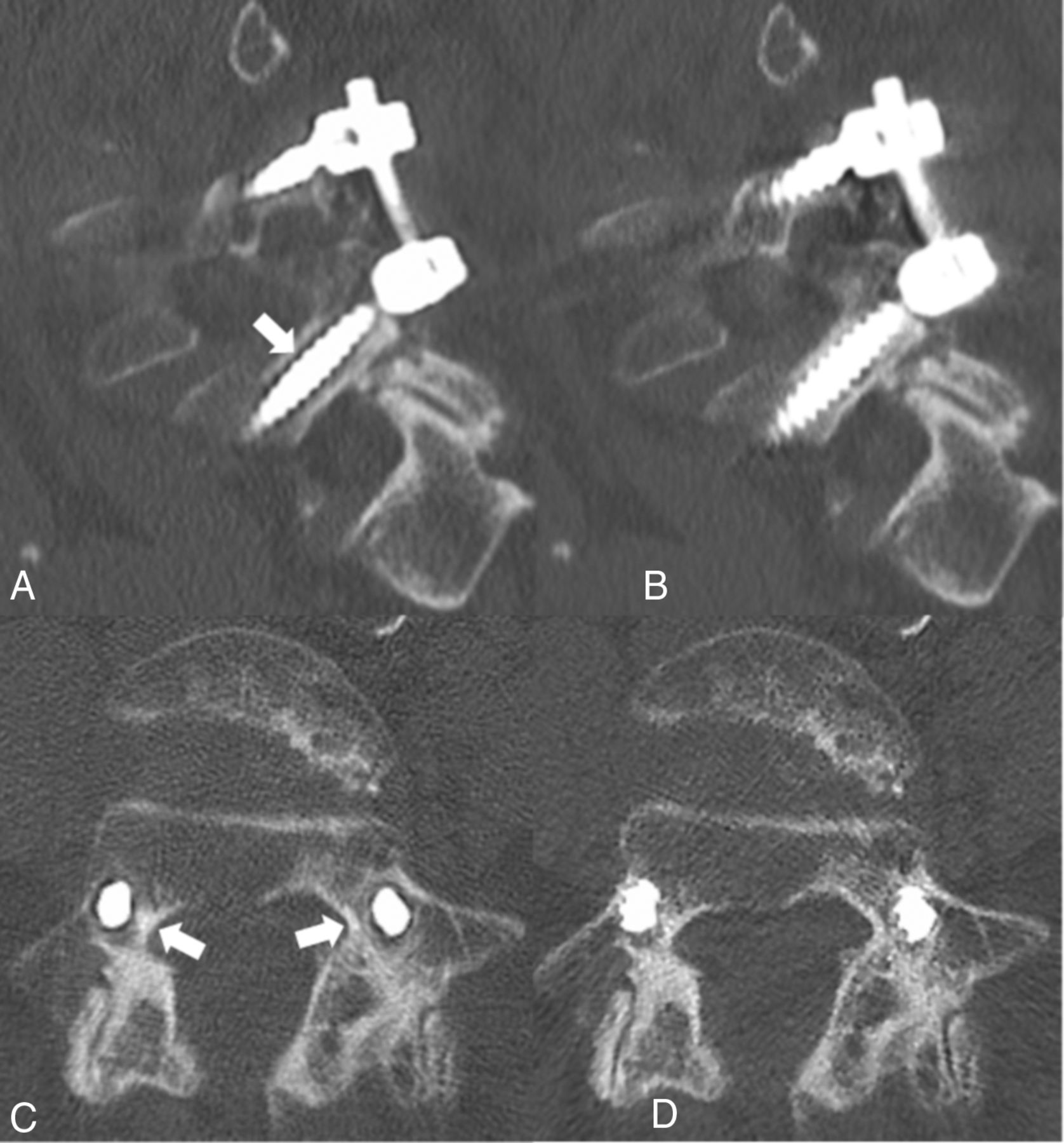
Perihardware lucency (Fig 2A, white arrow) is concerning because it usually signifies loosening of the hardware; however, this is not visualized in the non-MAR images (Fig 2B). More troublesome is that these artifacts persist in multiple planes as shown in the axial plane (Fig 2C, -D).

Fig 2.
  • Download figure
  • Open in new tab
  • Download powerpoint
Fig 2.

A, Sagittal CT image of posterior lumbar spinal fusion with the MAR technique applied shows lucency surrounding the L5 pedicle screw as indicated by the white arrow. B, Image A without the MAR protocol applied, C, Axial CT image shows bilateral pedicle screws with the MAR technique applied, demonstrating lucency surrounding the bilateral pedicle screws (white arrows). If the MAR images were viewed alone, these lucencies could be mistaken for hardware loosening. D, Image C without the MAR protocol applied.

Pedicle Screw Lucency.

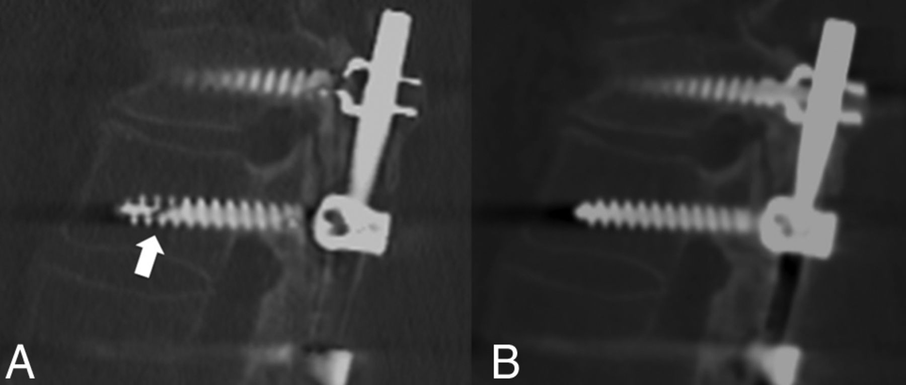
Pedicle screw lucency occurs when the implanted hardware fatigues and fractures (Fig 3A, white arrow), which is not seen on the non-MAR image (Fig 3B). This artifact is not uniform throughout the MAR series, and adjacent images demonstrate that adjacent screws are unaffected.

Fig 3.
  • Download figure
  • Open in new tab
  • Download powerpoint
Fig 3.

A, Sagittal CT image of posterior spinal fusion of the MAR protocol demonstrating lucency through the pedicle screw (white arrow). This lucency clearly mimics a fracture, hence could be called a “pseudofracture” if non-MAR images are not available. B, Image without the MAR application shows a normal appearance of the pedicle screw.

Subarachnoid Material on Myelography.

Factitious subarachnoid material can be seen on myelograms when MAR is applied, particularly in areas of concentrated intrathecal contrast. There is hypodense material in the dorsal spinal canal (Fig 4A, white arrow), which is not seen on the non-MAR image (Fig 4B). This artifact could be mistaken for arachnoiditis, tumor, or layering debris and may lead to unnecessary further work-up.

Fig 4.
  • Download figure
  • Open in new tab
  • Download powerpoint
Fig 4.

A, Sagittal MAR CT image with fusion hardware in the lumbar and sacral spine demonstrates apparent hypodense material (white arrow) in the dorsal spinal canal not seen in the non-MAR image. This filling defect could be mistaken for arachnoiditis, tumor, or layering debris and lead to unnecessary further work-up. B, Non-MAR image without evidence of material within the spinal canal.

Intraosseous Cement Abnormal Distribution.

Postvertebroplasty cement configuration can appear irregular and fragmented with indistinct margins (Fig 5A, white arrow) and an internal heterogeneous pattern of vertebral body filling; however, in the non-MAR image (Fig 5B), the bone cement has a normal uniform distribution with clearly defined margins.

Fig 5.
  • Download figure
  • Open in new tab
  • Download powerpoint
Fig 5.

A, Axial CT image with MAR applied demonstrating an irregular fragmented appearance of bone cement with indistinct margins (white arrow). B, Non-MAR counterpart image with normal appearance of bone cement with accurate representation of vertebral filling.

Conclusions

MAR is a useful imaging reconstruction technique that can minimize metal artifacts, thus improving soft-tissue visualization and diagnostic confidence in the setting of spinal hardware;12 however, it is critical to understand the generated artifacts to render a correct interpretation. One limitation of this article is that we exclusively used GE Healthcare scanners. While MAR techniques of different vendors may have overlap in method (ie, projection modification is used by GE Healthcare and Philips Healthcare), each vendor ultimately has its own proprietary algorithm.17 All can potentially simulate undersampling due to oversmoothing by the MAR algorithm in areas where dense objects interface with bone, as shown in previous phantom modeling demonstrating distortion of hardware.17

At our institution, we include a set of standard images along with the MAR postprocessed images (from a single CT acquisition) on the PACS to allow confident identification of MAR artifacts and true pathology or postoperative complication. Given the widespread use of MAR techniques and the implication of misinterpreting artifacts, it is crucial that radiologists recognize these artifacts exist.

Footnotes

  • Disclosures: Allison M. Grayev—UNRELATED: Royalties: McGraw Hill, Comments: Author.

  • Paper previously presented at: Zachary Clark Radiology Research Symposium, May 4, 2019, Madison, Wisconsin; and Annual Meeting of the American Society of Neuroradiology, May 18–23, 2019, Boston, Massachusetts.

References

  1. 1.↵
    1. McDermott KW,
    2. Freeman WJ,
    3. Elixhauser A
    . Overview of Operating Room Procedures During Inpatient Stays in U.S. Hospitals, 2014: Statistical Brief #233. In: Healthcare Cost and Utilization Project (HCUP) Statistical Briefs. Rockville: Agency for Healthcare Research and Quality; 2017
  2. 2.↵
    1. Weiss AJ,
    2. Elixhauser A,
    3. Andrews RM
    . Characteristics of Operating Room Procedures in U.S. Hospitals, 2011: Statistical Brief #170. In: Healthcare Cost and Utilization Project (HCUP) Statistical Briefs. Rockville: Agency for Healthcare Research and Quality; 2014
  3. 3.↵
    1. Rajaee SS,
    2. Bae HW,
    3. Kanim LEA, et al
    . Spinal fusion in the United States: analysis of trends from 1998 to 2008. Spine 2012;37:67–76 doi:10.1097/BRS.0b013e31820cccfb pmid:21311399
    CrossRefPubMed
  4. 4.↵
    1. Skolasky RL,
    2. Wegener ST,
    3. Maggard AM, et al
    . The impact of reduction of pain after lumbar spine surgery: the relationship between changes in pain and physical function and disability. Spine 2014;39:1426–32 doi:10.1097/BRS.0000000000000428 pmid:24859574
    CrossRefPubMed
  5. 5.↵
    1. Chan C,
    2. Peng P
    . Failed back surgery syndrome. Pain Med 2011;12:577–606 doi:10.1111/j.1526-4637.2011.01089.x pmid:21463472
    CrossRefPubMed
  6. 6.↵
    1. Young PM,
    2. Berquist TH,
    3. Bancroft LW, et al
    . Complications of spinal instrumentation. Radiographics 2007;27:775–89 doi:10.1148/rg.273065055 pmid:17495292
    CrossRefPubMed
  7. 7.↵
    1. Baber Z,
    2. Erdek MA
    . Failed back surgery syndrome: current perspectives. J Pain Res 2016;9:979–87 doi:10.2147/JPR.S92776 pmid:27853391
    CrossRefPubMed
  8. 8.↵
    1. Barrett JF,
    2. Keat N
    . Artifacts in CT: recognition and avoidance. Radiographics 2004;24:1679–91 doi:10.1148/rg.246045065 pmid:15537976
    CrossRefPubMed
  9. 9.↵
    1. Stradiotti P,
    2. Curti A,
    3. Castellazzi G, et al
    . Metal-related artifacts in instrumented spine: techniques for reducing artifacts in CT and MRI: state of the art. Eur Spine J 2009;18:102–08 doi:10.1007/s00586-009-0998-5 pmid:19437043
    CrossRefPubMed
  10. 10.↵
    1. White LM,
    2. Buckwalter KA
    . Technical considerations: CT and MR imaging in the postoperative orthopedic patient. Semin Musculoskelet Radiol 2002;06:5–17 doi:10.1055/s-2002-23160 pmid:11917267
    CrossRefPubMed
  11. 11.↵
    1. Gjesteby L,
    2. Man BD,
    3. Jin Y, et al
    . Metal artifact reduction in CT: where are we after four decades? IEEE Access 2016;4:5826–49 doi:10.1109/ACCESS.2016.2608621
    CrossRef
  12. 12.↵
    1. Kotsenas AL,
    2. Michalak GJ,
    3. DeLone DR, et al
    . CT metal artifact reduction in the spine: can an iterative reconstruction technique improve visualization? AJNR Am J Neuroradiol 2015;36:2184–90 doi:10.3174/ajnr.A4416 pmid:26251433
    Abstract/FREE Full Text
  13. 13.↵
    1. Zhou P,
    2. Zhang C,
    3. Gao Z, et al
    . Evaluation of the quality of CT images acquired with smart metal artifact reduction software. Open Life Science 2018;13:155–62 doi:10.1515/biol-2018-0021
    CrossRef
  14. 14.↵
    GE Healthcare. GE Healthcare. Smart Metal Artifact Reduction (MAR). 2013 https://www.gehealthcare.com/products/computed-tomography/radiation-therapy-planning/metal-artifact-reduction. Accessed May 30, 2019
  15. 15.↵
    1. Katsura M,
    2. Sato J,
    3. Akahane M, et al
    . Current and novel techniques for metal artifact reduction at CT: practical guide for radiologists. Radiographics 2018;38:450–61 doi:10.1148/rg.2018170102 pmid:29528826
    CrossRefPubMed
  16. 16.↵
    1. Wang Y,
    2. Qian B,
    3. Li B, et al
    . Metal artifacts reduction using monochromatic images from spectral CT: evaluation of pedicle screws in patients with scoliosis. Eur J Radiol 2013;82:e360–66 doi:10.1016/j.ejrad.2013.02.024 pmid:23518146
    CrossRefPubMed
  17. 17.↵
    1. Huang JY,
    2. Kerns JR,
    3. Nute JL, et al
    . An evaluation of three commercially available metal artifact reduction methods for CT imaging. Phys Med Biol 2015;60:1047–67 doi:10.1088/0031-9155/60/3/1047 pmid:25585685
    CrossRefPubMed
  • Received June 6, 2019.
  • Accepted after revision August 16, 2019.
  • © 2019 by American Journal of Neuroradiology
View Abstract
PreviousNext
Back to top

In this issue

American Journal of Neuroradiology: 40 (11)
American Journal of Neuroradiology
Vol. 40, Issue 11
1 Nov 2019
  • Table of Contents
  • Index by author
  • Complete Issue (PDF)
Advertisement
Print
Download PDF
Email Article

Thank you for your interest in spreading the word on American Journal of Neuroradiology.

NOTE: We only request your email address so that the person you are recommending the page to knows that you wanted them to see it, and that it is not junk mail. We do not capture any email address.

Enter multiple addresses on separate lines or separate them with commas.
Unintended Consequences: Review of New Artifacts Introduced by Iterative Reconstruction CT Metal Artifact Reduction in Spine Imaging
(Your Name) has sent you a message from American Journal of Neuroradiology
(Your Name) thought you would like to see the American Journal of Neuroradiology web site.
CAPTCHA
This question is for testing whether or not you are a human visitor and to prevent automated spam submissions.
Cite this article
D.R. Wayer, N.Y. Kim, B.J. Otto, A.M. Grayev, A.D. Kuner
Unintended Consequences: Review of New Artifacts Introduced by Iterative Reconstruction CT Metal Artifact Reduction in Spine Imaging
American Journal of Neuroradiology Nov 2019, 40 (11) 1973-1975; DOI: 10.3174/ajnr.A6238

Citation Manager Formats

  • BibTeX
  • Bookends
  • EasyBib
  • EndNote (tagged)
  • EndNote 8 (xml)
  • Medlars
  • Mendeley
  • Papers
  • RefWorks Tagged
  • Ref Manager
  • RIS
  • Zotero
0 Responses
Respond to this article
Share
Bookmark this article
Unintended Consequences: Review of New Artifacts Introduced by Iterative Reconstruction CT Metal Artifact Reduction in Spine Imaging
D.R. Wayer, N.Y. Kim, B.J. Otto, A.M. Grayev, A.D. Kuner
American Journal of Neuroradiology Nov 2019, 40 (11) 1973-1975; DOI: 10.3174/ajnr.A6238
del.icio.us logo Twitter logo Facebook logo Mendeley logo
  • Tweet Widget
  • Facebook Like
  • Google Plus One
Purchase

Jump to section

  • Article
    • Abstract
    • ABBREVIATION:
    • Conclusions
    • Footnotes
    • References
  • Figures & Data
  • Info & Metrics
  • Responses
  • References
  • PDF

Related Articles

  • No related articles found.
  • PubMed
  • Google Scholar

Cited By...

  • Atlas for the CT Syndesmophyte Score (CTSS) in patients with axial spondyloarthritis
  • Crossref
  • Google Scholar

This article has not yet been cited by articles in journals that are participating in Crossref Cited-by Linking.

More in this TOC Section

  • Optimization of Photon Counting CT Myelography
  • Characteristics of SIH Type I Culprit Lesions
  • Management Outcomes For VO Spine Biopsy
Show more Spine Imaging and Spine Image-Guided Interventions

Similar Articles

Advertisement

Indexed Content

  • Current Issue
  • Accepted Manuscripts
  • Article Preview
  • Past Issues
  • Editorials
  • Editor's Choice
  • Fellows' Journal Club
  • Letters to the Editor
  • Video Articles

Cases

  • Case Collection
  • Archive - Case of the Week
  • Archive - Case of the Month
  • Archive - Classic Case

More from AJNR

  • Trainee Corner
  • Imaging Protocols
  • MRI Safety Corner
  • Book Reviews

Multimedia

  • AJNR Podcasts
  • AJNR Scantastics

Resources

  • Turnaround Time
  • Submit a Manuscript
  • Submit a Video Article
  • Submit an eLetter to the Editor/Response
  • Manuscript Submission Guidelines
  • Statistical Tips
  • Fast Publishing of Accepted Manuscripts
  • Graphical Abstract Preparation
  • Imaging Protocol Submission
  • Evidence-Based Medicine Level Guide
  • Publishing Checklists
  • Author Policies
  • Become a Reviewer/Academy of Reviewers
  • News and Updates

About Us

  • About AJNR
  • Editorial Board
  • Editorial Board Alumni
  • Alerts
  • Permissions
  • Not an AJNR Subscriber? Join Now
  • Advertise with Us
  • Librarian Resources
  • Feedback
  • Terms and Conditions
  • AJNR Editorial Board Alumni

American Society of Neuroradiology

  • Not an ASNR Member? Join Now

© 2025 by the American Society of Neuroradiology All rights, including for text and data mining, AI training, and similar technologies, are reserved.
Print ISSN: 0195-6108 Online ISSN: 1936-959X

Powered by HighWire