Advertisement

Main menu

  • Home
  • Content
    • Current Issue
    • Accepted Manuscripts
    • Article Preview
    • Past Issue Archive
    • Video Articles
    • AJNR Case Collection
    • Case of the Week Archive
    • Case of the Month Archive
    • Classic Case Archive
  • Special Collections
    • AJNR Awards
    • Low-Field MRI
    • Alzheimer Disease
    • ASNR Foundation Special Collection
    • Photon-Counting CT
    • View All
  • Multimedia
    • AJNR Podcasts
    • AJNR SCANtastic
    • Trainee Corner
    • MRI Safety Corner
    • Imaging Protocols
  • For Authors
    • Submit a Manuscript
    • Submit a Video Article
    • Submit an eLetter to the Editor/Response
    • Manuscript Submission Guidelines
    • Statistical Tips
    • Fast Publishing of Accepted Manuscripts
    • Graphical Abstract Preparation
    • Imaging Protocol Submission
    • Author Policies
  • About Us
    • About AJNR
    • Editorial Board
    • Editorial Board Alumni
  • More
    • Become a Reviewer/Academy of Reviewers
    • Subscribers
    • Permissions
    • Alerts
    • Feedback
    • Advertisers
    • ASNR Home

User menu

  • Alerts
  • Log in

Search

  • Advanced search
American Journal of Neuroradiology

American Journal of Neuroradiology

ASHNR American Society of Functional Neuroradiology ASHNR American Society of Pediatric Neuroradiology ASSR
  • Alerts
  • Log in

Advanced Search

  • Home
  • Content
    • Current Issue
    • Accepted Manuscripts
    • Article Preview
    • Past Issue Archive
    • Video Articles
    • AJNR Case Collection
    • Case of the Week Archive
    • Case of the Month Archive
    • Classic Case Archive
  • Special Collections
    • AJNR Awards
    • Low-Field MRI
    • Alzheimer Disease
    • ASNR Foundation Special Collection
    • Photon-Counting CT
    • View All
  • Multimedia
    • AJNR Podcasts
    • AJNR SCANtastic
    • Trainee Corner
    • MRI Safety Corner
    • Imaging Protocols
  • For Authors
    • Submit a Manuscript
    • Submit a Video Article
    • Submit an eLetter to the Editor/Response
    • Manuscript Submission Guidelines
    • Statistical Tips
    • Fast Publishing of Accepted Manuscripts
    • Graphical Abstract Preparation
    • Imaging Protocol Submission
    • Author Policies
  • About Us
    • About AJNR
    • Editorial Board
    • Editorial Board Alumni
  • More
    • Become a Reviewer/Academy of Reviewers
    • Subscribers
    • Permissions
    • Alerts
    • Feedback
    • Advertisers
    • ASNR Home
  • Follow AJNR on Twitter
  • Visit AJNR on Facebook
  • Follow AJNR on Instagram
  • Join AJNR on LinkedIn
  • RSS Feeds

AJNR Awards, New Junior Editors, and more. Read the latest AJNR updates

Research ArticleAdult Brain

Role of 3D Pseudocontinuous Arterial Spin-Labeling Perfusion in the Diagnosis and Follow-Up in Patients with Herpes Simplex Encephalitis

R. Li, P.-A. Shi, T.-F. Liu, Y. Li, Y. Wang, K. Wu, X.-J. Chen, H.-F. Xiao, Y.-L. Wang, L. Ma and X. Lou
American Journal of Neuroradiology November 2019, 40 (11) 1901-1907; DOI: https://doi.org/10.3174/ajnr.A6279
R. Li
aFrom the School of Medicine (R.L., L.M.), Nankai University, Tianjin, China
bDepartment of Radiology (R.L., T.-F.L., Y.L., Y.W., K.W., X.-J.C., H.-F.X., Y.-L.W., L.M., X.L.), Chinese People's Liberation Army General Hospital, Beijing, China
  • Find this author on Google Scholar
  • Find this author on PubMed
  • Search for this author on this site
  • ORCID record for R. Li
P.-A. Shi
cDepartment of Endocrinology (P.-A.S.), Cangzhou Central Hospital, Cangzhou, Hebei, China
  • Find this author on Google Scholar
  • Find this author on PubMed
  • Search for this author on this site
  • ORCID record for P.-A. Shi
T.-F. Liu
bDepartment of Radiology (R.L., T.-F.L., Y.L., Y.W., K.W., X.-J.C., H.-F.X., Y.-L.W., L.M., X.L.), Chinese People's Liberation Army General Hospital, Beijing, China
  • Find this author on Google Scholar
  • Find this author on PubMed
  • Search for this author on this site
  • ORCID record for T.-F. Liu
Y. Li
bDepartment of Radiology (R.L., T.-F.L., Y.L., Y.W., K.W., X.-J.C., H.-F.X., Y.-L.W., L.M., X.L.), Chinese People's Liberation Army General Hospital, Beijing, China
  • Find this author on Google Scholar
  • Find this author on PubMed
  • Search for this author on this site
  • ORCID record for Y. Li
Y. Wang
bDepartment of Radiology (R.L., T.-F.L., Y.L., Y.W., K.W., X.-J.C., H.-F.X., Y.-L.W., L.M., X.L.), Chinese People's Liberation Army General Hospital, Beijing, China
  • Find this author on Google Scholar
  • Find this author on PubMed
  • Search for this author on this site
  • ORCID record for Y. Wang
K. Wu
bDepartment of Radiology (R.L., T.-F.L., Y.L., Y.W., K.W., X.-J.C., H.-F.X., Y.-L.W., L.M., X.L.), Chinese People's Liberation Army General Hospital, Beijing, China
  • Find this author on Google Scholar
  • Find this author on PubMed
  • Search for this author on this site
  • ORCID record for K. Wu
X.-J. Chen
bDepartment of Radiology (R.L., T.-F.L., Y.L., Y.W., K.W., X.-J.C., H.-F.X., Y.-L.W., L.M., X.L.), Chinese People's Liberation Army General Hospital, Beijing, China
  • Find this author on Google Scholar
  • Find this author on PubMed
  • Search for this author on this site
  • ORCID record for X.-J. Chen
H.-F. Xiao
bDepartment of Radiology (R.L., T.-F.L., Y.L., Y.W., K.W., X.-J.C., H.-F.X., Y.-L.W., L.M., X.L.), Chinese People's Liberation Army General Hospital, Beijing, China
  • Find this author on Google Scholar
  • Find this author on PubMed
  • Search for this author on this site
  • ORCID record for H.-F. Xiao
Y.-L. Wang
bDepartment of Radiology (R.L., T.-F.L., Y.L., Y.W., K.W., X.-J.C., H.-F.X., Y.-L.W., L.M., X.L.), Chinese People's Liberation Army General Hospital, Beijing, China
  • Find this author on Google Scholar
  • Find this author on PubMed
  • Search for this author on this site
  • ORCID record for Y.-L. Wang
L. Ma
aFrom the School of Medicine (R.L., L.M.), Nankai University, Tianjin, China
bDepartment of Radiology (R.L., T.-F.L., Y.L., Y.W., K.W., X.-J.C., H.-F.X., Y.-L.W., L.M., X.L.), Chinese People's Liberation Army General Hospital, Beijing, China
  • Find this author on Google Scholar
  • Find this author on PubMed
  • Search for this author on this site
  • ORCID record for L. Ma
X. Lou
bDepartment of Radiology (R.L., T.-F.L., Y.L., Y.W., K.W., X.-J.C., H.-F.X., Y.-L.W., L.M., X.L.), Chinese People's Liberation Army General Hospital, Beijing, China
  • Find this author on Google Scholar
  • Find this author on PubMed
  • Search for this author on this site
  • ORCID record for X. Lou
  • Article
  • Figures & Data
  • Info & Metrics
  • Responses
  • References
  • PDF
Loading

Article Figures & Data

Figures

  • Tables
  • Fig 1.
    • Download figure
    • Open in new tab
    • Download powerpoint
    Fig 1.

    Flow chart showing the selection of the final study population. A total of 40 patients who underwent both conventional MR imaging and 3D-pCASL were clinically suspected of having encephalitis. Finally, 17 cases with HSE were included in this study.
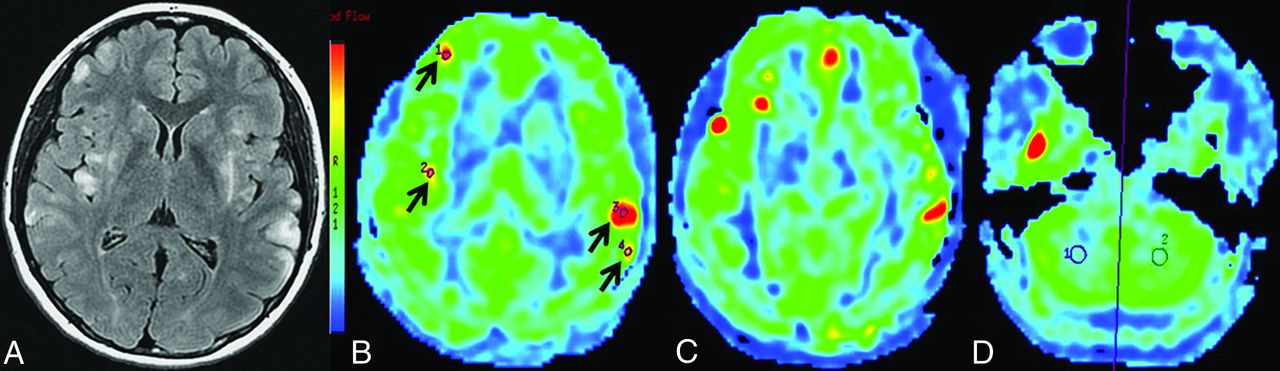
  • Fig 2.
    • Download figure
    • Open in new tab
    • Download powerpoint
    Fig 2.

    Case 11, a 20-year-old female patient at the acute stage (5 days after the onset of symptoms). A, Axial T2 FLAIR shows multiple lesions located in right frontal and bilateral insular lobes, as well as the bilateral temporal lobes. B, 3D-pCASL demonstrates increased CBF, consistent with involved regions on axial T2 FLAIR (black arrows). C, Other lesions also show hyperperfusion in different affected areas. D, ROI site selection for bilateral cerebellum CBF on the pCASL CBF color map.

  • Fig 3.
    • Download figure
    • Open in new tab
    • Download powerpoint
    Fig 3.

    Case 12, a 47 -year-old female patient at the subacute stage (15 days after the onset of symptoms). A, Axial T2WI shows hyperintensities in bilateral frontal, temporal, and insular lobes and the cingulate gyrus. B, Axial T1WI demonstrates slight hypointensities. C, Slight hyperintensities are observed on coronal T2 FLAIR. D, 3D-pCASL shows high perfusion (black arrows) in the corresponding involved areas.

  • Fig 4.
    • Download figure
    • Open in new tab
    • Download powerpoint
    Fig 4.

    Case 1, a 17-year-old male patient. Serial T2WI (A, E, and I), T1WI (B, F, and J), and coronal T2 FLAIR (C, G, and K) demonstrate abnormal signals on the left temporal lobe and hippocampus. Serial follow-up 3D-pCASL perfusion imaging was performed, which reveals dynamic changes in the involved area on the 11th (D), 24th (H), and 30th day (L), respectively (black arrows). Meanwhile, the patient's condition markedly improved with effective therapeutic intervention.

  • Fig 5.
    • Download figure
    • Open in new tab
    • Download powerpoint
    Fig 5.

    A, The boxplot of nCBF values in acute-, subacute-, and chronic-stage lesions and the control group, respectively. B, Time course of mean CBF changes for lesions at different time points in 4 follow-up HSE cases. The mean CBF value of the lesions gradually decreased after treatment in 4 case series.

Tables

  • Figures
    • View popup
    Table 1:

    Summary of the demographic and clinical features of consecutive patients with HSE

    Case No.Age (yr)SexClinical PresentationTime (Days)a
    117MHeadache, fever, sudden unconscious attack, seizure11/24/30
    258MHeadache, left lower extremity weakness and involuntary movement4/14/20
    335MHeadache, nausea, vomiting, and memory deterioration6/42
    449FHeadache and fever with irrelevant answer2/17/29
    521FHeadache, fever, slow response, and memory deterioration14
    627MPersistent vertigo with sudden onset of left lower extremity weakness26
    732MHeadache, fever, and seizure7
    828MHeadache, dizziness, and memory deterioration2
    944FMemory deterioration, slow response7
    1050MHeadache, fever, dizziness, paroxysmal loss of consciousness, seizure3
    1120FFever, disturbance of consciousness with limb seizure5
    1247FHeadache, fever, behavioral and psychological disorder15
    1347MHeadache, fever, dizziness20
    1426MHeadache and fever with paroxysmal limb seizure15
    1525MRecurrent fever, paraphasia, memory deterioration, and limb seizure85
    1646MHeadache, fever, convulsion with psychological and behavioral disorder60
    1764MHeadache, dizziness, memory deterioration, paroxysmal loss of consciousness, and seizure6
    • ↵a The time from symptom onset to 3D-pCASL evaluation.

    • View popup
    Table 2:

    Initial conventional MR imaging and ASL findings after admission in patients with HSE

    Case No.Lesion LocationaT2WIT1WICEPerfusionb
    1L. TSlight hyper/hypoSlight hypo/hyperNAObvious hyper
    2L. T, ISlight hyperSlight hypo/hyperGyriform enhancementObvious hyper
    3L. T, ISlight hyperSlight hypo/hyperPatchy enhancementObvious hyper
    4R. F, TSlight hyperSlight hypoNoneObvious hyper
    5L. F, I, and Bil. TSlight hyper/hyperSlight hypo/hyperGyriform enhancementObvious hyper
    6Bil. T, and R. PSlight hyper/hyperSlight hypo/hyperNoneSlight hypo
    7L. F, P, O, and Bil. I, TSlight hyperIsoPatchy enhancementSlight hyper/hyper
    8Bil. T, and L. ISlight hyperSlight hypoNoneObvious hyper
    9Bil. F, T, ISlight hyperSlight hypoNoneObvious hyper
    10Bil. F and cingulate gyrusSlight hyperIsoNoneObvious hyper
    11R. F, and Bil. T, ISlight hyperIsoNoneObvious hyper (multiple lesions)
    12Bil. F, T, I, and cingulate gyrusSlight hyperSlight hypoPatchy enhancementObvious hyper
    13R. T, and Bil. ISlight hyperSlight hypoSlight meningeal enhancementRegional hyper
    14L. T, ISlight hypo/hyperSlight hypo/hyperGyriform enhancementRegional hyper
    15L. T, ISlight hyper/hyperSlight hypo/hypoSlight gyriform enhancementObvious hypo
    16R. T, ISlight hyper/hyperSlight hypo/hypoSlight enhancementObvious hypo
    17R. T, ISlight hyperSlight hypoNoneObvious hyper
    • Note:— R indicates right; L, left; Bil., bilateral; F, frontal lobe; T, temporal lobe; P, parietal lobe; O, occipital lobe; I, insular lobe; hyper, hyperintensity or hyperperfusion; hypo, hypointensity or hyperperfusion; Iso, isointensity; CE, contrast enhancement; NA, not applicable.

    • ↵a Abnormal signals on T2 FLAIR.

    • ↵b Perfusion performance on the first ASL examination.

    • View popup
    Table 3:

    Mean CBF and mean nCBF values in the lesions at different stages and control group findings

    Stage (Days)No. of CasesRange of CBF Values (mL/100g/min)Mean CBF Values (mL/100g/min)Mean nCBF Values
    Acute stage (≤14)1159.5–162.3121.5 ± 33.82.68 ± 0.54
    Subacute stage (15–25)666.1–126.598.3 ± 22.82.42 ± 0.52
    Chronic stage (≥26)623.0–63.133.9 ± 15.00.87 ± 0.30
    Control groupa1547.9–71.958.8 ± 7.01.33 ± 0.08
    • ↵a The temporal cortex perfusion as a control reference.

PreviousNext
Back to top

In this issue

American Journal of Neuroradiology: 40 (11)
American Journal of Neuroradiology
Vol. 40, Issue 11
1 Nov 2019
  • Table of Contents
  • Index by author
  • Complete Issue (PDF)
Advertisement
Print
Download PDF
Email Article

Thank you for your interest in spreading the word on American Journal of Neuroradiology.

NOTE: We only request your email address so that the person you are recommending the page to knows that you wanted them to see it, and that it is not junk mail. We do not capture any email address.

Enter multiple addresses on separate lines or separate them with commas.
Role of 3D Pseudocontinuous Arterial Spin-Labeling Perfusion in the Diagnosis and Follow-Up in Patients with Herpes Simplex Encephalitis
(Your Name) has sent you a message from American Journal of Neuroradiology
(Your Name) thought you would like to see the American Journal of Neuroradiology web site.
CAPTCHA
This question is for testing whether or not you are a human visitor and to prevent automated spam submissions.
Cite this article
R. Li, P.-A. Shi, T.-F. Liu, Y. Li, Y. Wang, K. Wu, X.-J. Chen, H.-F. Xiao, Y.-L. Wang, L. Ma, X. Lou
Role of 3D Pseudocontinuous Arterial Spin-Labeling Perfusion in the Diagnosis and Follow-Up in Patients with Herpes Simplex Encephalitis
American Journal of Neuroradiology Nov 2019, 40 (11) 1901-1907; DOI: 10.3174/ajnr.A6279

Citation Manager Formats

  • BibTeX
  • Bookends
  • EasyBib
  • EndNote (tagged)
  • EndNote 8 (xml)
  • Medlars
  • Mendeley
  • Papers
  • RefWorks Tagged
  • Ref Manager
  • RIS
  • Zotero
0 Responses
Respond to this article
Share
Bookmark this article
Role of 3D Pseudocontinuous Arterial Spin-Labeling Perfusion in the Diagnosis and Follow-Up in Patients with Herpes Simplex Encephalitis
R. Li, P.-A. Shi, T.-F. Liu, Y. Li, Y. Wang, K. Wu, X.-J. Chen, H.-F. Xiao, Y.-L. Wang, L. Ma, X. Lou
American Journal of Neuroradiology Nov 2019, 40 (11) 1901-1907; DOI: 10.3174/ajnr.A6279
del.icio.us logo Twitter logo Facebook logo Mendeley logo
  • Tweet Widget
  • Facebook Like
  • Google Plus One
Purchase

Jump to section

  • Article
    • Abstract
    • ABBREVIATIONS:
    • MATERIALS AND METHODS
    • RESULTS
    • DISCUSSION
    • CONCLUSIONS
    • References
  • Figures & Data
  • Info & Metrics
  • Responses
  • References
  • PDF

Related Articles

  • No related articles found.
  • PubMed
  • Google Scholar

Cited By...

  • Regarding "Brain Perfusion Alterations on 3D Pseudocontinuous Arterial Spin-Labeling MR Imaging in Patients with Autoimmune Encephalitis: A Case Series and Literature Review"
  • Brain Perfusion Alterations on 3D Pseudocontinuous Arterial Spin-Labeling MR Imaging in Patients with Autoimmune Encephalitis: A Case Series and Literature Review
  • Crossref
  • Google Scholar

This article has not yet been cited by articles in journals that are participating in Crossref Cited-by Linking.

More in this TOC Section

Adult Brain

  • Diagnostic Neuroradiology of Monoclonal Antibodies
  • Clinical Outcomes After Chiari I Decompression
  • Segmentation of Brain Metastases with BLAST
Show more Adult Brain

Functional

  • Glutaric Aciduria Type 1: DK vs. Conventional MRI
  • Kurtosis and Epileptogenic Tubers: A Pilot Study
  • Multiparametric MRI in PEDS Pontine Glioma
Show more Functional

Similar Articles

Advertisement

Indexed Content

  • Current Issue
  • Accepted Manuscripts
  • Article Preview
  • Past Issues
  • Editorials
  • Editor's Choice
  • Fellows' Journal Club
  • Letters to the Editor
  • Video Articles

Cases

  • Case Collection
  • Archive - Case of the Week
  • Archive - Case of the Month
  • Archive - Classic Case

More from AJNR

  • Trainee Corner
  • Imaging Protocols
  • MRI Safety Corner
  • Book Reviews

Multimedia

  • AJNR Podcasts
  • AJNR Scantastics

Resources

  • Turnaround Time
  • Submit a Manuscript
  • Submit a Video Article
  • Submit an eLetter to the Editor/Response
  • Manuscript Submission Guidelines
  • Statistical Tips
  • Fast Publishing of Accepted Manuscripts
  • Graphical Abstract Preparation
  • Imaging Protocol Submission
  • Evidence-Based Medicine Level Guide
  • Publishing Checklists
  • Author Policies
  • Become a Reviewer/Academy of Reviewers
  • News and Updates

About Us

  • About AJNR
  • Editorial Board
  • Editorial Board Alumni
  • Alerts
  • Permissions
  • Not an AJNR Subscriber? Join Now
  • Advertise with Us
  • Librarian Resources
  • Feedback
  • Terms and Conditions
  • AJNR Editorial Board Alumni

American Society of Neuroradiology

  • Not an ASNR Member? Join Now

© 2025 by the American Society of Neuroradiology All rights, including for text and data mining, AI training, and similar technologies, are reserved.
Print ISSN: 0195-6108 Online ISSN: 1936-959X

Powered by HighWire