Advertisement

Main menu

  • Home
  • Content
    • Current Issue
    • Accepted Manuscripts
    • Article Preview
    • Past Issue Archive
    • Video Articles
    • AJNR Case Collection
    • Case of the Week Archive
    • Case of the Month Archive
    • Classic Case Archive
  • Special Collections
    • AJNR Awards
    • Low-Field MRI
    • Alzheimer Disease
    • ASNR Foundation Special Collection
    • Photon-Counting CT
    • View All
  • Multimedia
    • AJNR Podcasts
    • AJNR SCANtastic
    • Trainee Corner
    • MRI Safety Corner
    • Imaging Protocols
  • For Authors
    • Submit a Manuscript
    • Submit a Video Article
    • Submit an eLetter to the Editor/Response
    • Manuscript Submission Guidelines
    • Statistical Tips
    • Fast Publishing of Accepted Manuscripts
    • Graphical Abstract Preparation
    • Imaging Protocol Submission
    • Author Policies
  • About Us
    • About AJNR
    • Editorial Board
    • Editorial Board Alumni
  • More
    • Become a Reviewer/Academy of Reviewers
    • Subscribers
    • Permissions
    • Alerts
    • Feedback
    • Advertisers
    • ASNR Home

User menu

  • Alerts
  • Log in

Search

  • Advanced search
American Journal of Neuroradiology

American Journal of Neuroradiology

ASHNR American Society of Functional Neuroradiology ASHNR American Society of Pediatric Neuroradiology ASSR
  • Alerts
  • Log in

Advanced Search

  • Home
  • Content
    • Current Issue
    • Accepted Manuscripts
    • Article Preview
    • Past Issue Archive
    • Video Articles
    • AJNR Case Collection
    • Case of the Week Archive
    • Case of the Month Archive
    • Classic Case Archive
  • Special Collections
    • AJNR Awards
    • Low-Field MRI
    • Alzheimer Disease
    • ASNR Foundation Special Collection
    • Photon-Counting CT
    • View All
  • Multimedia
    • AJNR Podcasts
    • AJNR SCANtastic
    • Trainee Corner
    • MRI Safety Corner
    • Imaging Protocols
  • For Authors
    • Submit a Manuscript
    • Submit a Video Article
    • Submit an eLetter to the Editor/Response
    • Manuscript Submission Guidelines
    • Statistical Tips
    • Fast Publishing of Accepted Manuscripts
    • Graphical Abstract Preparation
    • Imaging Protocol Submission
    • Author Policies
  • About Us
    • About AJNR
    • Editorial Board
    • Editorial Board Alumni
  • More
    • Become a Reviewer/Academy of Reviewers
    • Subscribers
    • Permissions
    • Alerts
    • Feedback
    • Advertisers
    • ASNR Home
  • Follow AJNR on Twitter
  • Visit AJNR on Facebook
  • Follow AJNR on Instagram
  • Join AJNR on LinkedIn
  • RSS Feeds

AJNR Awards, New Junior Editors, and more. Read the latest AJNR updates

Research ArticlePediatric Neuroimaging
Open Access

MRI Features of Histologically Diagnosed Supratentorial Primitive Neuroectodermal Tumors and Pineoblastomas in Correlation with Molecular Diagnoses and Outcomes: A Report from the Children's Oncology Group ACNS0332 Trial

A. Jaju, E.I. Hwang, M. Kool, D. Capper, L. Chavez, S. Brabetz, C. Billups, Y. Li, M. Fouladi, R.J. Packer, S.M. Pfister, J.M. Olson and L.A. Heier
American Journal of Neuroradiology November 2019, 40 (11) 1796-1803; DOI: https://doi.org/10.3174/ajnr.A6253
A. Jaju
aFrom the Department of Radiology (A.J.), Ann and Robert H Lurie Children's Hospital of Chicago, Chicago, Illinois
bNorthwestern University Feinberg School of Medicine (A.J.), Chicago, Illinois
  • Find this author on Google Scholar
  • Find this author on PubMed
  • Search for this author on this site
  • ORCID record for A. Jaju
E.I. Hwang
cBrain Tumor Institute (E.I.H., R.J.P.), Children's National Health System, Washington, DC
  • Find this author on Google Scholar
  • Find this author on PubMed
  • Search for this author on this site
  • ORCID record for E.I. Hwang
M. Kool
dDepartment of Pediatric Neurooncology (M.K., S.B., S.M.P.), German Cancer Research Center, Heidelberg, Baden-Württemberg, Germany
  • Find this author on Google Scholar
  • Find this author on PubMed
  • Search for this author on this site
  • ORCID record for M. Kool
D. Capper
eDepartment of Pediatric Neuropathology (D.C.), University Hospital Heidelberg, Heidelberg, Baden-Württemberg, Germany
  • Find this author on Google Scholar
  • Find this author on PubMed
  • Search for this author on this site
  • ORCID record for D. Capper
L. Chavez
fDepartment of Medicine (L.C.), University of California San Diego, La Jolla, California
  • Find this author on Google Scholar
  • Find this author on PubMed
  • Search for this author on this site
  • ORCID record for L. Chavez
S. Brabetz
dDepartment of Pediatric Neurooncology (M.K., S.B., S.M.P.), German Cancer Research Center, Heidelberg, Baden-Württemberg, Germany
  • Find this author on Google Scholar
  • Find this author on PubMed
  • Search for this author on this site
  • ORCID record for S. Brabetz
C. Billups
gDepartment of Biostatistics (C.B., Y.L.), St. Jude Children's Research Hospital, Memphis, Tennessee
  • Find this author on Google Scholar
  • Find this author on PubMed
  • Search for this author on this site
  • ORCID record for C. Billups
Y. Li
gDepartment of Biostatistics (C.B., Y.L.), St. Jude Children's Research Hospital, Memphis, Tennessee
  • Find this author on Google Scholar
  • Find this author on PubMed
  • Search for this author on this site
  • ORCID record for Y. Li
M. Fouladi
hBrain Tumor Center (M.F.), Cincinnati Children's Hospital, Cincinnati, Ohio
  • Find this author on Google Scholar
  • Find this author on PubMed
  • Search for this author on this site
  • ORCID record for M. Fouladi
R.J. Packer
cBrain Tumor Institute (E.I.H., R.J.P.), Children's National Health System, Washington, DC
  • Find this author on Google Scholar
  • Find this author on PubMed
  • Search for this author on this site
  • ORCID record for R.J. Packer
S.M. Pfister
dDepartment of Pediatric Neurooncology (M.K., S.B., S.M.P.), German Cancer Research Center, Heidelberg, Baden-Württemberg, Germany
  • Find this author on Google Scholar
  • Find this author on PubMed
  • Search for this author on this site
  • ORCID record for S.M. Pfister
J.M. Olson
iFred Hurtchinson Cancer Research Center (J.M.O.), Seattle Children's Hospital, Seattle, Washington
  • Find this author on Google Scholar
  • Find this author on PubMed
  • Search for this author on this site
  • ORCID record for J.M. Olson
L.A. Heier
jDepartment of Radiology (L.A.H.), New York Presbyterian Hospital, New York, New York
  • Find this author on Google Scholar
  • Find this author on PubMed
  • Search for this author on this site
  • ORCID record for L.A. Heier
  • Article
  • Figures & Data
  • Info & Metrics
  • Responses
  • References
  • PDF
Loading

Article Figures & Data

Figures

  • Tables
  • Fig 1.
    • Download figure
    • Open in new tab
    • Download powerpoint
    Fig 1.

    Schematic representation of the external surface of cerebral hemispheres (A) shows the locations of hemispheric tumors, with the sizes of the shaded circles proportional to the number of tumors in each lobe. A midline sagittal section of the brain (B) shows pineal and nonpineal tumors, with pie diagrams representing the subgroups.

  • Fig 2.
    • Download figure
    • Open in new tab
    • Download powerpoint
    Fig 2.

    Kaplan-Meier curves show event-free survival distributions by tumor size (A), margins (B), presence of edema (C), and percentage enhancement (D) for all patients (n = 56).
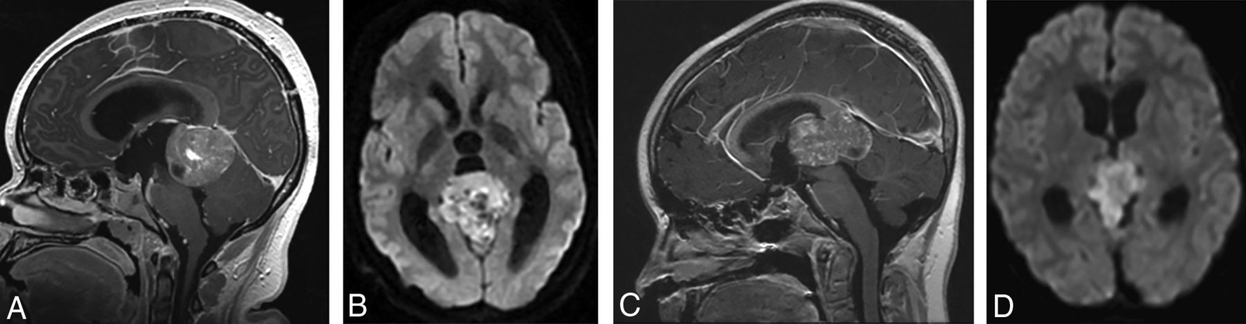
  • Fig 3.
    • Download figure
    • Open in new tab
    • Download powerpoint
    Fig 3.

    (A and B), A 15-year-old girl with a molecular diagnosis of pineoblastoma. Sagittal T1-weighted postcontrast (A) and axial diffusion-weighted (B) MR images demonstrate a mass centered in the pineal region with diffuse heterogeneous enhancement, small cystic foci, and diffusion restriction. (C and D), A 9-year-old girl with a molecular diagnosis of ATRT_MYC. Sagittal T1-weighted postcontrast (C) and axial diffusion-weighted (D) images demonstrate a similar mass centered in the pineal region with diffuse heterogeneous enhancement, small cystic foci, and diffusion restriction. Note the similarities in imaging appearance between the 2 examples.
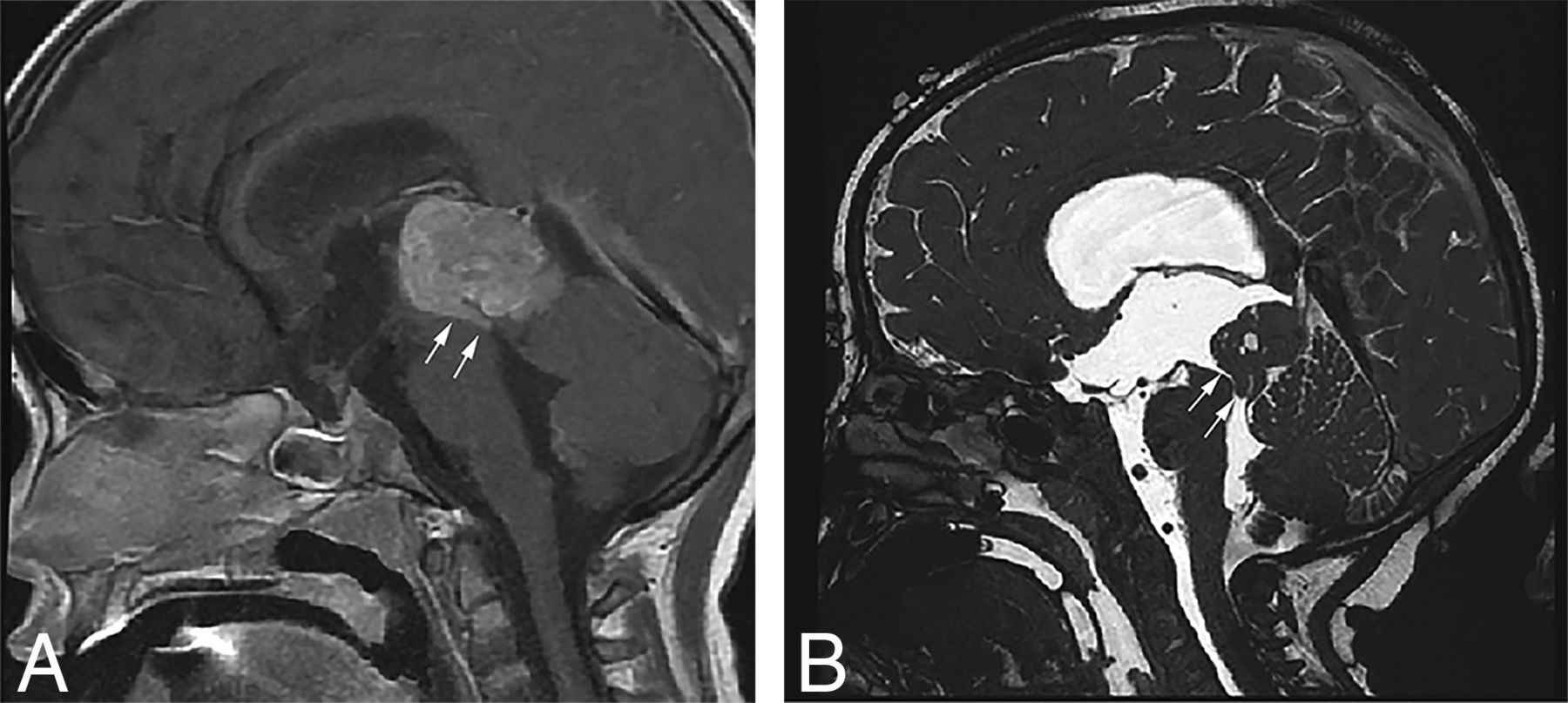
  • Fig 4.
    • Download figure
    • Open in new tab
    • Download powerpoint
    Fig 4.

    Sagittal T1-weighted postcontrast (A) and sagittal high-resolution balanced steady-state gradient-echo (B) images from 2 different patients with molecularly proved pineoblastomas demonstrating a tail-like aqueductal extension (white arrows).

  • Fig 5.
    • Download figure
    • Open in new tab
    • Download powerpoint
    Fig 5.

    (A and B), A 4-year-old girl with a molecular diagnosis of high-grade glioma (GBM_MYCN). Axial DWI (A) and sagittal postcontrast T1-weighted (B) images demonstrate a large mass centered in the left frontal lobe with prominent necrotic/cystic areas centrally and diffusion restriction and moderate heterogeneous enhancement of the solid component. (C and D), A 4-year-old boy with a molecular diagnosis of ependymoma (EP_RELA). Axial DWI (C) and sagittal postcontrast T1-weighted (D) images demonstrate a large mass centered deep in the left hemisphere with prominent necrotic/cystic areas centrally and diffusion restriction and moderate heterogeneous enhancement of the solid component. Note the similarities in age and imaging appearance between these 2 patients with different molecular diagnoses.

  • Fig 6.
    • Download figure
    • Open in new tab
    • Download powerpoint
    Fig 6.

    Two different patients with a molecular diagnosis of ET. (A and B), A 10-year-old girl with axial T2-weighted (A) and axial postcontrast T1-weighted (B) images has a large mass centered in the left deep nuclei with a prominent cystic component and moderate enhancement of the solid component. The tumor subclass was CNS_NB_FOXR2. Please note the similarities with high-grade glioma and ependymoma illustrated in Fig 5. (C and D), A 5-year-old girl with axial T2-weighted (C) and sagittal postcontrast T1-weighted (D) images has a large solid mass centered in the right lateral ventricle with minimal-to-no enhancement. The tumor subclass was ETMR. Of note, both of these tumors demonstrated diffusion restriction (not shown).

Tables

  • Figures
    • View popup
    Table 1:

    Molecular diagnoses for tumors with both imaging and methylation profiles available (n = 56)a

    PBL/ETNo.Non-ETNo.
    PBL27GBM_G34d8
    CNS_NB_FOXR2b3GBM_MYCNd5
    ETMRb1DMG_K27d2
    HGNET_MN1b1GBM_MIDd2
    MB_WNTb1EP_RELA2
    CNS ET, NOSb1
    ATRT_SHHc2
    ATRT_MYCc1
    Total3719
    • Note:—CNS_NB_FOXR2 indicates CNS neuroblastoma with FOXR2 activation; HGNET_MN1, CNS high-grade neuroepithelial tumor with MN1 alteration; MB_WNT: medulloblastoma with wingless (Wnt) activation; ATRT_SHH, atypical teratoid/rhabdoid tumor with sonic hedgehog (shh) activation; ATRT_MYC, atypical teratoid/rhabdoid tumor, subclass MYC; GBM_G34, glioblastoma, Isocitrate dehydrogenase (IDH) wild-type, H3.3 G34 mutant; GBM_MYCN, glioblastoma, IDH wild-type, subclass MYCN; DMG_K27, diffuse midline glioma H3K27M mutant; GBM-MID, glioblastoma, IDH wild-type, subclass midline; EP-RELA, ependymoma with positive RELA fusion.

    • ↵a For further analysis, the following have been combined into single groups:

    • ↵b As ET, other.

    • ↵c As ATRT.

    • ↵d As HGG.

    • View popup
    Table 2:

    Molecular diagnoses for tumors by pineal and nonpineal locations

    PinealNonpineal
    PBL261
    ET, other07
    ATRT12
    HGG116
    EP02
    Total2828
    • View popup
    Table 3:

    MR imaging features by tumor group (all patients n = 56)

    GroupP Value
    PBL/ETNon-ETAll Patients
    No.%No.%No.%
    Size (cm)<.001
        Median3.6–6.2–4.3–
        Minimum1.1–2.7–1.1–
        Maximum9.1–9.3–9.3–
    % Enhancement.17a, .80b
        None12.70011.8
        0–2538.1315.8610.7
        25–75616.2736.81323.2
        >752773.0947.43664.3
    Margins<.001
        Well-defined37100.01368.45089.3
        Ill-defined00631.6610.7
    Presence of edema<.001c
        Absent3286.5526.33766.1
        <2 cm from tumor margin410.81368.41730.4
        >2 cm from tumor margin12.715.323.6
    Presence of cyst/necrosis.22
        Absent1232.4315.81526.8
        Present2567.61684.24173.2
    Presence of calcification or hemorrhage.26
        Absent1643.2526.32137.5
        Present2156.81473.73562.5
    DWI–
        Bright2875.71578.94376.8
        Dark12.70011.8
        Intermediate513.5315.8814.3
        Artifact or not available38.115.347.1
    Metastasis–
        Intracranial12.70011.8
        Spinal513.50058.9
        Intracranial and spinal616.200610.7
        None2567.619100.04478.6
        All patients37100.019100.056100.0
    • Note:—– indicates no data available.

    • a Comparison of none versus 0%–25% versus 25%–75% versus >75%.

    • b Comparison of >75% versus ≤75%.

    • c Comparison of absent versus present.

    • View popup
    Table 4:

    MR imaging features by tumor group after excluding pineoblastomas (n = 29)

    GroupP Value
    ETNon-ETAll Patients
    No.%No.%No.%
    Size (cm).95
        Median5.7–6.2–6.1–
        Minimum3.6–2.7–2.7–
        Maximum9.1–9.3–9.3–
    % Enhancement.68a
        None000000
        0–25220315.8517.2
        25–75550736.81241.4
        >75330947.41241.4
    Margins.068
        Well-defined10100.01368.42379.3
        Ill-defined00631.6620.7
    Presence of edema.24b
        Absent550526.31034.5
        <2 cm from tumor margin4401368.41758.6
        >2 cm from tumor margin11015.326.9
    Presence of cyst/necrosis.53
        Absent00315.8310.3
        Present101001684.22689.7
    Presence of calcification or hemorrhage.11
        Absent660526.31137.9
        Present4401473.71862.1
    DWI–
        Bright9901578.92482.8
        Dark000000
        Intermediate00315.8310.3
        Artifact or not available11015.326.9
    Metastasis
        Intracranial000000
        Spinal000000
        Intracranial and spinal000000
        None1010019100.029100
        All patients10100.019100.029100.0
    • Note:— – indicates no data available.

    • a Comparison of none versus 0%–25% versus 25%–75% versus >75%.

    • b Comparison of absent versus present.

PreviousNext
Back to top

In this issue

American Journal of Neuroradiology: 40 (11)
American Journal of Neuroradiology
Vol. 40, Issue 11
1 Nov 2019
  • Table of Contents
  • Index by author
  • Complete Issue (PDF)
Advertisement
Print
Download PDF
Email Article

Thank you for your interest in spreading the word on American Journal of Neuroradiology.

NOTE: We only request your email address so that the person you are recommending the page to knows that you wanted them to see it, and that it is not junk mail. We do not capture any email address.

Enter multiple addresses on separate lines or separate them with commas.
MRI Features of Histologically Diagnosed Supratentorial Primitive Neuroectodermal Tumors and Pineoblastomas in Correlation with Molecular Diagnoses and Outcomes: A Report from the Children's Oncology Group ACNS0332 Trial
(Your Name) has sent you a message from American Journal of Neuroradiology
(Your Name) thought you would like to see the American Journal of Neuroradiology web site.
CAPTCHA
This question is for testing whether or not you are a human visitor and to prevent automated spam submissions.
Cite this article
A. Jaju, E.I. Hwang, M. Kool, D. Capper, L. Chavez, S. Brabetz, C. Billups, Y. Li, M. Fouladi, R.J. Packer, S.M. Pfister, J.M. Olson, L.A. Heier
MRI Features of Histologically Diagnosed Supratentorial Primitive Neuroectodermal Tumors and Pineoblastomas in Correlation with Molecular Diagnoses and Outcomes: A Report from the Children's Oncology Group ACNS0332 Trial
American Journal of Neuroradiology Nov 2019, 40 (11) 1796-1803; DOI: 10.3174/ajnr.A6253

Citation Manager Formats

  • BibTeX
  • Bookends
  • EasyBib
  • EndNote (tagged)
  • EndNote 8 (xml)
  • Medlars
  • Mendeley
  • Papers
  • RefWorks Tagged
  • Ref Manager
  • RIS
  • Zotero
0 Responses
Respond to this article
Share
Bookmark this article
MRI Features of Histologically Diagnosed Supratentorial Primitive Neuroectodermal Tumors and Pineoblastomas in Correlation with Molecular Diagnoses and Outcomes: A Report from the Children's Oncology Group ACNS0332 Trial
A. Jaju, E.I. Hwang, M. Kool, D. Capper, L. Chavez, S. Brabetz, C. Billups, Y. Li, M. Fouladi, R.J. Packer, S.M. Pfister, J.M. Olson, L.A. Heier
American Journal of Neuroradiology Nov 2019, 40 (11) 1796-1803; DOI: 10.3174/ajnr.A6253
del.icio.us logo Twitter logo Facebook logo Mendeley logo
  • Tweet Widget
  • Facebook Like
  • Google Plus One
Purchase

Jump to section

  • Article
    • Abstract
    • ABBREVIATIONS:
    • MATERIALS AND METHODS
    • RESULTS
    • DISCUSSION
    • CONCLUSIONS
    • Footnotes
    • References
  • Figures & Data
  • Info & Metrics
  • Responses
  • References
  • PDF

Related Articles

  • Erratum
  • PubMed
  • Google Scholar

Cited By...

  • MR Imaging and Clinical Characteristics of Diffuse Glioneuronal Tumor with Oligodendroglioma-like Features and Nuclear Clusters
  • Radiomics Can Distinguish Pediatric Supratentorial Embryonal Tumors, High-Grade Gliomas, and Ependymomas
  • Crossref
  • Google Scholar

This article has not yet been cited by articles in journals that are participating in Crossref Cited-by Linking.

More in this TOC Section

  • Frontal Paraventricular Cysts
  • Sodium MRI in Pediatric Brain Tumors
  • FRACTURE MR in Congenital Vertebral Anomalies
Show more Pediatric Neuroimaging

Similar Articles

Advertisement

Indexed Content

  • Current Issue
  • Accepted Manuscripts
  • Article Preview
  • Past Issues
  • Editorials
  • Editor's Choice
  • Fellows' Journal Club
  • Letters to the Editor
  • Video Articles

Cases

  • Case Collection
  • Archive - Case of the Week
  • Archive - Case of the Month
  • Archive - Classic Case

More from AJNR

  • Trainee Corner
  • Imaging Protocols
  • MRI Safety Corner
  • Book Reviews

Multimedia

  • AJNR Podcasts
  • AJNR Scantastics

Resources

  • Turnaround Time
  • Submit a Manuscript
  • Submit a Video Article
  • Submit an eLetter to the Editor/Response
  • Manuscript Submission Guidelines
  • Statistical Tips
  • Fast Publishing of Accepted Manuscripts
  • Graphical Abstract Preparation
  • Imaging Protocol Submission
  • Evidence-Based Medicine Level Guide
  • Publishing Checklists
  • Author Policies
  • Become a Reviewer/Academy of Reviewers
  • News and Updates

About Us

  • About AJNR
  • Editorial Board
  • Editorial Board Alumni
  • Alerts
  • Permissions
  • Not an AJNR Subscriber? Join Now
  • Advertise with Us
  • Librarian Resources
  • Feedback
  • Terms and Conditions
  • AJNR Editorial Board Alumni

American Society of Neuroradiology

  • Not an ASNR Member? Join Now

© 2025 by the American Society of Neuroradiology All rights, including for text and data mining, AI training, and similar technologies, are reserved.
Print ISSN: 0195-6108 Online ISSN: 1936-959X

Powered by HighWire