Advertisement

Main menu

  • Home
  • Content
    • Current Issue
    • Accepted Manuscripts
    • Article Preview
    • Past Issue Archive
    • Video Articles
    • AJNR Case Collection
    • Case of the Week Archive
    • Case of the Month Archive
    • Classic Case Archive
  • Special Collections
    • AJNR Awards
    • Low-Field MRI
    • Alzheimer Disease
    • ASNR Foundation Special Collection
    • Photon-Counting CT
    • View All
  • Multimedia
    • AJNR Podcasts
    • AJNR SCANtastic
    • Trainee Corner
    • MRI Safety Corner
    • Imaging Protocols
  • For Authors
    • Submit a Manuscript
    • Submit a Video Article
    • Submit an eLetter to the Editor/Response
    • Manuscript Submission Guidelines
    • Statistical Tips
    • Fast Publishing of Accepted Manuscripts
    • Graphical Abstract Preparation
    • Imaging Protocol Submission
    • Author Policies
  • About Us
    • About AJNR
    • Editorial Board
    • Editorial Board Alumni
  • More
    • Become a Reviewer/Academy of Reviewers
    • Subscribers
    • Permissions
    • Alerts
    • Feedback
    • Advertisers
    • ASNR Home

User menu

  • Alerts
  • Log in

Search

  • Advanced search
American Journal of Neuroradiology

American Journal of Neuroradiology

ASHNR American Society of Functional Neuroradiology ASHNR American Society of Pediatric Neuroradiology ASSR
  • Alerts
  • Log in

Advanced Search

  • Home
  • Content
    • Current Issue
    • Accepted Manuscripts
    • Article Preview
    • Past Issue Archive
    • Video Articles
    • AJNR Case Collection
    • Case of the Week Archive
    • Case of the Month Archive
    • Classic Case Archive
  • Special Collections
    • AJNR Awards
    • Low-Field MRI
    • Alzheimer Disease
    • ASNR Foundation Special Collection
    • Photon-Counting CT
    • View All
  • Multimedia
    • AJNR Podcasts
    • AJNR SCANtastic
    • Trainee Corner
    • MRI Safety Corner
    • Imaging Protocols
  • For Authors
    • Submit a Manuscript
    • Submit a Video Article
    • Submit an eLetter to the Editor/Response
    • Manuscript Submission Guidelines
    • Statistical Tips
    • Fast Publishing of Accepted Manuscripts
    • Graphical Abstract Preparation
    • Imaging Protocol Submission
    • Author Policies
  • About Us
    • About AJNR
    • Editorial Board
    • Editorial Board Alumni
  • More
    • Become a Reviewer/Academy of Reviewers
    • Subscribers
    • Permissions
    • Alerts
    • Feedback
    • Advertisers
    • ASNR Home
  • Follow AJNR on Twitter
  • Visit AJNR on Facebook
  • Follow AJNR on Instagram
  • Join AJNR on LinkedIn
  • RSS Feeds

AJNR Awards, New Junior Editors, and more. Read the latest AJNR updates

Research ArticleNeurointervention
Open Access

Osseous versus Nonosseous Spinal Epidural Arteriovenous Fistulas: Experiences of 13 Patients

Y. Song, S.H. Cho, D.W. Lee, J.J. Sheen, J.H. Shin and D.C. Suh
American Journal of Neuroradiology January 2019, 40 (1) 129-134; DOI: https://doi.org/10.3174/ajnr.A5904
Y. Song
aFrom the Departments of Radiology and Research Institute of Radiology (Y.S., D.W.L., J.J.S., J.H.S., D.C.S.), University of Ulsan, College of Medicine, Asan Medical Center, Seoul, Republic of Korea
  • Find this author on Google Scholar
  • Find this author on PubMed
  • Search for this author on this site
  • ORCID record for Y. Song
S.H. Cho
bDepartment of Neurosurgery (S.H.C.), Ulsan University College of Medicine, Asan Hospital, Gang-reung, Republic of Korea.
  • Find this author on Google Scholar
  • Find this author on PubMed
  • Search for this author on this site
  • ORCID record for S.H. Cho
D.W. Lee
aFrom the Departments of Radiology and Research Institute of Radiology (Y.S., D.W.L., J.J.S., J.H.S., D.C.S.), University of Ulsan, College of Medicine, Asan Medical Center, Seoul, Republic of Korea
  • Find this author on Google Scholar
  • Find this author on PubMed
  • Search for this author on this site
  • ORCID record for D.W. Lee
J.J. Sheen
aFrom the Departments of Radiology and Research Institute of Radiology (Y.S., D.W.L., J.J.S., J.H.S., D.C.S.), University of Ulsan, College of Medicine, Asan Medical Center, Seoul, Republic of Korea
  • Find this author on Google Scholar
  • Find this author on PubMed
  • Search for this author on this site
  • ORCID record for J.J. Sheen
J.H. Shin
aFrom the Departments of Radiology and Research Institute of Radiology (Y.S., D.W.L., J.J.S., J.H.S., D.C.S.), University of Ulsan, College of Medicine, Asan Medical Center, Seoul, Republic of Korea
  • Find this author on Google Scholar
  • Find this author on PubMed
  • Search for this author on this site
  • ORCID record for J.H. Shin
D.C. Suh
aFrom the Departments of Radiology and Research Institute of Radiology (Y.S., D.W.L., J.J.S., J.H.S., D.C.S.), University of Ulsan, College of Medicine, Asan Medical Center, Seoul, Republic of Korea
  • Find this author on Google Scholar
  • Find this author on PubMed
  • Search for this author on this site
  • ORCID record for D.C. Suh
  • Article
  • Figures & Data
  • Supplemental
  • Info & Metrics
  • Responses
  • References
  • PDF
Loading

Article Figures & Data

Figures

  • Fig 1.
    • Download figure
    • Open in new tab
    • Download powerpoint
    Fig 1.

    Schematic diagram of a spinal epidural arteriovenous fistula. An osseous SEDAVF shows bone involvement and compression of the nerve root or the spinal cord by bulging with cortical erosion (A). A nonosseous SEDAVF shows engorgement (fistulous sac or pouch) of the epidural vein (B) within the spinal canal compressing the spinal cord (C).
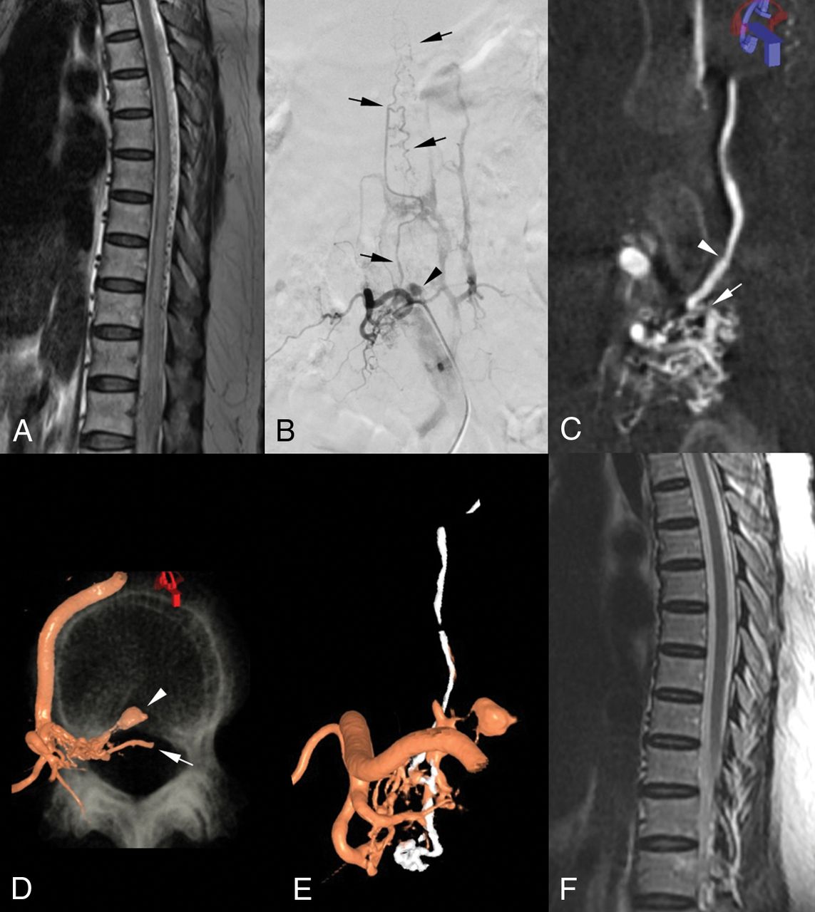
  • Fig 2.
    • Download figure
    • Open in new tab
    • Download powerpoint
    Fig 2.

    Spinal epidural arteriovenous fistula (perimedullary venous drainage) in a 63-year-old woman (case 4). A, Sagittal T2-weighted MR imaging reveals diffuse spinal cord edema up to the T5 level and multiple signal voids in the posterior aspect of the cord. B, Right lumbar arteriogram shows an arteriovenous fistula at the L2 level with multiple fine feeding arteries and early venous drainage to the epidural and paravertebral venous plexuses and to intradural veins (arrows). A small venous sac is visible around the fistula (arrowhead). C, 3D rotational angiogram shows the anatomy in detail where the intradural reflux originated (arrow). Note the focal narrowing of the vein where it penetrates the dura (arrowhead). D, Volume-rendering image demonstrates the epidural location of the fistula and the venous pouch (arrowhead). An intradural course of the radicular vein is also visible (arrow). E, Postembolization 3D angiogram confirms the presence of radiopaque glue in the fistula and along the intradural vein (white areas). F, Follow-up MR imaging 2 months later shows that the diffuse cord edema and the perimedullary vessels have disappeared.

  • Fig 3.
    • Download figure
    • Open in new tab
    • Download powerpoint
    Fig 3.

    Osseous spinal epidural arteriovenous fistula in a 57-year-old man (case 9). A, T2-weighted axial MR image reveals a well-defined lesion with dark signal intensity in the right epidural space at the C6 level. B, CT scan at the corresponding level shows adjacent bone destruction of the right lamina and spinous process of the C6 vertebra that resulted from the well-enhanced epidural lesion. C (right) and D (left), Deep cervical arteriograms. A large arteriovenous fistula with multiple arterial feeders is visible at the C6 level. E, 3D fusion image demonstrates that the feeders from both sides converge on a focal region—the epidural venous plexus and internal jugular veins—from which venous flow drains exclusively via extradural veins. F, After 2 sessions of transvenous coil embolization, the fistula flow has almost completely disappeared (G and H).

  • Fig 4.
    • Download figure
    • Open in new tab
    • Download powerpoint
    Fig 4.

    Outcomes in patients according to each scoring system and the presence of intradural reflux. The mean scores of the initial and follow-up evaluations are shown. Asterisks indicate statistically significant improvement at follow-up.

PreviousNext
Back to top

In this issue

American Journal of Neuroradiology: 40 (1)
American Journal of Neuroradiology
Vol. 40, Issue 1
1 Jan 2019
  • Table of Contents
  • Index by author
  • Complete Issue (PDF)
Advertisement
Print
Download PDF
Email Article

Thank you for your interest in spreading the word on American Journal of Neuroradiology.

NOTE: We only request your email address so that the person you are recommending the page to knows that you wanted them to see it, and that it is not junk mail. We do not capture any email address.

Enter multiple addresses on separate lines or separate them with commas.
Osseous versus Nonosseous Spinal Epidural Arteriovenous Fistulas: Experiences of 13 Patients
(Your Name) has sent you a message from American Journal of Neuroradiology
(Your Name) thought you would like to see the American Journal of Neuroradiology web site.
CAPTCHA
This question is for testing whether or not you are a human visitor and to prevent automated spam submissions.
Cite this article
Y. Song, S.H. Cho, D.W. Lee, J.J. Sheen, J.H. Shin, D.C. Suh
Osseous versus Nonosseous Spinal Epidural Arteriovenous Fistulas: Experiences of 13 Patients
American Journal of Neuroradiology Jan 2019, 40 (1) 129-134; DOI: 10.3174/ajnr.A5904

Citation Manager Formats

  • BibTeX
  • Bookends
  • EasyBib
  • EndNote (tagged)
  • EndNote 8 (xml)
  • Medlars
  • Mendeley
  • Papers
  • RefWorks Tagged
  • Ref Manager
  • RIS
  • Zotero
0 Responses
Respond to this article
Share
Bookmark this article
Osseous versus Nonosseous Spinal Epidural Arteriovenous Fistulas: Experiences of 13 Patients
Y. Song, S.H. Cho, D.W. Lee, J.J. Sheen, J.H. Shin, D.C. Suh
American Journal of Neuroradiology Jan 2019, 40 (1) 129-134; DOI: 10.3174/ajnr.A5904
del.icio.us logo Twitter logo Facebook logo Mendeley logo
  • Tweet Widget
  • Facebook Like
  • Google Plus One
Purchase

Jump to section

  • Article
    • Abstract
    • ABBREVIATIONS:
    • Materials and Methods
    • Results
    • Discussion
    • Conclusions
    • Footnotes
    • References
  • Figures & Data
  • Supplemental
  • Info & Metrics
  • Responses
  • References
  • PDF

Related Articles

  • PubMed
  • Google Scholar

Cited By...

  • No citing articles found.
  • Crossref
  • Google Scholar

This article has not yet been cited by articles in journals that are participating in Crossref Cited-by Linking.

More in this TOC Section

Neurointervention

  • Rescue Reentry in Carotid Near-Occlusion
  • Contour Neurovascular System: Five Year Follow Up
  • Effect of SARS-CoV2 on Endovascular Thrombectomy
Show more Neurointervention

Spine Imaging and Spine Image-Guided Interventions

  • Optimization of Photon Counting CT Myelography
  • Characteristics of SIH Type I Culprit Lesions
  • Management Outcomes For VO Spine Biopsy
Show more Spine Imaging and Spine Image-Guided Interventions

Similar Articles

Advertisement

Indexed Content

  • Current Issue
  • Accepted Manuscripts
  • Article Preview
  • Past Issues
  • Editorials
  • Editor's Choice
  • Fellows' Journal Club
  • Letters to the Editor
  • Video Articles

Cases

  • Case Collection
  • Archive - Case of the Week
  • Archive - Case of the Month
  • Archive - Classic Case

More from AJNR

  • Trainee Corner
  • Imaging Protocols
  • MRI Safety Corner
  • Book Reviews

Multimedia

  • AJNR Podcasts
  • AJNR Scantastics

Resources

  • Turnaround Time
  • Submit a Manuscript
  • Submit a Video Article
  • Submit an eLetter to the Editor/Response
  • Manuscript Submission Guidelines
  • Statistical Tips
  • Fast Publishing of Accepted Manuscripts
  • Graphical Abstract Preparation
  • Imaging Protocol Submission
  • Evidence-Based Medicine Level Guide
  • Publishing Checklists
  • Author Policies
  • Become a Reviewer/Academy of Reviewers
  • News and Updates

About Us

  • About AJNR
  • Editorial Board
  • Editorial Board Alumni
  • Alerts
  • Permissions
  • Not an AJNR Subscriber? Join Now
  • Advertise with Us
  • Librarian Resources
  • Feedback
  • Terms and Conditions
  • AJNR Editorial Board Alumni

American Society of Neuroradiology

  • Not an ASNR Member? Join Now

© 2025 by the American Society of Neuroradiology All rights, including for text and data mining, AI training, and similar technologies, are reserved.
Print ISSN: 0195-6108 Online ISSN: 1936-959X

Powered by HighWire