
Article Figures & Data
Figures
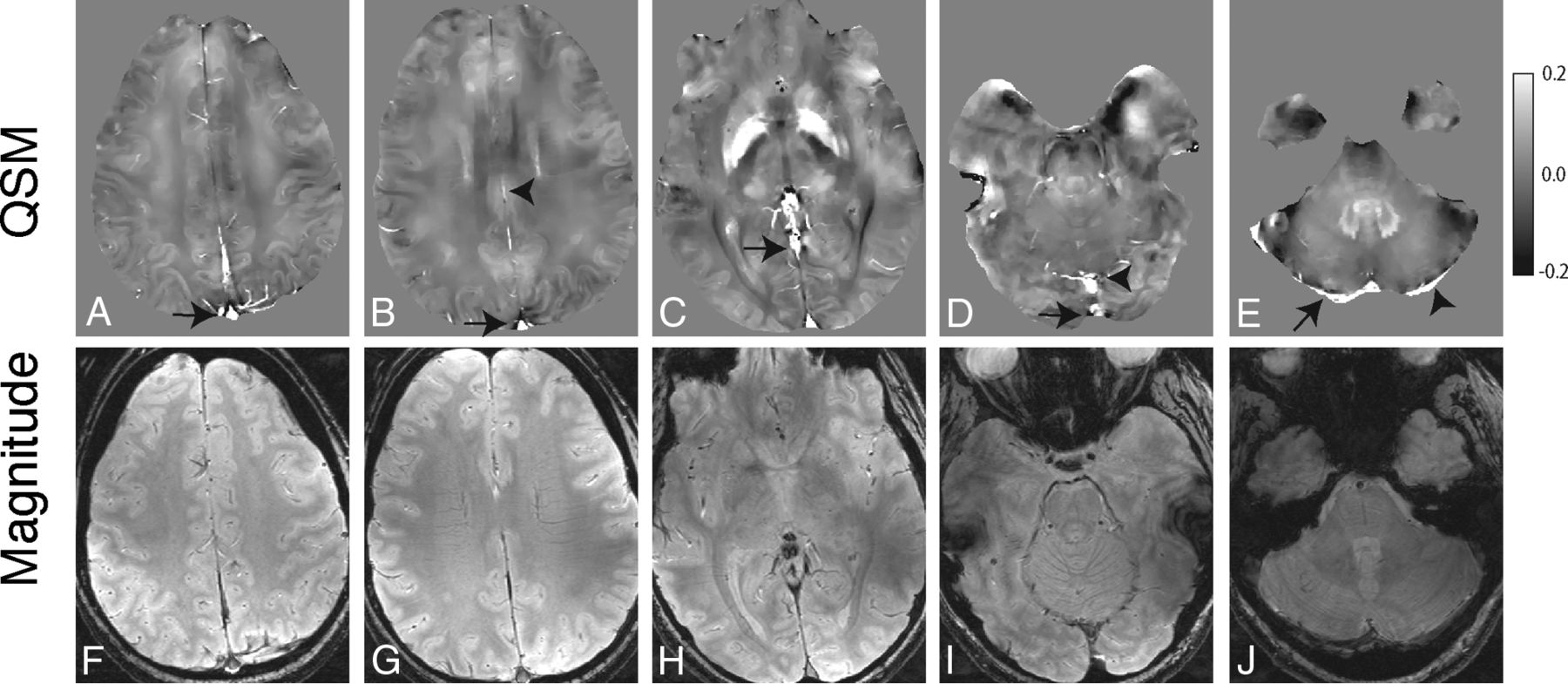
- Fig 1.
Axial QSM (A–E) and single-echo gradient-echo magnitude (F–J) images from 1 in vivo subject demonstrating representative venous segments from which we obtained ROI measurements: superior aspect of the superior sagittal sinus (arrow, A), inferior aspect of the superior sagittal sinus (arrow, B) and inferior sagittal sinus (arrowhead, B), anterior aspect of the straight sinus (arrow, C), inferior aspect of the superior sagittal sinus (arrow, D) and posterior aspect of the straight sinus (arrowhead, D), right transverse sinus (arrow, E), and left transverse sinus (arrowhead, E).
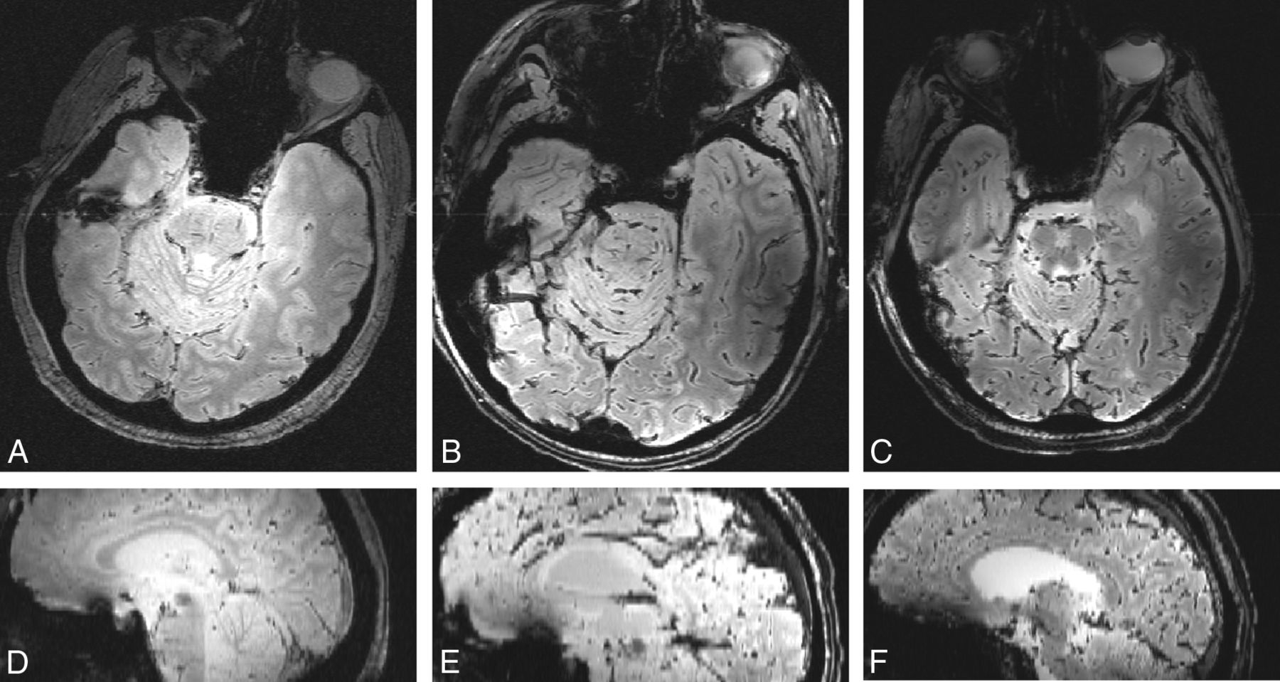
- Fig 2.
Axial T2*-weighted images and isotropically interpolated sagittal reformats using the single-echo gradient-echo sequence. Postmortem subjects 1 (A and D), 2 (B and E), and 3 (C and F) demonstrate head positioning in these planes (axial and sagittal, respectively).
- Fig 3.
Scatterplot with linear regression of 2 postmortem subjects comparing QSM and R2* ROI measurements within venous segments. A, Subject 1. B, Subject 3.
- Fig 4.
Axial R2* maps (A–C) and corresponding QSM (D–F) images depicting representative venous segments (right and left transverse sinus, A and D; inferior aspect of superior sagittal sinus and anterior aspect of straight sinus. B and E; superior aspect of superior sagittal sinus and inferior sagittal sinus, C and F) in postmortem subject 3 (A, C, D, F) and subject 2 (B and E). For the R2* images, values of >500 seconds−1 were removed to reduce visual washout from bright R2* intravenous voxels and from bone/air.


Tables
- Table 1:
Comparison among R2*, susceptibility measurements, and calculated hematocrit values in intracranial veins in 3 postmortem subjects
Venous Segment Subject 1 Subject 2 Subject 3 Susceptibility (ppm) Hematocrit R2* (1/s) Susceptibility (ppm) Hematocrit Susceptibility (ppm) Hematocrit R2* (1/s) Superior aspect of superior sagittal sinus 0.963 0.319 141 0.677 0.225 0.927 0.308 155 Inferior aspect of superior sagittal sinus NA NA NA 0.782 0.259 0.985 0.327 164 Left transverse sinus 1.749 0.580 291 0.780 0.259 1.142 0.379 176 Right transverse sinus 1.135 0.377 277 1.045 0.347 0.785 0.260 161 Anterior aspect of straight sinus 0.661 0.219 94 0.643 0.213 0.681 0.226 104 Posterior aspect of straight sinus 0.972 0.322 182 0.841 0.279 0.939 0.312 177 Inferior sagittal sinus 0.523 0.174 90 0.307 0.102 0.447 0.148 76 Note:—NA indicates not available.
- Table 2:
Percentage decrease in hematocrit between contiguous venous segments in postmortem subjects
Venous Segments Compared Subject 1a Subject 2b Subject 3c Superior aspect of superior sagittal sinus/inferior aspect of superior sagittal sinus NA 13.4 5.8 Anterior aspect of straight sinus/posterior aspect of straight sinus 32.0 23.5 27.4 - Table 3:
Susceptibility measurements (ppm) from intracranial venous segments in 10 healthy subjects
Venous Segment Subject Average ± SD 1 2 3 4 5 6 7 8 9 10 Superior aspect of superior sagittal sinus 0.202 0.212 0.179 0.263 0.188 0.239 0.205 0.172 0.234 0.227 0.212 ± 0.029 Inferior aspect of superior sagittal sinus 0.270 0.238 0.249 0.285 0.211 0.215 0.228 0.230 0.261 0.251 0.244 ± 0.024 Left transverse sinus 0.343 0.255 0.329 0.389 0.263 0.280 0.292 0.311 0.278 0.327 0.307 ± 0.041 Right transverse sinus 0.346 0.228 0.306 0.366 0.290 0.267 0.252 0.252 0.285 0.297 0.289 ± 0.043 Anterior aspect of straight sinus 0.393 0.286 0.369 0.329 0.287 0.287 0.290 0.326 0.325 0.272 0.316 ± 0.040 Posterior aspect of straight sinus 0.403 0.280 0.286 0.334 0.184 0.187 0.201 0.270 0.295 0.250 0.269 ± 0.069 Inferior sagittal sinus 0.190 0.145 0.194 0.295 0.184 0.173 0.172 0.227 0.226 0.155 0.196 ± 0.044 - Table 4:
Calculated percentage SvO2 for intracranial venous segments in 10 healthy subjects
Venous Segment Subject Average ± SD 1 2 3 4 5 6 7 8 9 10 Superior aspect of superior sagittal sinus 74.0 73.3 75.7 69.5 75.0 71.3 73.8 76.2 71.7 72.1 73.3 ± 2.1 Inferior aspect of superior sagittal sinus 69.0 71.3 70.6 67.9 73.4 73.0 72.1 71.9 69.7 70.4 70.9 ± 1.8 Left transverse sinus 63.6 70.1 64.6 60.2 69.5 68.3 67.4 66.0 68.4 64.8 66.3 ± 3.1 Right transverse sinus 63.4 72.1 66.4 61.9 67.5 69.2 70.3 70.3 67.9 67.0 67.6 ± 3.2 Anterior aspect of straight sinus 60.0 67.8 61.7 64.7 67.7 67.7 67.5 64.9 65.0 68.8 65.6 ± 2.9 Posterior aspect of straight sinus 59.2 68.3 67.8 64.3 75.3 75.1 74.0 69.0 67.1 70.5 69.1 ± 5.1 Inferior sagittal sinus 74.9 78.2 74.6 67.1 75.3 76.1 76.2 72.1 72.2 77.5 74.4 ± 3.2


Jump to section
Related Articles
Cited By...
- No citing articles found.