Advertisement

Main menu

  • Home
  • Content
    • Current Issue
    • Accepted Manuscripts
    • Article Preview
    • Past Issue Archive
    • Video Articles
    • AJNR Case Collection
    • Case of the Week Archive
    • Case of the Month Archive
    • Classic Case Archive
  • Special Collections
    • AJNR Awards
    • Low-Field MRI
    • Alzheimer Disease
    • ASNR Foundation Special Collection
    • Photon-Counting CT
    • View All
  • Multimedia
    • AJNR Podcasts
    • AJNR SCANtastic
    • Trainee Corner
    • MRI Safety Corner
    • Imaging Protocols
  • For Authors
    • Submit a Manuscript
    • Submit a Video Article
    • Submit an eLetter to the Editor/Response
    • Manuscript Submission Guidelines
    • Statistical Tips
    • Fast Publishing of Accepted Manuscripts
    • Graphical Abstract Preparation
    • Imaging Protocol Submission
    • Author Policies
  • About Us
    • About AJNR
    • Editorial Board
    • Editorial Board Alumni
  • More
    • Become a Reviewer/Academy of Reviewers
    • Subscribers
    • Permissions
    • Alerts
    • Feedback
    • Advertisers
    • ASNR Home

User menu

  • Alerts
  • Log in

Search

  • Advanced search
American Journal of Neuroradiology

American Journal of Neuroradiology

ASHNR American Society of Functional Neuroradiology ASHNR American Society of Pediatric Neuroradiology ASSR
  • Alerts
  • Log in

Advanced Search

  • Home
  • Content
    • Current Issue
    • Accepted Manuscripts
    • Article Preview
    • Past Issue Archive
    • Video Articles
    • AJNR Case Collection
    • Case of the Week Archive
    • Case of the Month Archive
    • Classic Case Archive
  • Special Collections
    • AJNR Awards
    • Low-Field MRI
    • Alzheimer Disease
    • ASNR Foundation Special Collection
    • Photon-Counting CT
    • View All
  • Multimedia
    • AJNR Podcasts
    • AJNR SCANtastic
    • Trainee Corner
    • MRI Safety Corner
    • Imaging Protocols
  • For Authors
    • Submit a Manuscript
    • Submit a Video Article
    • Submit an eLetter to the Editor/Response
    • Manuscript Submission Guidelines
    • Statistical Tips
    • Fast Publishing of Accepted Manuscripts
    • Graphical Abstract Preparation
    • Imaging Protocol Submission
    • Author Policies
  • About Us
    • About AJNR
    • Editorial Board
    • Editorial Board Alumni
  • More
    • Become a Reviewer/Academy of Reviewers
    • Subscribers
    • Permissions
    • Alerts
    • Feedback
    • Advertisers
    • ASNR Home
  • Follow AJNR on Twitter
  • Visit AJNR on Facebook
  • Follow AJNR on Instagram
  • Join AJNR on LinkedIn
  • RSS Feeds

AJNR Awards, New Junior Editors, and more. Read the latest AJNR updates

Research ArticleAdult Brain

Visualization and Classification of Deeply Seated Collateral Networks in Moyamoya Angiopathy with 7T MRI

T. Matsushige, M. Kraemer, T. Sato, P. Berlit, M. Forsting, M.E. Ladd, R. Jabbarli, U. Sure, N. Khan, M. Schlamann and K.H. Wrede
American Journal of Neuroradiology July 2018, 39 (7) 1248-1254; DOI: https://doi.org/10.3174/ajnr.A5700
T. Matsushige
aFrom the Department of Neurosurgery (T.M., T.S., R.J., U.S., K.H.W.), University Hospital Essen, University Duisburg-Essen, Essen, Germany
bDepartment of Neurosurgery (T.M.), Graduate School of Biomedical and Health Sciences, Hiroshima University, Hiroshima, Japan
cErwin L. Hahn Institute for Magnetic Resonance Imaging (T.M., T.S., M.E.L., K.H.W.), University Duisburg-Essen, Essen, Germany
  • Find this author on Google Scholar
  • Find this author on PubMed
  • Search for this author on this site
  • ORCID record for T. Matsushige
M. Kraemer
dDepartment of Neurology (M.K., P.B.), Alfried Krupp Hospital, Essen, Germany
eDepartment of Neurology (M.K.), University Hospital Duesseldorf, Duesseldorf, Germany
  • Find this author on Google Scholar
  • Find this author on PubMed
  • Search for this author on this site
  • ORCID record for M. Kraemer
T. Sato
aFrom the Department of Neurosurgery (T.M., T.S., R.J., U.S., K.H.W.), University Hospital Essen, University Duisburg-Essen, Essen, Germany
cErwin L. Hahn Institute for Magnetic Resonance Imaging (T.M., T.S., M.E.L., K.H.W.), University Duisburg-Essen, Essen, Germany
fDepartment of Neurosurgery (T.S.), Fukushima Medical University, Fukushima, Japan
  • Find this author on Google Scholar
  • Find this author on PubMed
  • Search for this author on this site
  • ORCID record for T. Sato
P. Berlit
dDepartment of Neurology (M.K., P.B.), Alfried Krupp Hospital, Essen, Germany
  • Find this author on Google Scholar
  • Find this author on PubMed
  • Search for this author on this site
  • ORCID record for P. Berlit
M. Forsting
gDepartment of Diagnostic and Interventional Radiology and Neuroradiology (M.F., M.S.), University Hospital Essen, Essen, Germany
  • Find this author on Google Scholar
  • Find this author on PubMed
  • Search for this author on this site
  • ORCID record for M. Forsting
M.E. Ladd
cErwin L. Hahn Institute for Magnetic Resonance Imaging (T.M., T.S., M.E.L., K.H.W.), University Duisburg-Essen, Essen, Germany
hMedical Physics in Radiology (M.E.L.), German Cancer Research Center, Heidelberg, Germany
iFaculty of Physics and Astronomy and Faculty of Medicine (M.E.L.), University of Heidelberg, Heidelberg, Germany
  • Find this author on Google Scholar
  • Find this author on PubMed
  • Search for this author on this site
  • ORCID record for M.E. Ladd
R. Jabbarli
aFrom the Department of Neurosurgery (T.M., T.S., R.J., U.S., K.H.W.), University Hospital Essen, University Duisburg-Essen, Essen, Germany
  • Find this author on Google Scholar
  • Find this author on PubMed
  • Search for this author on this site
  • ORCID record for R. Jabbarli
U. Sure
aFrom the Department of Neurosurgery (T.M., T.S., R.J., U.S., K.H.W.), University Hospital Essen, University Duisburg-Essen, Essen, Germany
  • Find this author on Google Scholar
  • Find this author on PubMed
  • Search for this author on this site
  • ORCID record for U. Sure
N. Khan
jMoyamoya Center, Division of Pediatric Neurosurgery (N.K.), Department of Surgery, University Children's Hospital Zurich, Zurich, Switzerland
  • Find this author on Google Scholar
  • Find this author on PubMed
  • Search for this author on this site
  • ORCID record for N. Khan
M. Schlamann
gDepartment of Diagnostic and Interventional Radiology and Neuroradiology (M.F., M.S.), University Hospital Essen, Essen, Germany
kDepartment of Neuroradiology (M.S.), University Hospital Cologne, Cologne, Germany.
  • Find this author on Google Scholar
  • Find this author on PubMed
  • Search for this author on this site
  • ORCID record for M. Schlamann
K.H. Wrede
aFrom the Department of Neurosurgery (T.M., T.S., R.J., U.S., K.H.W.), University Hospital Essen, University Duisburg-Essen, Essen, Germany
cErwin L. Hahn Institute for Magnetic Resonance Imaging (T.M., T.S., M.E.L., K.H.W.), University Duisburg-Essen, Essen, Germany
  • Find this author on Google Scholar
  • Find this author on PubMed
  • Search for this author on this site
  • ORCID record for K.H. Wrede
  • Article
  • Figures & Data
  • Supplemental
  • Info & Metrics
  • Responses
  • References
  • PDF
Loading

Article Figures & Data

Figures

  • Tables
  • Fig 1.
    • Download figure
    • Open in new tab
    • Download powerpoint
    Fig 1.

    Schematic illustrations of deeply seated collateral networks are shown in coronal (A) and sagittal (B) MR imaging views. Six pathways of collateral vessels according to perfusing territories can be divided into collateral networks to cortical vessels (remote, type a and b) and to major trunk vessels (local, types c–f). Anastomoses between striate arteries or choroidal arteries and cortical arteries directly or via medullary arteries (a); septal transcallosal anastomoses between choroidal arteries with pericallosal arteries (b); anastomoses between choroidal arteries or the posterior communicating artery and thalamostriate arteries (c); intrastriatal anastomosis among striatal arteries (d); intrathalamic anastomosis among thalamic arteries (e); and focal Moyamoya vessels in the basal cistern (f).

  • Fig 2.
    • Download figure
    • Open in new tab
    • Download powerpoint
    Fig 2.

    Case 1. Selective right internal carotid angiography (anteroposterior, A; lateral view, B) shows anastomotic connections to cortical arteries from the anterior choroidal artery (black arrowheads, type b) as well as from thalamostriate arteries (white arrows, type a). Collateral networks are excellently visualized in MIP from TOF-MRA with slab thickness of 20 mm (white arrows and black arrowheads) (C). Transcallosal connections to cortical arteries (white arrowheads) not detected by DSA are poorly visualized on MPRAGE (D).

  • Fig 3.
    • Download figure
    • Open in new tab
    • Download powerpoint
    Fig 3.

    Case 8. Selective right internal carotid angiography (anteroposterior, A; right 45°oblique view, B) shows anastomosis between the posterior communicating artery and the thalamostriate arteries (arrows, type c). MIP from TOF-MRA (C) demonstrates this anastomosis clearly as well as connection with the anterior choroidal artery (asterisk). The latter anastomosis could neither be detected in DSA nor visualized by MIP from MPRAGE (D).
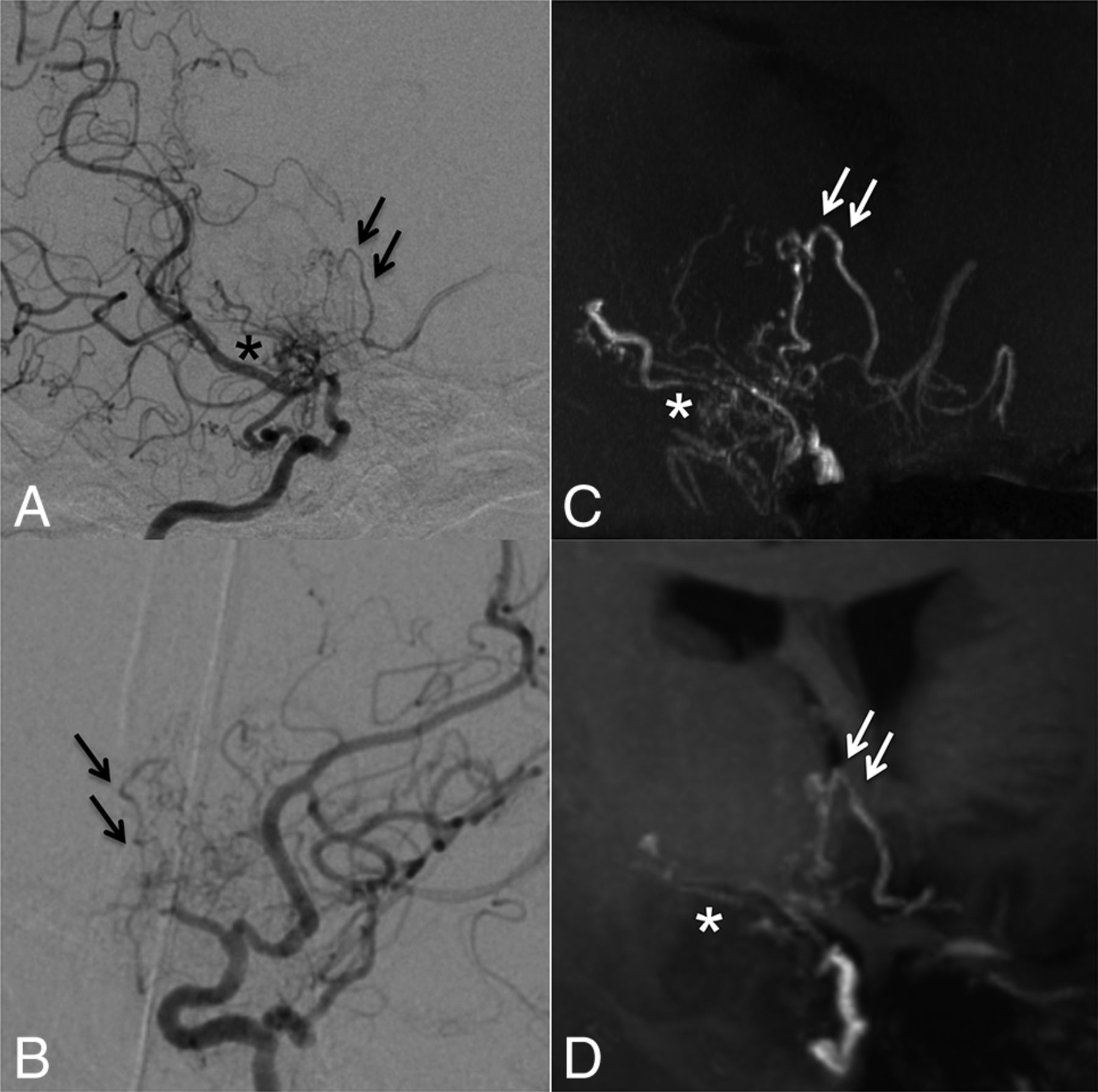
  • Fig 4.
    • Download figure
    • Open in new tab
    • Download powerpoint
    Fig 4.

    Case 5. Selective right internal carotid angiography (right 45° oblique, A; left 45° oblique view, B) shows intrastriatal anastomoses among thalamostriate arteries running through the periventricular space (arrows, type d). MIP from TOF-MRA (C) excellently demonstrates these networks as well as focal connections among Moyamoya vessels in the basal cistern (asterisk, type f). MIP from MPRAGE (D) moderately depicts the network; however, basal Moyamoya vessels are poorly visualized.
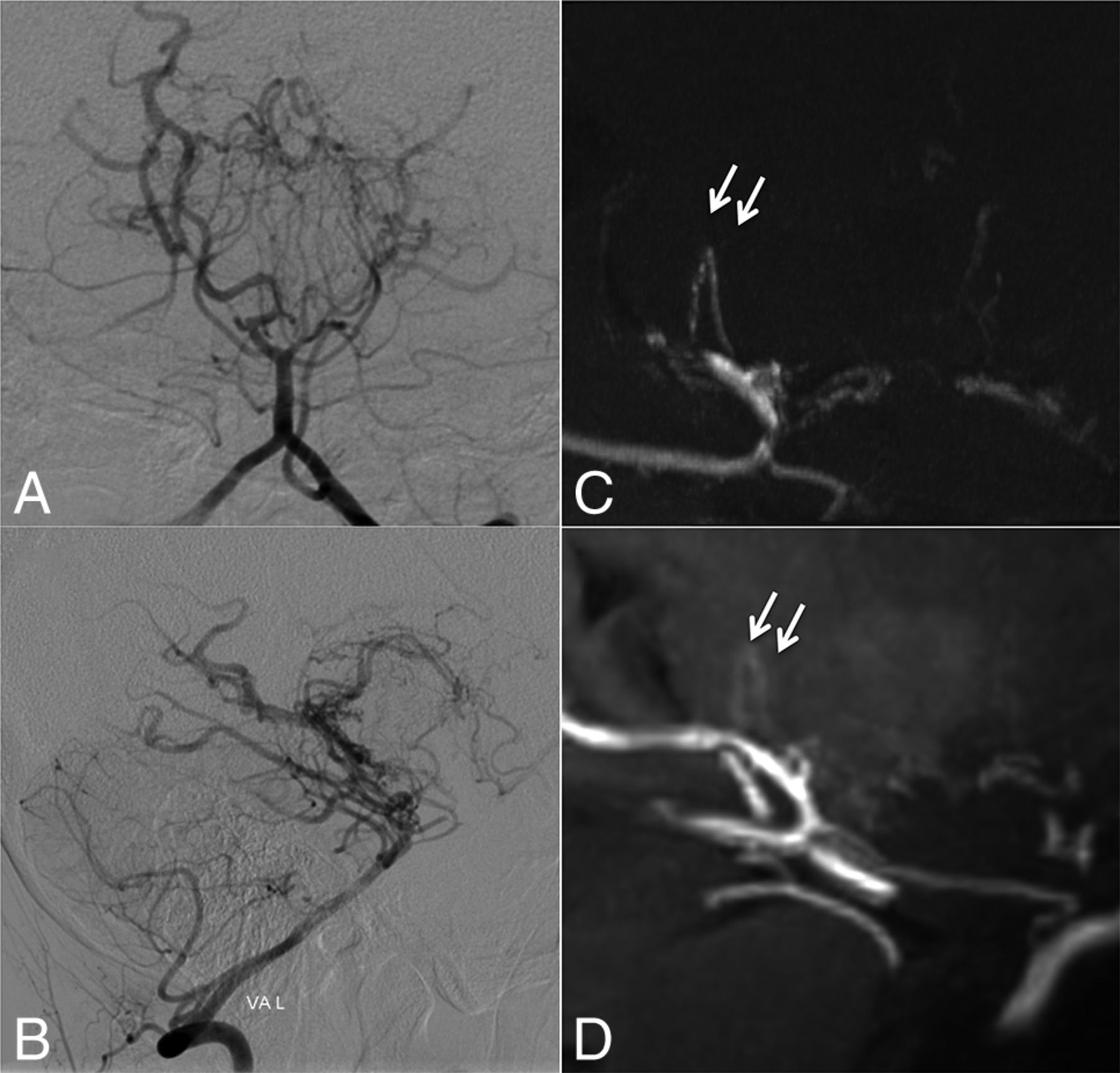
  • Fig 5.
    • Download figure
    • Open in new tab
    • Download powerpoint
    Fig 5.

    Case 6. Selective left vertebral angiography (anteroposterior, A; lateral view, B) shows no marked anastomosis. The MIP from TOF-MRA (C) demonstrates excellent visualization of inner thalamic anastomosis among posterior thalamostriate arteries (white arrows, type e), whereas the visualization is only moderate on MIP from MPRAGE (D).

  • Fig 6.
    • Download figure
    • Open in new tab
    • Download powerpoint
    Fig 6.

    Case 10. Selective right vertebral angiography (anteroposterior, A; lateral view, B) shows a tiny, barely visible anastomosis (black arrows) between the posterior thalamostriate artery and a medullary-cortical artery. MIP from TOF-MRA (C) and MIP from MPRAGE (D) do not delineate the anastomosis. The white arrow (C) indicates the supposed anatomic location of the anastomosis.

Tables

  • Figures
  • Comparison of 7T MRA and DSA regarding the number of detected collateral networks and image quality for anastomoses to major trunk vessels and collateral pathways to cortical vessels

    TypeConnecting Vessels in Collateral NetworksDSATOF-MRAMPRAGE
    Collateral networks connecting to cortical arteries
        aStriate/choroidal arteries and medullary-cortical arteries11168
        bStriate/choroidal arteries and the pericallosal arteries794
    Total182512
    Image quality (n = 18)a (mean) (SEM) (range)4.0 (0.26, 3–5)4.8 (0.10, 3–5)2.4 (0.35, 1–5)
    Collateral networks connecting to major trunk vessels
        cChoroidal arteries or PcomA and thalamostriate arteries21199
        dIntrastriatal arteries212216
        eIntrathalamic arteries886
        fMoyamoya vessels in the basal cistern323230
    Total828161
    Image quality (n = 82)a (mean) (SEM) (range)4.6 (0.07, 2–5)4.4 (0.12, 3–5)2.7 (0.13, 1–5)
    • Note:—PcomA indicates posterior communicating artery.

    • ↵a Image quality was evaluated in collateral networks detected by DSA as a clinical standard using a 5-point scale.

PreviousNext
Back to top

In this issue

American Journal of Neuroradiology: 39 (7)
American Journal of Neuroradiology
Vol. 39, Issue 7
1 Jul 2018
  • Table of Contents
  • Index by author
  • Complete Issue (PDF)
Advertisement
Print
Download PDF
Email Article

Thank you for your interest in spreading the word on American Journal of Neuroradiology.

NOTE: We only request your email address so that the person you are recommending the page to knows that you wanted them to see it, and that it is not junk mail. We do not capture any email address.

Enter multiple addresses on separate lines or separate them with commas.
Visualization and Classification of Deeply Seated Collateral Networks in Moyamoya Angiopathy with 7T MRI
(Your Name) has sent you a message from American Journal of Neuroradiology
(Your Name) thought you would like to see the American Journal of Neuroradiology web site.
CAPTCHA
This question is for testing whether or not you are a human visitor and to prevent automated spam submissions.
Cite this article
T. Matsushige, M. Kraemer, T. Sato, P. Berlit, M. Forsting, M.E. Ladd, R. Jabbarli, U. Sure, N. Khan, M. Schlamann, K.H. Wrede
Visualization and Classification of Deeply Seated Collateral Networks in Moyamoya Angiopathy with 7T MRI
American Journal of Neuroradiology Jul 2018, 39 (7) 1248-1254; DOI: 10.3174/ajnr.A5700

Citation Manager Formats

  • BibTeX
  • Bookends
  • EasyBib
  • EndNote (tagged)
  • EndNote 8 (xml)
  • Medlars
  • Mendeley
  • Papers
  • RefWorks Tagged
  • Ref Manager
  • RIS
  • Zotero
0 Responses
Respond to this article
Share
Bookmark this article
Visualization and Classification of Deeply Seated Collateral Networks in Moyamoya Angiopathy with 7T MRI
T. Matsushige, M. Kraemer, T. Sato, P. Berlit, M. Forsting, M.E. Ladd, R. Jabbarli, U. Sure, N. Khan, M. Schlamann, K.H. Wrede
American Journal of Neuroradiology Jul 2018, 39 (7) 1248-1254; DOI: 10.3174/ajnr.A5700
del.icio.us logo Twitter logo Facebook logo Mendeley logo
  • Tweet Widget
  • Facebook Like
  • Google Plus One
Purchase

Jump to section

  • Article
    • Abstract
    • ABBREVIATIONS:
    • Materials and Methods
    • Results
    • Discussion
    • Conclusions
    • Footnotes
    • References
  • Figures & Data
  • Supplemental
  • Info & Metrics
  • Responses
  • References
  • PDF

Related Articles

  • PubMed
  • Google Scholar

Cited By...

  • Transcranial Ultrasound Localization Microscopy in Moya Moya patients using a clinical ultrasound system
  • Report from the society of magnetic resonance angiography: clinical applications of 7T neurovascular MR in the assessment of intracranial vascular disease
  • Cortical Distribution of Fragile Periventricular Anastomotic Collateral Vessels in Moyamoya Disease: An Exploratory Cross-Sectional Study of Japanese Patients with Moyamoya Disease
  • Crossref
  • Google Scholar

This article has not yet been cited by articles in journals that are participating in Crossref Cited-by Linking.

More in this TOC Section

  • Diagnostic Neuroradiology of Monoclonal Antibodies
  • Cerebral ADC Changes in Fabry Disease
  • ML for Glioma Molecular Subtype Prediction
Show more Adult Brain

Similar Articles

Advertisement

Indexed Content

  • Current Issue
  • Accepted Manuscripts
  • Article Preview
  • Past Issues
  • Editorials
  • Editor's Choice
  • Fellows' Journal Club
  • Letters to the Editor
  • Video Articles

Cases

  • Case Collection
  • Archive - Case of the Week
  • Archive - Case of the Month
  • Archive - Classic Case

More from AJNR

  • Trainee Corner
  • Imaging Protocols
  • MRI Safety Corner
  • Book Reviews

Multimedia

  • AJNR Podcasts
  • AJNR Scantastics

Resources

  • Turnaround Time
  • Submit a Manuscript
  • Submit a Video Article
  • Submit an eLetter to the Editor/Response
  • Manuscript Submission Guidelines
  • Statistical Tips
  • Fast Publishing of Accepted Manuscripts
  • Graphical Abstract Preparation
  • Imaging Protocol Submission
  • Evidence-Based Medicine Level Guide
  • Publishing Checklists
  • Author Policies
  • Become a Reviewer/Academy of Reviewers
  • News and Updates

About Us

  • About AJNR
  • Editorial Board
  • Editorial Board Alumni
  • Alerts
  • Permissions
  • Not an AJNR Subscriber? Join Now
  • Advertise with Us
  • Librarian Resources
  • Feedback
  • Terms and Conditions
  • AJNR Editorial Board Alumni

American Society of Neuroradiology

  • Not an ASNR Member? Join Now

© 2025 by the American Society of Neuroradiology All rights, including for text and data mining, AI training, and similar technologies, are reserved.
Print ISSN: 0195-6108 Online ISSN: 1936-959X

Powered by HighWire