
Article Figures & Data
Figures
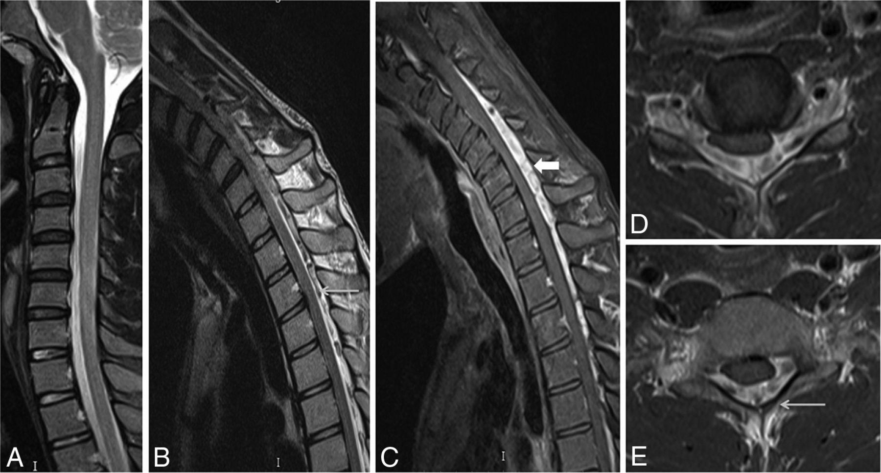
- Fig 1.
A 22-year-old man with wasting and weakness of the right hand and forearm muscles with cold paresis. Neutral position sagittal T2-weighted MR image (A) shows a normal appearance of the cervical cord. Flexion MR T2-weighted image (B) shows an enlarged posterior epidural space with multiple flow voids (arrow). Postgadolinium fat-suppressed sagittal T1-weighted flexion MR image (C) shows an enhancing epidural venous plexus extending from the C3 to T3 vertebral levels (block arrow). Axial postgadolinium T1 fat-suppressed images (D and E) show the enhancing posterior epidural venous plexus with flow voids within (arrow) and asymmetric flattening of the right hemicord.
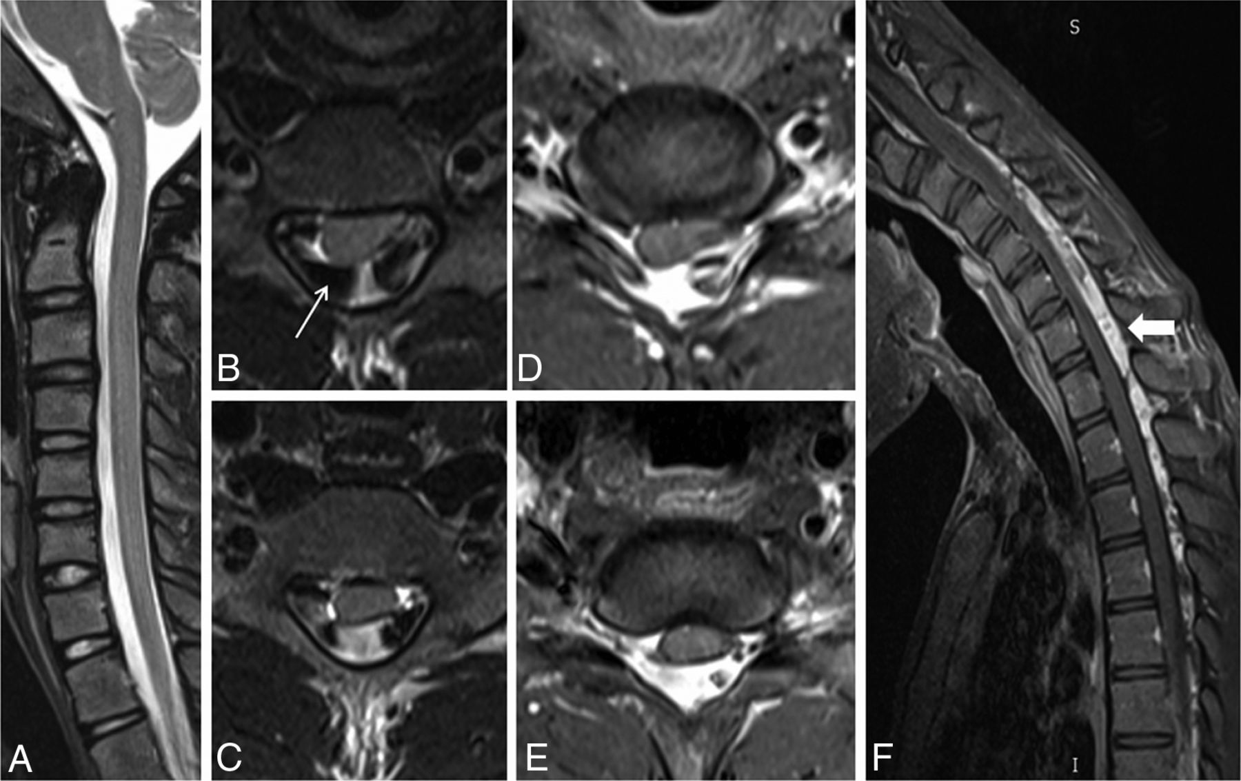
- Fig 2.
A 20-year-old male patient with weakness and wasting of the left hand muscles. Neutral position sagittal T2-weighted image (A) shows the normal appearance of the cervical cord. Axial T2-weighted flexion MR images (B and C) and postgadolinium T1 fat-suppressed images (D and E) show widening of the LDS with anterior displacement of the posterior dura and asymmetric cord atrophy, more on the left side, along with multiple flow voids within the posterior epidural space (arrow). Postgadolinium T1 fat-suppressed flexion MR sagittal image (F) shows an enhancing posterior epidural venous plexus extending from the C4 to T4 vertebral level (block arrow).
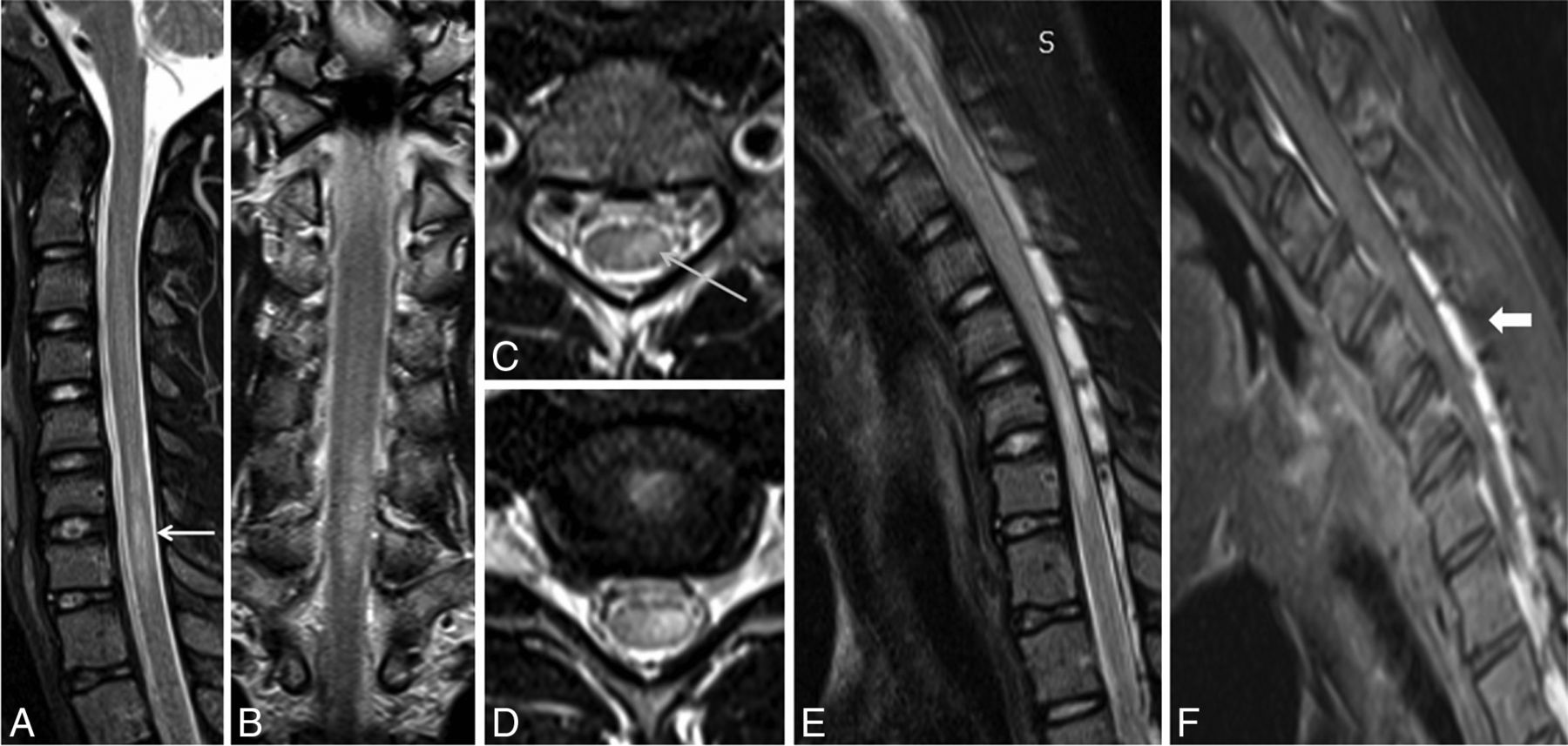
- Fig 3.
A 21-year-old man with asymmetric wasting of the bilateral hand muscles. Neutral MR T2-weighted sagittal and coronal (A and B) images show lower cervical cord atrophy with segmental hyperintensities in the cervical cord at the C6 and C7 vertebral levels (white arrow). Axial T2-weighted images (C and D) show asymmetric hyperintensities, more pronounced in the left half of the cervical cord (arrow). Flexion cervical MR STIR image (E) shows an enlarged posterior epidural space, which is seen as an enhancing posterior epidural venous plexus on the postgadolinium T1 fat-suppressed sagittal image (F) (block arrow).
- Fig 4.
Scatterplot showing the various LDS measurements by the 2 radiologists.
- Fig 5.
Histogram showing decrement in AP/TR cord diameter ratio during flexion cervical MR imaging in the 45 patients with Hirayama disease.
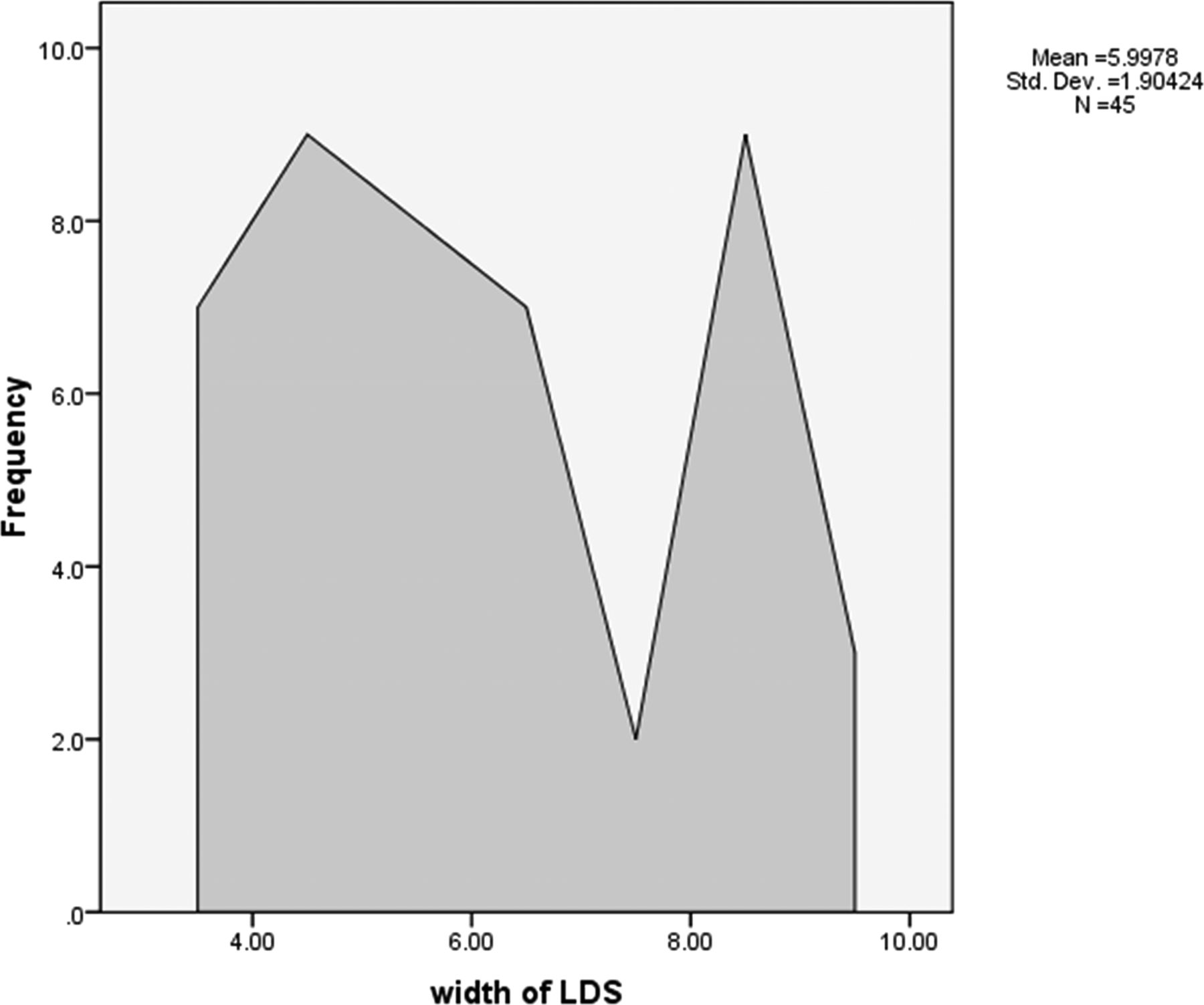
- Fig 6.
Frequency polygon showing the laminodural space measurements (in millimeters) during flexion cervical MR imaging in the 45 patients with HD.


Tables
Vertebral Levels Minimum Cord Diameter (mm) Maximum Cord Diameter (mm) Mean C2 6.27 8.11 7.13 ± 0.43 C3 5.56 7.84 6.91 ± 0.63 C4 4.99 7.54 6.50 ± 0.62 C5 4.58 7.60 6.06 ± 0.71 C6 3.97 6.98 5.64 ± 0.73 C7 4.15 6.91 5.61 ± 0.71 T1 4.75 7.73 5.93 ± 0.56 - Table 2:
Summarized average results of measured parameters of 2 radiologists during neutral and flexion MRI in 45 patients with Hirayama disease
Minimum Maximum Mean SD Distance of LDS 3.00 9.80 5.9978 1.90424 Spinal canal diameter in neutral MRI 10.80 15.30 12.7756 .99457 Spinal canal diameter in flexion MRI 10.90 15.50 12.9644 1.01604 AP cord diameter at neutral MRI 3.30 7.40 5.5378 1.00029 TR cord diameter at neutral MRI 7.50 14.40 12.1911 1.32593 AP cord diameter at flexion MRI 2.50 6.60 4.8089 .96903 TR cord diameter at flexion MRI 9.20 16.20 14.1022 1.39569 Ratio of LDS/spinal canal diameter in flexion MRI 0.24 0.74 0.461 0.14 Ratio of AP/TR diameter of cord in flexion MRI 0.17 0.59 0.3455 0.08634 Ratio of AP/TR diameter of cord in neutral MRI 0.26 0.72 0.4587 0.09899 Decrement of AP/TR diameter of cord during flexion MRI 0.03 0.26 0.118 0.06 Note:—Measurements are in millimeters.


