Advertisement

Main menu

  • Home
  • Content
    • Current Issue
    • Accepted Manuscripts
    • Article Preview
    • Past Issue Archive
    • Video Articles
    • AJNR Case Collection
    • Case of the Week Archive
    • Case of the Month Archive
    • Classic Case Archive
  • Special Collections
    • AJNR Awards
    • Low-Field MRI
    • Alzheimer Disease
    • ASNR Foundation Special Collection
    • Photon-Counting CT
    • View All
  • Multimedia
    • AJNR Podcasts
    • AJNR SCANtastic
    • Trainee Corner
    • MRI Safety Corner
    • Imaging Protocols
  • For Authors
    • Submit a Manuscript
    • Submit a Video Article
    • Submit an eLetter to the Editor/Response
    • Manuscript Submission Guidelines
    • Statistical Tips
    • Fast Publishing of Accepted Manuscripts
    • Graphical Abstract Preparation
    • Imaging Protocol Submission
    • Author Policies
  • About Us
    • About AJNR
    • Editorial Board
    • Editorial Board Alumni
  • More
    • Become a Reviewer/Academy of Reviewers
    • Subscribers
    • Permissions
    • Alerts
    • Feedback
    • Advertisers
    • ASNR Home

User menu

  • Alerts
  • Log in

Search

  • Advanced search
American Journal of Neuroradiology

American Journal of Neuroradiology

ASHNR American Society of Functional Neuroradiology ASHNR American Society of Pediatric Neuroradiology ASSR
  • Alerts
  • Log in

Advanced Search

  • Home
  • Content
    • Current Issue
    • Accepted Manuscripts
    • Article Preview
    • Past Issue Archive
    • Video Articles
    • AJNR Case Collection
    • Case of the Week Archive
    • Case of the Month Archive
    • Classic Case Archive
  • Special Collections
    • AJNR Awards
    • Low-Field MRI
    • Alzheimer Disease
    • ASNR Foundation Special Collection
    • Photon-Counting CT
    • View All
  • Multimedia
    • AJNR Podcasts
    • AJNR SCANtastic
    • Trainee Corner
    • MRI Safety Corner
    • Imaging Protocols
  • For Authors
    • Submit a Manuscript
    • Submit a Video Article
    • Submit an eLetter to the Editor/Response
    • Manuscript Submission Guidelines
    • Statistical Tips
    • Fast Publishing of Accepted Manuscripts
    • Graphical Abstract Preparation
    • Imaging Protocol Submission
    • Author Policies
  • About Us
    • About AJNR
    • Editorial Board
    • Editorial Board Alumni
  • More
    • Become a Reviewer/Academy of Reviewers
    • Subscribers
    • Permissions
    • Alerts
    • Feedback
    • Advertisers
    • ASNR Home
  • Follow AJNR on Twitter
  • Visit AJNR on Facebook
  • Follow AJNR on Instagram
  • Join AJNR on LinkedIn
  • RSS Feeds

AJNR Awards, New Junior Editors, and more. Read the latest AJNR updates

Research ArticleSpine Imaging and Spine Image-Guided Interventions

Cervical Cord Atrophy and Long-Term Disease Progression in Patients with Primary-Progressive Multiple Sclerosis

F.X. Aymerich, C. Auger, J. Alonso, M. Alberich, J. Sastre-Garriga, M. Tintoré, X. Montalban and A. Rovira
American Journal of Neuroradiology February 2018, 39 (2) 399-404; DOI: https://doi.org/10.3174/ajnr.A5495
F.X. Aymerich
aFrom the Magnetic Resonance Unit (F.X.A., C.A., J.A., M.A., A.R.), Department of Radiology
cDepartment of Automatic Control (F.X.A.), Universitat Politècnica de Catalunya–Barcelona Tech, Barcelona, Spain.
  • Find this author on Google Scholar
  • Find this author on PubMed
  • Search for this author on this site
  • ORCID record for F.X. Aymerich
C. Auger
aFrom the Magnetic Resonance Unit (F.X.A., C.A., J.A., M.A., A.R.), Department of Radiology
  • Find this author on Google Scholar
  • Find this author on PubMed
  • Search for this author on this site
  • ORCID record for C. Auger
J. Alonso
aFrom the Magnetic Resonance Unit (F.X.A., C.A., J.A., M.A., A.R.), Department of Radiology
  • Find this author on Google Scholar
  • Find this author on PubMed
  • Search for this author on this site
  • ORCID record for J. Alonso
M. Alberich
aFrom the Magnetic Resonance Unit (F.X.A., C.A., J.A., M.A., A.R.), Department of Radiology
  • Find this author on Google Scholar
  • Find this author on PubMed
  • Search for this author on this site
  • ORCID record for M. Alberich
J. Sastre-Garriga
bCentre d'Esclerosi Múltiple de Catalunya (J.S.-G., M.T., X.M.), Department of Neurology/Neuroimmunology, Hospital Universitari Vall d'Hebron, Universitat Autònoma de Barcelona, Barcelona, Spain
  • Find this author on Google Scholar
  • Find this author on PubMed
  • Search for this author on this site
  • ORCID record for J. Sastre-Garriga
M. Tintoré
bCentre d'Esclerosi Múltiple de Catalunya (J.S.-G., M.T., X.M.), Department of Neurology/Neuroimmunology, Hospital Universitari Vall d'Hebron, Universitat Autònoma de Barcelona, Barcelona, Spain
  • Find this author on Google Scholar
  • Find this author on PubMed
  • Search for this author on this site
  • ORCID record for M. Tintoré
X. Montalban
bCentre d'Esclerosi Múltiple de Catalunya (J.S.-G., M.T., X.M.), Department of Neurology/Neuroimmunology, Hospital Universitari Vall d'Hebron, Universitat Autònoma de Barcelona, Barcelona, Spain
  • Find this author on Google Scholar
  • Find this author on PubMed
  • Search for this author on this site
  • ORCID record for X. Montalban
A. Rovira
aFrom the Magnetic Resonance Unit (F.X.A., C.A., J.A., M.A., A.R.), Department of Radiology
  • Find this author on Google Scholar
  • Find this author on PubMed
  • Search for this author on this site
  • ORCID record for A. Rovira
  • Article
  • Figures & Data
  • Info & Metrics
  • Responses
  • References
  • PDF
Loading

Article Figures & Data

Figures

  • Tables
  • Fig 1.
    • Download figure
    • Open in new tab
    • Download powerpoint
    Fig 1.

    An example of the cord surface estimation obtained using the active surface method. The upper left image shows the location of the landmarks (red markers) that can be visualized in this sagittal slice. These landmarks were manually placed in the axial slices at the center area of the cord with a distance between them of approximately 10 mm. The upper right image shows the cord outline estimation (red lines). The lower axial slices show some examples of the spinal cord segmentation obtained (region within red contour).
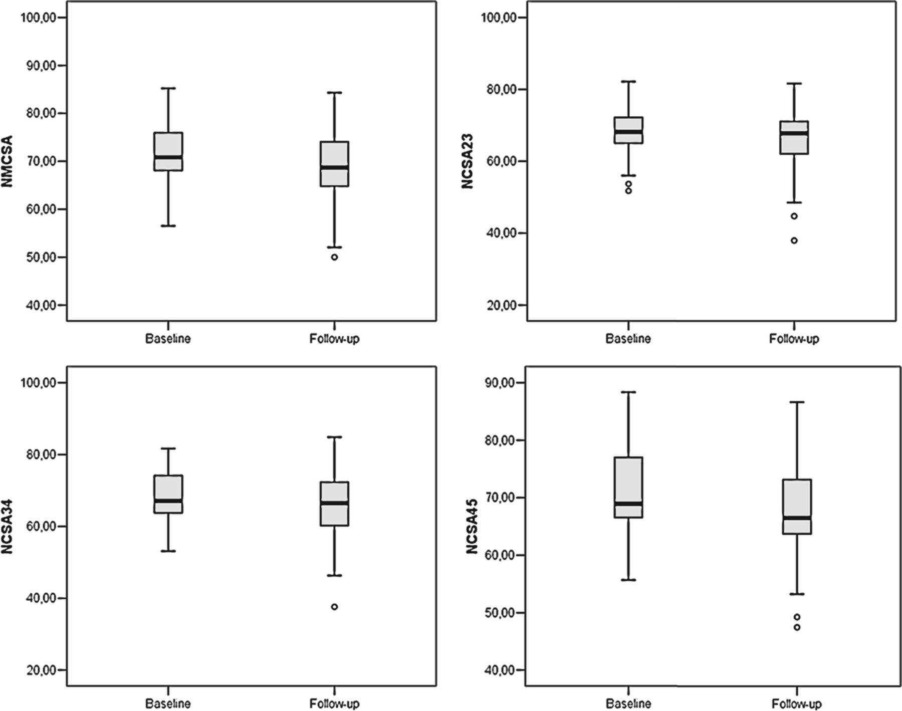
  • Fig 2.
    • Download figure
    • Open in new tab
    • Download powerpoint
    Fig 2.

    Box-and-whisker plots of normalized cross-sectional areas: NMCSA (upper left), NCSA23 (upper right), NCSA34 (lower left), and NCSA45 (lower right).

Tables

  • Figures
    • View popup
    Table 1:

    Main baseline demographic, clinical, and conventional MRI characteristics

    Patients with MS
    Median age (range) (yr)51 (33–61)
    Men/women19:12
    Mean disease duration at baseline (range) (yr)11.74 (2–33)
    Median EDSS score at baseline (range)5.5 (3.0–6.5)
    Mean brain T2LV at baseline (SD) (mL)18.12 (20.63)
    Mean brain T1LV at baseline (SD) (mL)7.41 (8.47)
    Mean BPF at baseline (SD)73.13% (5.86%)
    • View popup
    Table 2:

    Evolution of the EDSS

    Baseline2 Years14 Years
    Median EDSS score (range)5.5 (3.0–6.5)6.0 (4.0–8.5)7.5 (4.0–9.5)
    • View popup
    Table 3:

    MRI characteristics at follow-up

    Follow-Up
    Mean brain T2LV (SD) (mL)21.56 (20.65)
    Mean brain T1LV (SD) (mL)9.53 (9.32)
    Mean BPF (SD)70.33% (5.84%)
    • View popup
    Table 4:

    Longitudinal cross-sectional area measurements

    NMCSANCSA23NCSA34NCSA45
    Mean at baseline (SD) (mm2)71.49 (6.37)67.76 (7.26)68.21 (7.39)71.51 (8.27)
    Mean at follow-up (SD) (mm2)68.12 (8.91)65.40 (10.27)65.23 (10.61)67.95 (9.97)
    Cord area change averaged by year (SD) (%)−0.77 (1.14)−0.62 (1.20)−0.74 (1.47)−0.77 (1.61)
    P value.001.006.009.006
PreviousNext
Back to top

In this issue

American Journal of Neuroradiology: 39 (2)
American Journal of Neuroradiology
Vol. 39, Issue 2
1 Feb 2018
  • Table of Contents
  • Index by author
  • Complete Issue (PDF)
Advertisement
Print
Download PDF
Email Article

Thank you for your interest in spreading the word on American Journal of Neuroradiology.

NOTE: We only request your email address so that the person you are recommending the page to knows that you wanted them to see it, and that it is not junk mail. We do not capture any email address.

Enter multiple addresses on separate lines or separate them with commas.
Cervical Cord Atrophy and Long-Term Disease Progression in Patients with Primary-Progressive Multiple Sclerosis
(Your Name) has sent you a message from American Journal of Neuroradiology
(Your Name) thought you would like to see the American Journal of Neuroradiology web site.
CAPTCHA
This question is for testing whether or not you are a human visitor and to prevent automated spam submissions.
Cite this article
F.X. Aymerich, C. Auger, J. Alonso, M. Alberich, J. Sastre-Garriga, M. Tintoré, X. Montalban, A. Rovira
Cervical Cord Atrophy and Long-Term Disease Progression in Patients with Primary-Progressive Multiple Sclerosis
American Journal of Neuroradiology Feb 2018, 39 (2) 399-404; DOI: 10.3174/ajnr.A5495

Citation Manager Formats

  • BibTeX
  • Bookends
  • EasyBib
  • EndNote (tagged)
  • EndNote 8 (xml)
  • Medlars
  • Mendeley
  • Papers
  • RefWorks Tagged
  • Ref Manager
  • RIS
  • Zotero
0 Responses
Respond to this article
Share
Bookmark this article
Cervical Cord Atrophy and Long-Term Disease Progression in Patients with Primary-Progressive Multiple Sclerosis
F.X. Aymerich, C. Auger, J. Alonso, M. Alberich, J. Sastre-Garriga, M. Tintoré, X. Montalban, A. Rovira
American Journal of Neuroradiology Feb 2018, 39 (2) 399-404; DOI: 10.3174/ajnr.A5495
del.icio.us logo Twitter logo Facebook logo Mendeley logo
  • Tweet Widget
  • Facebook Like
  • Google Plus One
Purchase

Jump to section

  • Article
    • Abstract
    • ABBREVIATIONS:
    • Materials and Methods
    • Results
    • Discussion
    • Conclusions
    • Acknowledgments
    • Footnotes
    • References
  • Figures & Data
  • Info & Metrics
  • Responses
  • References
  • PDF

Related Articles

  • No related articles found.
  • PubMed
  • Google Scholar

Cited By...

  • Clinically relevant cranio-caudal patterns of cervical cord atrophy evolution in MS
  • Crossref
  • Google Scholar

This article has not yet been cited by articles in journals that are participating in Crossref Cited-by Linking.

More in this TOC Section

  • Optimization of Photon Counting CT Myelography
  • Characteristics of SIH Type I Culprit Lesions
  • Management Outcomes For VO Spine Biopsy
Show more Spine Imaging and Spine Image-Guided Interventions

Similar Articles

Advertisement

Indexed Content

  • Current Issue
  • Accepted Manuscripts
  • Article Preview
  • Past Issues
  • Editorials
  • Editor's Choice
  • Fellows' Journal Club
  • Letters to the Editor
  • Video Articles

Cases

  • Case Collection
  • Archive - Case of the Week
  • Archive - Case of the Month
  • Archive - Classic Case

More from AJNR

  • Trainee Corner
  • Imaging Protocols
  • MRI Safety Corner
  • Book Reviews

Multimedia

  • AJNR Podcasts
  • AJNR Scantastics

Resources

  • Turnaround Time
  • Submit a Manuscript
  • Submit a Video Article
  • Submit an eLetter to the Editor/Response
  • Manuscript Submission Guidelines
  • Statistical Tips
  • Fast Publishing of Accepted Manuscripts
  • Graphical Abstract Preparation
  • Imaging Protocol Submission
  • Evidence-Based Medicine Level Guide
  • Publishing Checklists
  • Author Policies
  • Become a Reviewer/Academy of Reviewers
  • News and Updates

About Us

  • About AJNR
  • Editorial Board
  • Editorial Board Alumni
  • Alerts
  • Permissions
  • Not an AJNR Subscriber? Join Now
  • Advertise with Us
  • Librarian Resources
  • Feedback
  • Terms and Conditions
  • AJNR Editorial Board Alumni

American Society of Neuroradiology

  • Not an ASNR Member? Join Now

© 2025 by the American Society of Neuroradiology All rights, including for text and data mining, AI training, and similar technologies, are reserved.
Print ISSN: 0195-6108 Online ISSN: 1936-959X

Powered by HighWire