
Article Figures & Data
Figures
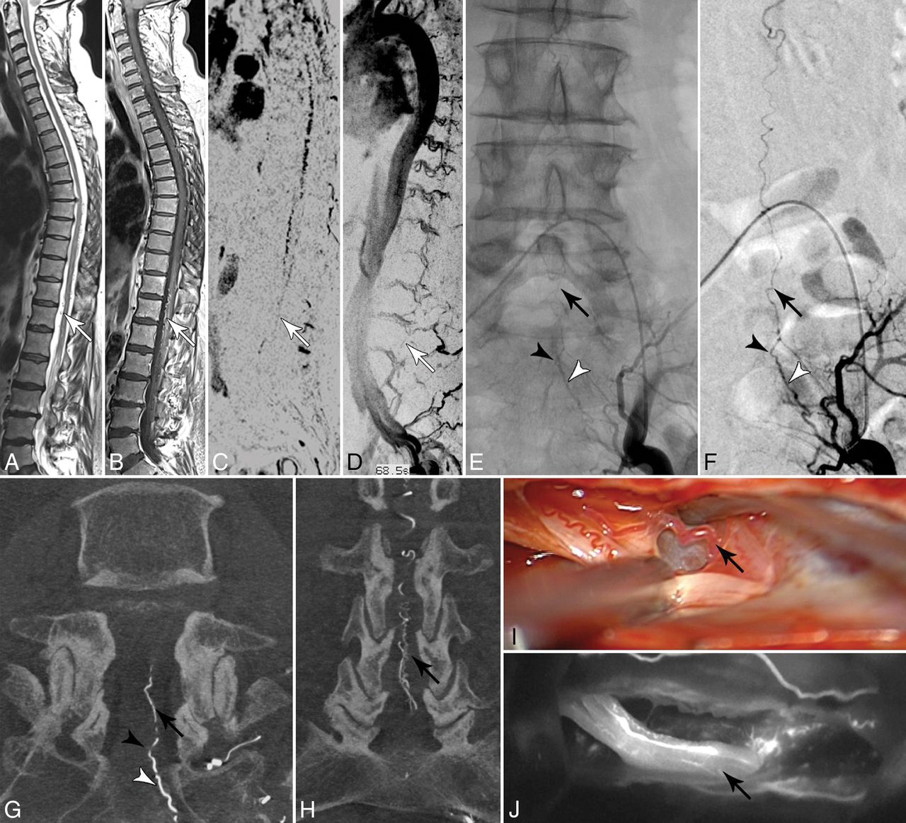
- FIGURE.
A and B, Sagittal T2- and contrast-enhanced T1-weighted images reveal congestive myelopathy and dilated perimedullary veins (white arrows). C and D, Spinal CE-MRA shows dilated radicular veins in the lumbar region suspicious for an SDAVF in the lumbosacral region (white arrows). E–H, DSA examinations identify the fistula in the dural sleeve of the left S2 root (black arrowhead) supplied via the lateral sacral artery (white arrowhead). Note the upward draining sacral radicular vein (black arrow). I and J, Intraoperative indocyanine green angiography confirms the intradural course of the arterialized draining vein (black arrow) embedded at the ventral side of the S2 nerve root.

Tables
- Table 1:
Clinical presentation of patients with deep lumbosacral spinal dural arteriovenous fistulas
Case No. Age (yr)/Sex Duration of Symptoms (mo) Symptoms at Time of Diagnosis AL-Score Previous Diagnosis and Treatment 1 66, M 24 Paraparesis 3/5a, sensory transverse lesion L5, sphincter dysfunction 3 Lumbar disc prolapse: discectomy L4–5 2 56, M 36 Paraparesis 3/5, sensory transverse lesion L2 3 3 63, M 24 Paraparesis 2–3/5, sensory transverse lesion L1, sphincter dysfunction 4 4 60, M 30 Paraparesis 4/5, sensory transverse lesion L4, sphincter dysfunction 3 Lumbar disc prolapse: discectomy L3–4, L4–5 5 71, M 14 Distal accentuated paraparesis 3/5, mild hypesthesia of the right leg 3 6 66, M 18 Paraparesis 3/5, sensory transverse lesion L5, sphincter dysfunction 4 Lumbar spinal stenosis: dorsal decompression L1–2 7 55, M 2 Paraplegia and anesthesia below T8, sphincter dysfunction 5 8 67, M 26 Paraparesis 4/5, sensory transverse lesion T12, sphincter dysfunction 2 Intramedullary tumor: biopsy 9 73, M 9 Paraparesis 3/5, sensory transverse lesion T5, sphincter dysfunction 4 Intramedullary tumor: biopsy 10 70, M 3 Paraparesis 3/5, sensory transverse lesion S1, sphincter dysfunction 4 11 69, M 6 Paraparesis 1–2/5, sensory transverse lesion L4, sphincter dysfunction 5 12 67, F 12 No paresis, hypesthesia below T11, ataxia 1 13 63, M 1 Paraparesis 3/5, sphincter dysfunction 3 14 61, M 11 Distal accentuated paraparesis 3/5, hypesthesia of the right leg, ataxia, sphincter dysfunction 3 Lumbar spinal stenosis: dorsal decompression and discectomies L4–5, L5–S1 15 67, F 9 Paraparesis 4/5, dysesthesia below T12, paresthesia on the dorsum of the left foot 2 Lumbar disc prolapse: discectomy L5–S1 16 63, M ND No paresis, sensory transverse lesion L1, ataxia, sphincter dysfunction 1 Repeat insufficient embolizations of lsSDAVF 17 55, M 10 Monoparesis left foot 1 Lumbar stenosis: dorsal decompression 18 78, M 4 Spastic paraparesis 3/5, paresthesia in both feet, sphincter dysfunction 4 19 74, M 24 Paresis of the left leg 4/5, diffuse paresthesia of the lower extremities 2 20 53, F 6 No paresis, diffuse paresthesia of the lower extremities, mild ataxia, mild sphincter dysfunction 1 Note:—AL-Score indicates Aminoff-Logue disability score; ND, no data.
↵a Muscle strength grade.
Case No. MRI/MRA DSA Shunt Location T2/T1 Hyperintensity (Extension) Contrast Enhancement (Extension) Perimedullary Vein Enlargement (Extension) Prominent FV Prominent Lumbar Vein DSA until Diagnosis Arterial Feeder 1 S1 R T7-conus T9–T10 Mild, T7–T8 No No 4 Iliolumbar artery R 2 L5 R T9–T11 T5–T11 Mild, T5–T11 No Yes 4 Iliolumbar artery R 3 S1 L T8-conus Absent Absent Yes No 4 Middle sacral artery L 4 S1 R T9-conus Absent Mild, T10–T12 Yes No 3 Iliolumbar artery R 5 S1 R T3-conus ND ND Yes No 3 Iliolumbar artery R 6 L5 L T8-conus T9-conus Mild, T10–T12 No Yes 2 L5 L 7 S1 R T6-conus T8–T12 Mild, T7–T11 No Yes 2 Lateral sacral artery L 8 S2 R T10–T12 T11–T12 Mild, T11–T12 Yes No 3 Lateral sacral artery R 9 L5 L T4-conus T9–T12 Absent No Yes 3 L4 L 10 S1 R T4-conus Absent Severe, T6-conus Yes No 2 Lateral sacral artery R 11 S1 R T6-conus T12–L1 Mild, T6-conus No Yes 5 Iliolumbar artery bilateral 12 S3 R T10-conus T12 Severe, T7-conus Yes No 3 Lateral sacral artery bilateral 13 S1 R T2-conus T12-conus Mild, T8-conus Yes No 4 Iliolumbar artery R 14 S2 L T5-conus T7-conus Mild, T8–L3 Yes No 4 Iliolumbar artery bilateral 15 S1 L T5-conus Absent Severe, T7-conus Yes No 2 Lateral sacral artery L 16 S2 L Absent T10–11 Absent Yes No 2 Iliolumbar artery L 17 S1 L T5-conus T4–L1 Mild, T3–T4 Yes No 2 Iliolumbar artery bilateral 18 S2 L T8–T12 T8–T11 Mild, T9–T12 Yes No 5 Iliolumbar artery bilateral 19 L5 R T8-conus T8-conus Severe, T6–T12 No Yes 2 Iliolumbar artery R 20 S2 R T12-conus T9-conus Mild, T7–T11 No Yes 1 Lateral sacral artery Note:—R indicates right; L, left; ND, no data.


Jump to section
Related Articles
Cited By...
- Long-term outcome in a cohort of 36 patients with sacral dural arteriovenous fistulae after endovascular embolisation or microsurgery
- Spinal Dorsal Intradural Arteriovenous Fistulas: Natural History, Imaging, and Management
- Long-Term Outcome of Patients with Spinal Dural Arteriovenous Fistula: The Dilemma of Delayed Diagnosis
- Spinal Epidural Arteriovenous Fistula with Perimedullary Venous Reflux: Clinical and Neuroradiologic Features of an Underestimated Vascular Disorder
- Dilated Vein of the Filum Terminale on MRI: A Marker for Deep Lumbar and Sacral Dural and Epidural Arteriovenous Fistulas