Advertisement

Main menu

  • Home
  • Content
    • Current Issue
    • Accepted Manuscripts
    • Article Preview
    • Past Issue Archive
    • Video Articles
    • AJNR Case Collection
    • Case of the Week Archive
    • Case of the Month Archive
    • Classic Case Archive
  • Special Collections
    • AJNR Awards
    • Low-Field MRI
    • Alzheimer Disease
    • ASNR Foundation Special Collection
    • Photon-Counting CT
    • View All
  • Multimedia
    • AJNR Podcasts
    • AJNR SCANtastic
    • Trainee Corner
    • MRI Safety Corner
    • Imaging Protocols
  • For Authors
    • Submit a Manuscript
    • Submit a Video Article
    • Submit an eLetter to the Editor/Response
    • Manuscript Submission Guidelines
    • Statistical Tips
    • Fast Publishing of Accepted Manuscripts
    • Graphical Abstract Preparation
    • Imaging Protocol Submission
    • Author Policies
  • About Us
    • About AJNR
    • Editorial Board
    • Editorial Board Alumni
  • More
    • Become a Reviewer/Academy of Reviewers
    • Subscribers
    • Permissions
    • Alerts
    • Feedback
    • Advertisers
    • ASNR Home

User menu

  • Alerts
  • Log in

Search

  • Advanced search
American Journal of Neuroradiology

American Journal of Neuroradiology

ASHNR American Society of Functional Neuroradiology ASHNR American Society of Pediatric Neuroradiology ASSR
  • Alerts
  • Log in

Advanced Search

  • Home
  • Content
    • Current Issue
    • Accepted Manuscripts
    • Article Preview
    • Past Issue Archive
    • Video Articles
    • AJNR Case Collection
    • Case of the Week Archive
    • Case of the Month Archive
    • Classic Case Archive
  • Special Collections
    • AJNR Awards
    • Low-Field MRI
    • Alzheimer Disease
    • ASNR Foundation Special Collection
    • Photon-Counting CT
    • View All
  • Multimedia
    • AJNR Podcasts
    • AJNR SCANtastic
    • Trainee Corner
    • MRI Safety Corner
    • Imaging Protocols
  • For Authors
    • Submit a Manuscript
    • Submit a Video Article
    • Submit an eLetter to the Editor/Response
    • Manuscript Submission Guidelines
    • Statistical Tips
    • Fast Publishing of Accepted Manuscripts
    • Graphical Abstract Preparation
    • Imaging Protocol Submission
    • Author Policies
  • About Us
    • About AJNR
    • Editorial Board
    • Editorial Board Alumni
  • More
    • Become a Reviewer/Academy of Reviewers
    • Subscribers
    • Permissions
    • Alerts
    • Feedback
    • Advertisers
    • ASNR Home
  • Follow AJNR on Twitter
  • Visit AJNR on Facebook
  • Follow AJNR on Instagram
  • Join AJNR on LinkedIn
  • RSS Feeds

AJNR Awards, New Junior Editors, and more. Read the latest AJNR updates

Research ArticlePediatric Neuroimaging

Temporal Lobe Malformations in Achondroplasia: Expanding the Brain Imaging Phenotype Associated with FGFR3-Related Skeletal Dysplasias

S.A. Manikkam, K. Chetcuti, K.B. Howell, R. Savarirayan, A.M. Fink and S.A. Mandelstam
American Journal of Neuroradiology February 2018, 39 (2) 380-384; DOI: https://doi.org/10.3174/ajnr.A5468
S.A. Manikkam
aFrom the Departments of Medical Imaging (S.A. Manikkam, A.M.F., S.A. Mandelstam)
  • Find this author on Google Scholar
  • Find this author on PubMed
  • Search for this author on this site
  • ORCID record for S.A. Manikkam
K. Chetcuti
cDepartment of Radiology (K.C.), Alder Hey Children's Hospital, Liverpool, UK
  • Find this author on Google Scholar
  • Find this author on PubMed
  • Search for this author on this site
  • ORCID record for K. Chetcuti
K.B. Howell
bNeurology (K.B.H.), Royal Children's Hospital, Melbourne, Australia
dDepartments of Paediatrics (K.B.H., S.A. Mandelstam)
fMurdoch Children's Research Institute (K.B.H., R.S., A.M.F., S.A. Mandelstam), Melbourne, Australia
  • Find this author on Google Scholar
  • Find this author on PubMed
  • Search for this author on this site
  • ORCID record for K.B. Howell
R. Savarirayan
fMurdoch Children's Research Institute (K.B.H., R.S., A.M.F., S.A. Mandelstam), Melbourne, Australia
gVictorian Clinical Genetics Services (R.S.), Melbourne, Australia
  • Find this author on Google Scholar
  • Find this author on PubMed
  • Search for this author on this site
  • ORCID record for R. Savarirayan
A.M. Fink
aFrom the Departments of Medical Imaging (S.A. Manikkam, A.M.F., S.A. Mandelstam)
eRadiology (A.M.F., S.A. Mandelstam), University of Melbourne, Melbourne, Australia
fMurdoch Children's Research Institute (K.B.H., R.S., A.M.F., S.A. Mandelstam), Melbourne, Australia
  • Find this author on Google Scholar
  • Find this author on PubMed
  • Search for this author on this site
  • ORCID record for A.M. Fink
S.A. Mandelstam
aFrom the Departments of Medical Imaging (S.A. Manikkam, A.M.F., S.A. Mandelstam)
dDepartments of Paediatrics (K.B.H., S.A. Mandelstam)
eRadiology (A.M.F., S.A. Mandelstam), University of Melbourne, Melbourne, Australia
fMurdoch Children's Research Institute (K.B.H., R.S., A.M.F., S.A. Mandelstam), Melbourne, Australia
hFlorey Institute of Neuroscience and Mental Health (S.A. Mandelstam), Melbourne, Australia.
  • Find this author on Google Scholar
  • Find this author on PubMed
  • Search for this author on this site
  • ORCID record for S.A. Mandelstam
  • Article
  • Figures & Data
  • Supplemental
  • Info & Metrics
  • Responses
  • References
  • PDF
Loading

Article Figures & Data

Figures

  • Tables
  • Fig 1.
    • Download figure
    • Open in new tab
    • Download powerpoint
    Fig 1.

    A, Axial T1-weighted MR image in a 5-year-old boy with achondroplasia shows a deep transverse sulcus within the temporal lobe (arrow) and abnormal configuration of the temporal horns of the lateral ventricles. B, Axial T1-weighted MR image in a 5-year-old male control shows the normal configuration of the mesial temporal lobes and temporal horns.

  • Fig 2.
    • Download figure
    • Open in new tab
    • Download powerpoint
    Fig 2.

    A, Sagittal T2-weighted MR image in a 2-year-old boy with achondroplasia shows a sagittal cleft within the temporal lobe (white arrow). B, Sagittal T2-weighted MR image in a 2-year-old male control shows the normal appearance of the temporal lobe. C, Antenatal sagittal T2-weighted MR image of the child with achondroplasia in A demonstrates the prenatal appearance of the sagittal clefting within the temporal lobe at 30 weeks (black arrow). D, Antenatal sagittal T2-weighted MR image of a control fetus shows the normal appearance of the temporal lobe at the same gestation.
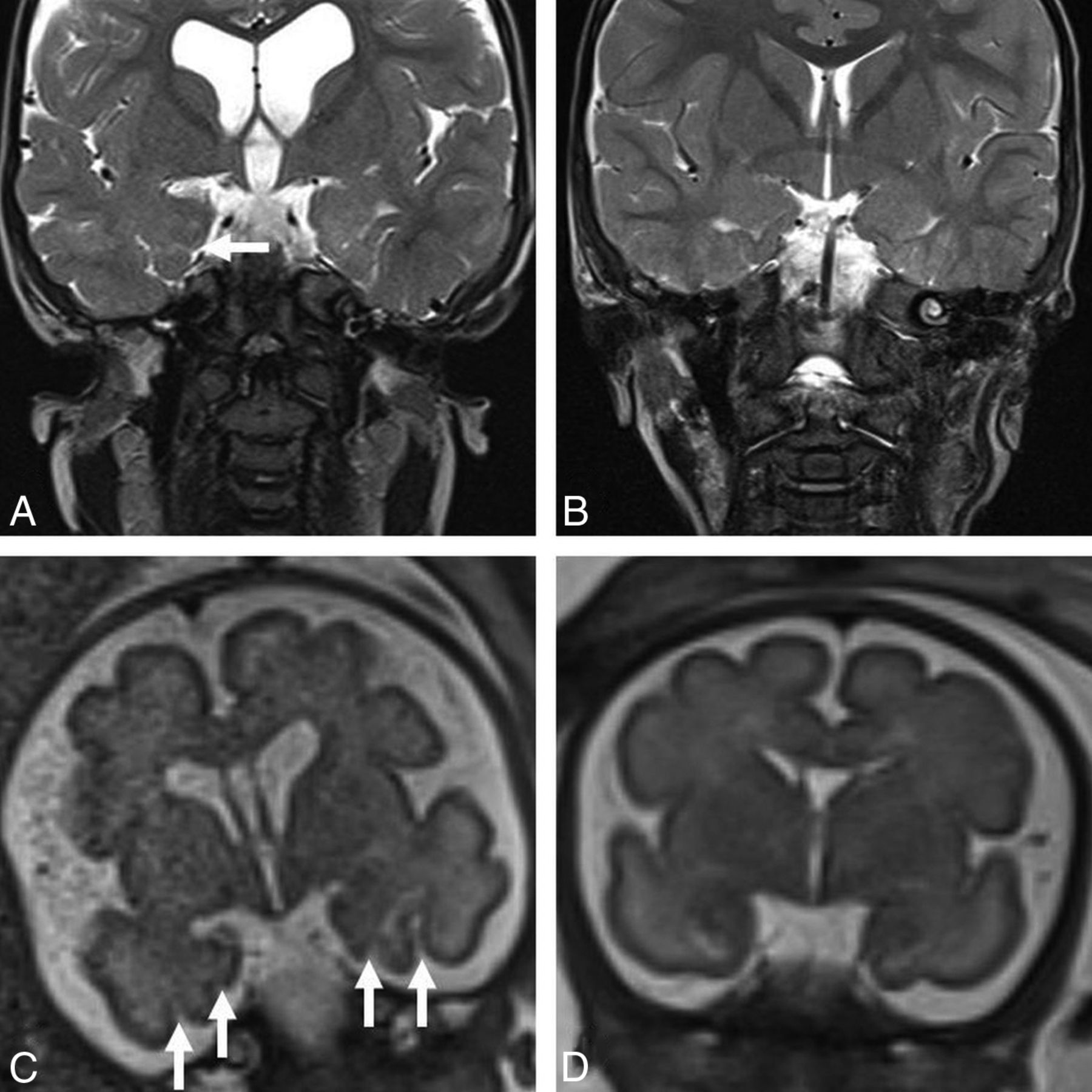
  • Fig 3.
    • Download figure
    • Open in new tab
    • Download powerpoint
    Fig 3.

    A, Coronal T2-weighted MR image in a 30-month-old boy with achondroplasia shows incomplete hippocampal inversion (arrow) and ventriculomegaly. B, Coronal T2-weighted MR image in a 30-month-old male control shows the normal appearance of the hippocampi.

  • Fig 4.
    • Download figure
    • Open in new tab
    • Download powerpoint
    Fig 4.

    A, Coronal T2-weighted MR image in a 30-month-old boy with achondroplasia shows oversulcation and loss of gray-white matter differentiation of the mesial temporal lobes (arrow) and ventriculomegaly. B, Coronal T2-weighted MR image in a 30-month-old male control shows the normal sulcation and gray-white matter differentiation of the temporal lobes. C, Antenatal coronal T2-weighted fetal MR image of the child with achondroplasia in A obtained at 30 weeks shows oversulcation of the mesial temporal lobes (white arrows) and mild ventriculomegaly. D, Antenatal coronal T2-weighted MR image of a control fetus shows the normal appearance of the temporal lobes at the same gestation.

  • Fig 5.
    • Download figure
    • Open in new tab
    • Download powerpoint
    Fig 5.

    A, Axial T1-weighted MR image in a 1-year-old girl with achondroplasia shows oversulcation of the calcar avis (arrow) and moderate ventriculomegaly. B, Axial T1-weighted MR image in a 1-year-old female control shows the normal sulcation pattern of the calcar avis.

Tables

  • Figures
  • Prevalence of MRI findings in children with achondroplasia

    MRI FindingPrevalence
    Deep transverse temporal sulcus (axial T1WI)13/13 (100%)
    Sagittal clefting in the medial temporal lobe (sagittal T2WI)13/13 (100%)
    Incomplete hippocampal inversion12/13 (92%)
    Ventriculomegaly12/13 (92%)
    Oversulcation of the mesial temporal lobe11/13 (85%)
    Extension of oversulcation to the calcar avis9/13 (69%)
    Loss of gray-white matter differentiation of the mesial temporal lobe5/7a (71%)
    Abnormal triangular shape of the temporal horn6/13 (46%)
    Megalencephaly5/13 (38%)
    Temporal lobe enlargement1/13 (8%)
    • ↵a In the 6 children younger than 36 months of age, loss of gray-white matter differentiation of the mesial temporal lobe could not be accurately assessed on available sequences due to the incomplete myelination.

PreviousNext
Back to top

In this issue

American Journal of Neuroradiology: 39 (2)
American Journal of Neuroradiology
Vol. 39, Issue 2
1 Feb 2018
  • Table of Contents
  • Index by author
  • Complete Issue (PDF)
Advertisement
Print
Download PDF
Email Article

Thank you for your interest in spreading the word on American Journal of Neuroradiology.

NOTE: We only request your email address so that the person you are recommending the page to knows that you wanted them to see it, and that it is not junk mail. We do not capture any email address.

Enter multiple addresses on separate lines or separate them with commas.
Temporal Lobe Malformations in Achondroplasia: Expanding the Brain Imaging Phenotype Associated with FGFR3-Related Skeletal Dysplasias
(Your Name) has sent you a message from American Journal of Neuroradiology
(Your Name) thought you would like to see the American Journal of Neuroradiology web site.
CAPTCHA
This question is for testing whether or not you are a human visitor and to prevent automated spam submissions.
Cite this article
S.A. Manikkam, K. Chetcuti, K.B. Howell, R. Savarirayan, A.M. Fink, S.A. Mandelstam
Temporal Lobe Malformations in Achondroplasia: Expanding the Brain Imaging Phenotype Associated with FGFR3-Related Skeletal Dysplasias
American Journal of Neuroradiology Feb 2018, 39 (2) 380-384; DOI: 10.3174/ajnr.A5468

Citation Manager Formats

  • BibTeX
  • Bookends
  • EasyBib
  • EndNote (tagged)
  • EndNote 8 (xml)
  • Medlars
  • Mendeley
  • Papers
  • RefWorks Tagged
  • Ref Manager
  • RIS
  • Zotero
0 Responses
Respond to this article
Share
Bookmark this article
Temporal Lobe Malformations in Achondroplasia: Expanding the Brain Imaging Phenotype Associated with FGFR3-Related Skeletal Dysplasias
S.A. Manikkam, K. Chetcuti, K.B. Howell, R. Savarirayan, A.M. Fink, S.A. Mandelstam
American Journal of Neuroradiology Feb 2018, 39 (2) 380-384; DOI: 10.3174/ajnr.A5468
del.icio.us logo Twitter logo Facebook logo Mendeley logo
  • Tweet Widget
  • Facebook Like
  • Google Plus One
Purchase

Jump to section

  • Article
    • Abstract
    • ABBREVIATION:
    • Discussion
    • References
  • Figures & Data
  • Supplemental
  • Info & Metrics
  • Responses
  • References
  • PDF

Related Articles

  • PubMed
  • Google Scholar

Cited By...

  • Pituitary Gland Duplication Syndrome: An International Imaging Analysis
  • Common Neuroimaging Findings in Bosch-Boonstra-Schaaf Optic Atrophy Syndrome
  • Macrocerebellum in Achondroplasia: A Further CNS Manifestation of FGFR3 Mutations?
  • Crossref
  • Google Scholar

This article has not yet been cited by articles in journals that are participating in Crossref Cited-by Linking.

More in this TOC Section

  • Frontal Paraventricular Cysts
  • Sodium MRI in Pediatric Brain Tumors
  • FRACTURE MR in Congenital Vertebral Anomalies
Show more Pediatric Neuroimaging

Similar Articles

Advertisement

Indexed Content

  • Current Issue
  • Accepted Manuscripts
  • Article Preview
  • Past Issues
  • Editorials
  • Editor's Choice
  • Fellows' Journal Club
  • Letters to the Editor
  • Video Articles

Cases

  • Case Collection
  • Archive - Case of the Week
  • Archive - Case of the Month
  • Archive - Classic Case

More from AJNR

  • Trainee Corner
  • Imaging Protocols
  • MRI Safety Corner
  • Book Reviews

Multimedia

  • AJNR Podcasts
  • AJNR Scantastics

Resources

  • Turnaround Time
  • Submit a Manuscript
  • Submit a Video Article
  • Submit an eLetter to the Editor/Response
  • Manuscript Submission Guidelines
  • Statistical Tips
  • Fast Publishing of Accepted Manuscripts
  • Graphical Abstract Preparation
  • Imaging Protocol Submission
  • Evidence-Based Medicine Level Guide
  • Publishing Checklists
  • Author Policies
  • Become a Reviewer/Academy of Reviewers
  • News and Updates

About Us

  • About AJNR
  • Editorial Board
  • Editorial Board Alumni
  • Alerts
  • Permissions
  • Not an AJNR Subscriber? Join Now
  • Advertise with Us
  • Librarian Resources
  • Feedback
  • Terms and Conditions
  • AJNR Editorial Board Alumni

American Society of Neuroradiology

  • Not an ASNR Member? Join Now

© 2025 by the American Society of Neuroradiology All rights, including for text and data mining, AI training, and similar technologies, are reserved.
Print ISSN: 0195-6108 Online ISSN: 1936-959X

Powered by HighWire