Advertisement

Main menu

  • Home
  • Content
    • Current Issue
    • Accepted Manuscripts
    • Article Preview
    • Past Issue Archive
    • Video Articles
    • AJNR Case Collection
    • Case of the Week Archive
    • Case of the Month Archive
    • Classic Case Archive
  • Special Collections
    • AJNR Awards
    • Low-Field MRI
    • Alzheimer Disease
    • ASNR Foundation Special Collection
    • Photon-Counting CT
    • View All
  • Multimedia
    • AJNR Podcasts
    • AJNR SCANtastic
    • Trainee Corner
    • MRI Safety Corner
    • Imaging Protocols
  • For Authors
    • Submit a Manuscript
    • Submit a Video Article
    • Submit an eLetter to the Editor/Response
    • Manuscript Submission Guidelines
    • Statistical Tips
    • Fast Publishing of Accepted Manuscripts
    • Graphical Abstract Preparation
    • Imaging Protocol Submission
    • Author Policies
  • About Us
    • About AJNR
    • Editorial Board
    • Editorial Board Alumni
  • More
    • Become a Reviewer/Academy of Reviewers
    • Subscribers
    • Permissions
    • Alerts
    • Feedback
    • Advertisers
    • ASNR Home

User menu

  • Alerts
  • Log in

Search

  • Advanced search
American Journal of Neuroradiology

American Journal of Neuroradiology

ASHNR American Society of Functional Neuroradiology ASHNR American Society of Pediatric Neuroradiology ASSR
  • Alerts
  • Log in

Advanced Search

  • Home
  • Content
    • Current Issue
    • Accepted Manuscripts
    • Article Preview
    • Past Issue Archive
    • Video Articles
    • AJNR Case Collection
    • Case of the Week Archive
    • Case of the Month Archive
    • Classic Case Archive
  • Special Collections
    • AJNR Awards
    • Low-Field MRI
    • Alzheimer Disease
    • ASNR Foundation Special Collection
    • Photon-Counting CT
    • View All
  • Multimedia
    • AJNR Podcasts
    • AJNR SCANtastic
    • Trainee Corner
    • MRI Safety Corner
    • Imaging Protocols
  • For Authors
    • Submit a Manuscript
    • Submit a Video Article
    • Submit an eLetter to the Editor/Response
    • Manuscript Submission Guidelines
    • Statistical Tips
    • Fast Publishing of Accepted Manuscripts
    • Graphical Abstract Preparation
    • Imaging Protocol Submission
    • Author Policies
  • About Us
    • About AJNR
    • Editorial Board
    • Editorial Board Alumni
  • More
    • Become a Reviewer/Academy of Reviewers
    • Subscribers
    • Permissions
    • Alerts
    • Feedback
    • Advertisers
    • ASNR Home
  • Follow AJNR on Twitter
  • Visit AJNR on Facebook
  • Follow AJNR on Instagram
  • Join AJNR on LinkedIn
  • RSS Feeds

AJNR Awards, New Junior Editors, and more. Read the latest AJNR updates

Research ArticleAdult Brain

Depiction of the Superior Petrosal Vein Complex by 3D Contrast-Enhanced MR Angiography

B. Bender, T.-K. Hauser, A. Korn, U. Klose, M. Tatagiba, U. Ernemann and F.H. Ebner
American Journal of Neuroradiology December 2018, 39 (12) 2249-2255; DOI: https://doi.org/10.3174/ajnr.A5864
B. Bender
aFrom the Department of Radiology (B.B., T.-K.H., A.K., U.K., U.E.), Diagnostic and Interventional Neuroradiology
  • Find this author on Google Scholar
  • Find this author on PubMed
  • Search for this author on this site
  • ORCID record for B. Bender
T.-K. Hauser
aFrom the Department of Radiology (B.B., T.-K.H., A.K., U.K., U.E.), Diagnostic and Interventional Neuroradiology
  • Find this author on Google Scholar
  • Find this author on PubMed
  • Search for this author on this site
  • ORCID record for T.-K. Hauser
A. Korn
aFrom the Department of Radiology (B.B., T.-K.H., A.K., U.K., U.E.), Diagnostic and Interventional Neuroradiology
cRadiologie in Hof (A.K.), Hof, Germany.
  • Find this author on Google Scholar
  • Find this author on PubMed
  • Search for this author on this site
  • ORCID record for A. Korn
U. Klose
aFrom the Department of Radiology (B.B., T.-K.H., A.K., U.K., U.E.), Diagnostic and Interventional Neuroradiology
  • Find this author on Google Scholar
  • Find this author on PubMed
  • Search for this author on this site
  • ORCID record for U. Klose
M. Tatagiba
bDepartment of Neurosurgery (M.T., F.H.E.), University Hospital Tübingen, Tübingen, Germany
  • Find this author on Google Scholar
  • Find this author on PubMed
  • Search for this author on this site
  • ORCID record for M. Tatagiba
U. Ernemann
aFrom the Department of Radiology (B.B., T.-K.H., A.K., U.K., U.E.), Diagnostic and Interventional Neuroradiology
  • Find this author on Google Scholar
  • Find this author on PubMed
  • Search for this author on this site
  • ORCID record for U. Ernemann
F.H. Ebner
cRadiologie in Hof (A.K.), Hof, Germany.
  • Find this author on Google Scholar
  • Find this author on PubMed
  • Search for this author on this site
  • ORCID record for F.H. Ebner
  • Article
  • Figures & Data
  • Supplemental
  • Info & Metrics
  • Responses
  • References
  • PDF
Loading

Article Figures & Data

Figures

  • Tables
  • Fig 1.
    • Download figure
    • Open in new tab
    • Download powerpoint
    Fig 1.

    Anatomic overview of the tributaries of the superior petrosal veins (reproduced with permission of Oxford University Press from Matsushima K, Matsushima T, Kuga Y, et al. Classification of the superior petrosal veins and sinus based on drainage pattern. Neurosurgery 2014;10 Suppl 2:357–67).11 A, Anterior view of the left petrosal cerebellar surface and anterolateral brain stem of an anatomic preparation with veins perfused with blue and arteries with red silicone. B, Colored multifusion image (T1 MPRAGE and conventional 3D digital subtraction angiography) with a similar view compared with A. The 4 drainage groups are red (petrosal group), blue (anterior pontomesencephalic group), green (posterior mesencephalic group), and yellow (tentorial group).

  • Fig 2.
    • Download figure
    • Open in new tab
    • Download powerpoint
    Fig 2.

    Transversal MIP at the level of the drainage of the SPV into the SPS (type II, internal acoustic meatus not shown). The tributary veins are clearly identifiable: lateral mesencephalic vein (asterisk, posterior mesencephalic group, anastomosis to the basal vein), transverse pontine vein (anterior pontomesencephalic group), and vein of cerebellopontine fissure (petrosal group). The artificial union of the SCA and the transverse pontine vein is clearly distinguishable on the original imaging data (asterisk, depending on the literature, also named pontotrigeminal vein and brachial tributary of the superior petrosal vein). BA indicates basilar artery; SCA, superior cerebellar artery; VCPF, vein of cerebellopontine fissure; infr. tent. segm. of anast, infratentorial segment of anastomosing.

  • Fig 3.
    • Download figure
    • Open in new tab
    • Download powerpoint
    Fig 3.

    Scatterplot of the diameter of the SPV versus the combined diameters of its anastomoses. To better visualize overlaying data points, we added a random offset between −0.025 and 0.025 to each diameter. SPVs with no visible anastomosis are shown as red circles, while x represents SPVs with at least 1 anastomosis (green indicates the diameter of the SPV ≤ 2 mm; blue, diameter of the SPV of > 2 mm). SPVs with a diameter of >2 mm are considered dangerous by Zhong et al.6
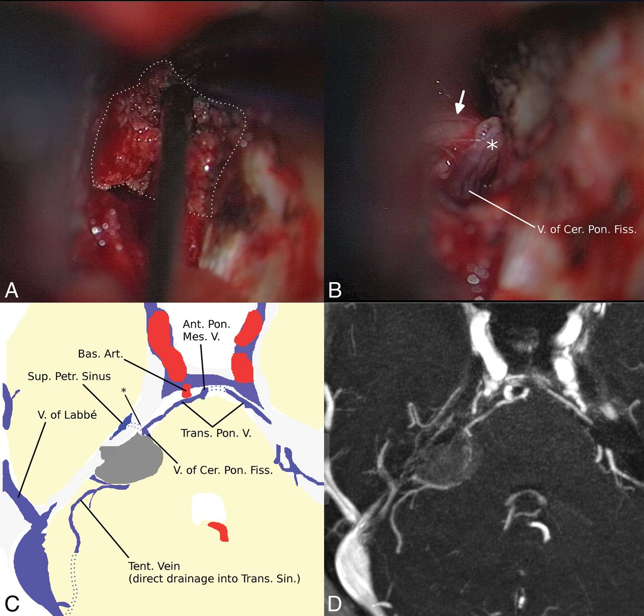
  • Fig 4.
    • Download figure
    • Open in new tab
    • Download powerpoint
    Fig 4.

    A, Intraoperative view shows that the venous anatomy is hidden for the neurosurgeon by the meningioma (white dots). B, After removal of the tumor, the neurosurgeon can see the medial superior petrosal vein (type III) and the trigeminal nerve (arrow). Two tributaries are encountered in concordance with the MR imaging. A schematic drawing (C) based on axial MIP reconstruction (D) of the preoperative 3D-MRA shows a compressed single SPV (asterisk in C) just ventral to the meningioma, with 2 tributaries and a main drainage via a pontine transverse vein and the anterior pontomesencephalic vein. Although the location was known, partial thermocoagulation (note the different color of the SPV marked with an asterisk in B) of the previously compressed SPV could not be prevented. The dorsal region of the cerebellum had direct drainage into the basal vein via a large lateral mesencephalic vein (not shown). V indicates vein; V. of Cer. Pon. Fiss, vein of the cerebellopontine fissure; Bas. Art., basal artery; Ant. Pon. Mes. V, anterior pontomesencephalic vein; Sup. Petr. Sinus, superior petrosal sinus; Trans. Pon. V., transverse pontine vein; Tent. Vein, tentorial vein; Trans. Sin, transverse sinus.

  • Fig 5.
    • Download figure
    • Open in new tab
    • Download powerpoint
    Fig 5.

    Intraoperative views of the tumor (A, white arrow) and after removal of the tumor (B and C): T1-weighted postcontrast scan (D) and axial (E) and sagittal (F) MIP reconstructions of the MR imaging. Nerves VII and VIII (asterisk) are seen directly below the tumor. A single large SPV (white arrow in B–D; purple colored in E,F) drains a large portion of the cerebellum and enters the SPS just in front of the tumor (type II). With a diameter of >2 mm and no visible collaterals in the MRA and intraoperatively, fibrous arachnoidea and tumor (arrowhead) sticking to the SPV were not removed due to the high risk of obliteration.

Tables

  • Figures
    • View popup
    Table 1:

    Results of the ROI analysis for objective image quality and the calculated correlation coefficients for interobserver agreementa

    Anatomic StructureMean Rater 1 (A.U.)Mean Rater 2 (A.U.)No.Pearson Correlation Coefficient RSignificance
    Noisea4 ± 14 ± 1250.818<.001
    Basilar artery1110 ± 2941170 ± 381250.852<.001
    Brain stem176 ± 24179 ± 24250.895<.001
    Vein of Galen1040 ± 3441020 ± 303230.910<.001
    Transverse sinus (R)905 ± 280896 ± 276250.982<.001
    Transverse sinus (L)878 ± 232902 ± 244250.982<.001
    Basal vein (R)812 ± 283783 ± 243190.910<.001
    Basal vein (L)847 ± 233839 ± 234240.936<.001
    SPS (R)561 ± 186601 ± 183250.852<.001
    SPS (L)537 ± 233612 ± 169250.741<.001
    • Note:—R indicates right; L, left; A.U., arbitrary units; No., number of cases.

    • ↵a For noise, the SD of the signal in the ROI is given; for all other ROIs, the mean signal is presented.

    • View popup
    Table 2:

    Results of the evaluation of the diameters of the main vesselsa

    Anatomic StructureMean Rater 1 (A.U.)Mean Rater 2 (A.U.)No.Pearson Correlation Coefficient RSignificance
    Basal vein (R)1.7 ± 0.41.6 ± 0.8240.563.003
    Basal vein (L)1.8 ± 0.41.7 ± 0.4250.659<.001
    Lateral mesencephalic vein (R)0.9 ± 0.50.9 ± 0.5190.753<.001
    Lateral mesencephalic vein (L)0.7 ± 0.70.8 ± 0.6200.828<.001
    Anterior pontomesencephalic vein0.6 ± 0.60.6 ± 0.5250.726<.001
    SPS (R)2.6 ± 0.52.5 ± 0.7250.663<.001
    SPS (L)2.6 ± 0.62.5 ± 0.6250.595.002
    • Note:—No. indicates the number of veins present; R, right; L, left; A.U., arbitrary units.

    • ↵a Missing veins were omitted for the calculation of the mean and rated as 0 mm for the correlation coefficient. Visible veins <0.5 mm were rated as 0.2 mm.

    • View popup
    Table 3:

    Position and size of 83 petrosal veins in 25 patients without intraoperative validation and in 8 patients with intraoperative validation

    Type of SPVNo. of SPVs in PatientsMean Diameter (Range) (mm) in Patients
    Without SurgeryWith SurgeryWithout SurgeryWith Surgery
    I, lateral13 (15.7%)1 (4.8%)2.0 (0.5–2.9)2.5 (2.5–2.5)
    II, intermediate41 (49.4%)10 (47.6%)2.4 (<0.5–5.6)2.1 (1.5–3.6)
    III, medial29 (34.9%)10 (47.6%)1.3 (<0.5–2.7)1.0 (<0.5–2.1)
    I–III83212.0 (<0.5–5.6)1.6 (<0.5–3.6)
    • View popup
    Table 4:

    Frequencies of superior petrosal vein drainage groups for each cerebellopontine angle (n = 50) and each superior petrosal vein (n = 83)

    Drainage GroupFrequency of Group/SideFrequency of Group/SPV
    LeftRightTotalLeftRightTotal
    Petrosal252449/50 sides (98%)313162/83 (74.7%)
    Posterior mesencephalic222244/50 sides (88%)222345/83 (54.2%)
    Anterior pontomesencephalic232144/50 sides (88%)272754/83 (65.1%)
    Tentorial141529/50 sides (58%)141529/83 (34.9%)
PreviousNext
Back to top

In this issue

American Journal of Neuroradiology: 39 (12)
American Journal of Neuroradiology
Vol. 39, Issue 12
1 Dec 2018
  • Table of Contents
  • Index by author
  • Complete Issue (PDF)
Advertisement
Print
Download PDF
Email Article

Thank you for your interest in spreading the word on American Journal of Neuroradiology.

NOTE: We only request your email address so that the person you are recommending the page to knows that you wanted them to see it, and that it is not junk mail. We do not capture any email address.

Enter multiple addresses on separate lines or separate them with commas.
Depiction of the Superior Petrosal Vein Complex by 3D Contrast-Enhanced MR Angiography
(Your Name) has sent you a message from American Journal of Neuroradiology
(Your Name) thought you would like to see the American Journal of Neuroradiology web site.
CAPTCHA
This question is for testing whether or not you are a human visitor and to prevent automated spam submissions.
Cite this article
B. Bender, T.-K. Hauser, A. Korn, U. Klose, M. Tatagiba, U. Ernemann, F.H. Ebner
Depiction of the Superior Petrosal Vein Complex by 3D Contrast-Enhanced MR Angiography
American Journal of Neuroradiology Dec 2018, 39 (12) 2249-2255; DOI: 10.3174/ajnr.A5864

Citation Manager Formats

  • BibTeX
  • Bookends
  • EasyBib
  • EndNote (tagged)
  • EndNote 8 (xml)
  • Medlars
  • Mendeley
  • Papers
  • RefWorks Tagged
  • Ref Manager
  • RIS
  • Zotero
0 Responses
Respond to this article
Share
Bookmark this article
Depiction of the Superior Petrosal Vein Complex by 3D Contrast-Enhanced MR Angiography
B. Bender, T.-K. Hauser, A. Korn, U. Klose, M. Tatagiba, U. Ernemann, F.H. Ebner
American Journal of Neuroradiology Dec 2018, 39 (12) 2249-2255; DOI: 10.3174/ajnr.A5864
del.icio.us logo Twitter logo Facebook logo Mendeley logo
  • Tweet Widget
  • Facebook Like
  • Google Plus One
Purchase

Jump to section

  • Article
    • Abstract
    • ABBREVIATIONS:
    • Materials and Methods
    • Results
    • Discussion
    • Conclusions
    • Footnotes
    • References
  • Figures & Data
  • Supplemental
  • Info & Metrics
  • Responses
  • References
  • PDF

Related Articles

  • PubMed
  • Google Scholar

Cited By...

  • No citing articles found.
  • Crossref
  • Google Scholar

This article has not yet been cited by articles in journals that are participating in Crossref Cited-by Linking.

More in this TOC Section

  • Diagnostic Neuroradiology of Monoclonal Antibodies
  • Clinical Outcomes After Chiari I Decompression
  • Segmentation of Brain Metastases with BLAST
Show more Adult Brain

Similar Articles

Advertisement

Indexed Content

  • Current Issue
  • Accepted Manuscripts
  • Article Preview
  • Past Issues
  • Editorials
  • Editor's Choice
  • Fellows' Journal Club
  • Letters to the Editor
  • Video Articles

Cases

  • Case Collection
  • Archive - Case of the Week
  • Archive - Case of the Month
  • Archive - Classic Case

More from AJNR

  • Trainee Corner
  • Imaging Protocols
  • MRI Safety Corner
  • Book Reviews

Multimedia

  • AJNR Podcasts
  • AJNR Scantastics

Resources

  • Turnaround Time
  • Submit a Manuscript
  • Submit a Video Article
  • Submit an eLetter to the Editor/Response
  • Manuscript Submission Guidelines
  • Statistical Tips
  • Fast Publishing of Accepted Manuscripts
  • Graphical Abstract Preparation
  • Imaging Protocol Submission
  • Evidence-Based Medicine Level Guide
  • Publishing Checklists
  • Author Policies
  • Become a Reviewer/Academy of Reviewers
  • News and Updates

About Us

  • About AJNR
  • Editorial Board
  • Editorial Board Alumni
  • Alerts
  • Permissions
  • Not an AJNR Subscriber? Join Now
  • Advertise with Us
  • Librarian Resources
  • Feedback
  • Terms and Conditions
  • AJNR Editorial Board Alumni

American Society of Neuroradiology

  • Not an ASNR Member? Join Now

© 2025 by the American Society of Neuroradiology All rights, including for text and data mining, AI training, and similar technologies, are reserved.
Print ISSN: 0195-6108 Online ISSN: 1936-959X

Powered by HighWire