
Article Figures & Data
Figures
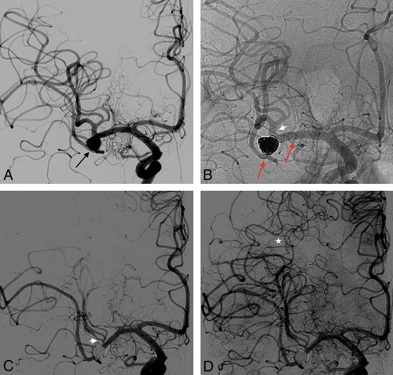
- Fig 1.
A, Procedural angiogram of a 63-year-old man showing a 9-mm left MCA aneurysm recanalized after treatment with a Woven EndoBridge (WEB; Sequent Medical, Aliso Viejo, California) device (black star). This image also shows the lenticulostriate arteries arising from the M1 (black arrow). B, The working projection demonstrates the implantation of 2 LEO stents (Y-configuration) into the M2 branches and a second WEB into the aneurysm. There is a small incomplete wall stent apposition in the superior M2 branch, probably related to a small arterial dissection (short black arrow). C, A 13-month DSA image shows total occlusion of the aneurysm and remodeling of the lenticulostriate arteries covered with 2 overlapped LEOs (red arrow). D, MR imaging reveals small and asymptomatic ischemic lesions in the left basal ganglia (white arrow).
- Fig 2.
A, Procedural angiogram of a 49-year-old male patient with a 7-mm unruptured aneurysm on the right MCA bifurcation (black arrow). B, A nonsubtracted angiographic image obtained during treatment demonstrates the successful SAC embolization of the aneurysm with a LEO stent (2.5 ×18 mm) deployed from the M1 to the inferior M2 segment (red arrows). The M2 superior branch is covered by the stent (short white arrow). C, A 20-month follow-up DSA reveals complete aneurysm occlusion and asymptomatic narrowing of the covered superior M2 branch. D, Blood flow compensation is provided by the collateral circulation (white star).
- Fig 3.
A, Procedural angiogram depicting a right, previously coiled, and recanalized ICA bifurcation aneurysm (black star), and a 3-mm PcomA aneurysm (black arrow). B, The ICA bifurcation aneurysm is treated with SAC with a LEO stent (3.5 × 18 mm) implanted from the ICA to the M1 (red arrow). The right A1 (white arrow) and the right anterior choroidal artery (short black arrow) are covered by the stent. The PcomA artery and the small related aneurysm are covered with the LEO without additional coils. The working (C) and frontal (D) projections during a 24-month follow-up DSA demonstrate a small residual neck of the ICA bifurcation aneurysm (RR II), flow remodeling of the covered A1 segment (white arrow), and occlusion of the PcomA artery and aneurysm (black arrow). The anterior choroidal artery is still patent (short black arrow).


Jump to section
Related Articles
Cited By...
- Flow-Diversion Treatment for Unruptured ICA Bifurcation Aneurysms with Unfavorable Morphology for Coiling
- Early experience treating intracranial aneurysms using Accero: a novel, fully visible, low profile braided stent with platinum-nitinol composite wire technology
- Distal anterior cerebral artery aneurysms treated with flow diversion: experience of a large-volume center and systematic review of the literature