Advertisement

Main menu

  • Home
  • Content
    • Current Issue
    • Accepted Manuscripts
    • Article Preview
    • Past Issue Archive
    • Video Articles
    • AJNR Case Collection
    • Case of the Week Archive
    • Case of the Month Archive
    • Classic Case Archive
  • Special Collections
    • AJNR Awards
    • Low-Field MRI
    • Alzheimer Disease
    • ASNR Foundation Special Collection
    • Photon-Counting CT
    • View All
  • Multimedia
    • AJNR Podcasts
    • AJNR SCANtastic
    • Trainee Corner
    • MRI Safety Corner
    • Imaging Protocols
  • For Authors
    • Submit a Manuscript
    • Submit a Video Article
    • Submit an eLetter to the Editor/Response
    • Manuscript Submission Guidelines
    • Statistical Tips
    • Fast Publishing of Accepted Manuscripts
    • Graphical Abstract Preparation
    • Imaging Protocol Submission
    • Author Policies
  • About Us
    • About AJNR
    • Editorial Board
    • Editorial Board Alumni
  • More
    • Become a Reviewer/Academy of Reviewers
    • Subscribers
    • Permissions
    • Alerts
    • Feedback
    • Advertisers
    • ASNR Home

User menu

  • Alerts
  • Log in

Search

  • Advanced search
American Journal of Neuroradiology

American Journal of Neuroradiology

ASHNR American Society of Functional Neuroradiology ASHNR American Society of Pediatric Neuroradiology ASSR
  • Alerts
  • Log in

Advanced Search

  • Home
  • Content
    • Current Issue
    • Accepted Manuscripts
    • Article Preview
    • Past Issue Archive
    • Video Articles
    • AJNR Case Collection
    • Case of the Week Archive
    • Case of the Month Archive
    • Classic Case Archive
  • Special Collections
    • AJNR Awards
    • Low-Field MRI
    • Alzheimer Disease
    • ASNR Foundation Special Collection
    • Photon-Counting CT
    • View All
  • Multimedia
    • AJNR Podcasts
    • AJNR SCANtastic
    • Trainee Corner
    • MRI Safety Corner
    • Imaging Protocols
  • For Authors
    • Submit a Manuscript
    • Submit a Video Article
    • Submit an eLetter to the Editor/Response
    • Manuscript Submission Guidelines
    • Statistical Tips
    • Fast Publishing of Accepted Manuscripts
    • Graphical Abstract Preparation
    • Imaging Protocol Submission
    • Author Policies
  • About Us
    • About AJNR
    • Editorial Board
    • Editorial Board Alumni
  • More
    • Become a Reviewer/Academy of Reviewers
    • Subscribers
    • Permissions
    • Alerts
    • Feedback
    • Advertisers
    • ASNR Home
  • Follow AJNR on Twitter
  • Visit AJNR on Facebook
  • Follow AJNR on Instagram
  • Join AJNR on LinkedIn
  • RSS Feeds

AJNR Awards, New Junior Editors, and more. Read the latest AJNR updates

Research ArticleSpine Imaging and Spine Image-Guided Interventions

Prospective Comparison of Changes in Lumbar Spine MRI Findings over Time between Individuals with Acute Low Back Pain and Controls: An Exploratory Study

J. Panagopoulos, J.S. Magnussen, J. Hush, C.G. Maher, M. Crites-Battie, J.G. Jarvik, T.S. Jensen and M.J. Hancock
American Journal of Neuroradiology September 2017, 38 (9) 1826-1832; DOI: https://doi.org/10.3174/ajnr.A5357
J. Panagopoulos
aFrom the Faculty of Medicine and Health Sciences (J.P., M.J.H., J.S.M., J.H.)
  • Find this author on Google Scholar
  • Find this author on PubMed
  • Search for this author on this site
  • ORCID record for J. Panagopoulos
J.S. Magnussen
aFrom the Faculty of Medicine and Health Sciences (J.P., M.J.H., J.S.M., J.H.)
bCentre for Physical Health (J.S.M., J.H., M.J.H.), Macquarie University, Sydney, New South Wales, Australia
  • Find this author on Google Scholar
  • Find this author on PubMed
  • Search for this author on this site
  • ORCID record for J.S. Magnussen
J. Hush
aFrom the Faculty of Medicine and Health Sciences (J.P., M.J.H., J.S.M., J.H.)
bCentre for Physical Health (J.S.M., J.H., M.J.H.), Macquarie University, Sydney, New South Wales, Australia
  • Find this author on Google Scholar
  • Find this author on PubMed
  • Search for this author on this site
  • ORCID record for J. Hush
C.G. Maher
cGeorge Institute of Public Health (C.G.M.), University of Sydney, Sydney, New South Wales Australia
  • Find this author on Google Scholar
  • Find this author on PubMed
  • Search for this author on this site
  • ORCID record for C.G. Maher
M. Crites-Battie
dDepartment of Physiotherapy (M.C.-B.), University of Alberta, Edmonton, Alberta, Canada
  • Find this author on Google Scholar
  • Find this author on PubMed
  • Search for this author on this site
  • ORCID record for M. Crites-Battie
J.G. Jarvik
eDepartments of Radiology, Neurological Surgery, and Health Services (J.G.J.), University of Washington, Seattle, Washington
  • Find this author on Google Scholar
  • Find this author on PubMed
  • Search for this author on this site
  • ORCID record for J.G. Jarvik
T.S. Jensen
fNordic Institute of Clinical Biomechanics (T.S.J.), Odense, Denmark
gSpine Centre of Southern Denmark (T.S.J.), Middelfart, Denmark
hInstitute for Regional Health Research (T.S.J.), University of Southern Denmark, Odense, Denmark.
  • Find this author on Google Scholar
  • Find this author on PubMed
  • Search for this author on this site
  • ORCID record for T.S. Jensen
M.J. Hancock
aFrom the Faculty of Medicine and Health Sciences (J.P., M.J.H., J.S.M., J.H.)
bCentre for Physical Health (J.S.M., J.H., M.J.H.), Macquarie University, Sydney, New South Wales, Australia
  • Find this author on Google Scholar
  • Find this author on PubMed
  • Search for this author on this site
  • ORCID record for M.J. Hancock
  • Article
  • Figures & Data
  • Supplemental
  • Info & Metrics
  • Responses
  • References
  • PDF
Loading

Article Figures & Data

Figures

  • Tables
  • Fig 1.
    • Download figure
    • Open in new tab
    • Download powerpoint
    Fig 1.

    Bar chart showing proportions of subjects and controls for whom a definite or probable change occurred in MR imaging findings with time. DD indicates disc degeneration; SI, signal intensity; DH, disc herniation; MC, Modic changes; Spondy/Retro, spondylolisthesis or retrolisthesis.

  • Fig 2.
    • Download figure
    • Open in new tab
    • Download powerpoint
    Fig 2.

    Images of an annular fissure from 1 participant with LBP showing an example of definite change.
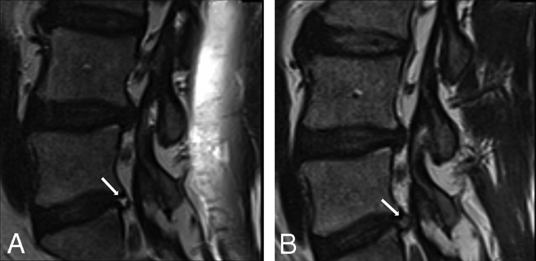
  • Fig 3.
    • Download figure
    • Open in new tab
    • Download powerpoint
    Fig 3.

    Images of disc herniation from 1 participant with LBP showing evidence of definite change.

  • Fig 4.
    • Download figure
    • Open in new tab
    • Download powerpoint
    Fig 4.

    Images of a high-intensity zone from 1 participant with LBP showing evidence of probable change.

Tables

  • Figures
    • View popup
    Table 1:

    MRI findings and the method of assessment at each spinal level

    MRI FindingMethod of Scoring at Each Spinal Level
    Disc degenerationScale of Pfirrmann et al; 1–513
    Signal intensity lossHypointense, intermediate, hyperintense13
    Disc height lossAbsent, mild, moderate, severe13
    Modic changesAccording to Fardon et al14 combined task force; present or absent
    Annular fissureAccording to Fardon et al14 combined task force; present or absent
    HIZAccording to April and Bogduk11 and Fardon et al14 combined task force; present or absent
    Disc herniationAccording to Fardon et al14 combined task force; normal, protrusion, extrusion, sequestration
    Canal stenosisNone, mild, moderate, severe12
    Spondylolisthesis or retrolisthesisPresent or absent12
    Edema (posterior elements)Present or absent
    Nerve root compressionNo contact, contact, contact and deviation, compression15
    Facet joint arthropathyNone, mild, moderate, severe12
    • View popup
    Table 2:

    Baseline characteristics and MRI findings of participants

    VariableLBP Participants (n = 20)Control Participants (n = 10)
    Female sex (No.) (%)9 (45%)5 (50%)
    Age (mean) (SD) (yr)37.4 (9.4)39.8 (9.4)
    ≥2 previous episodes (%)55%50%
    Pain intensity (mean) (SD) (NPRS)5.95 (1.47)0
    Duration of current episode (median) (IQR) (day)6.5 (3.3–9.5)0
    Disc degeneration ≥3 (No.) (%)16 (80%)6 (60%)
    Signal intensity loss (No.) (%)12 (60%)4 (40%)
    Disc height loss (No.) (%)2 (10%)2 (20%)
    Modic changes (No.) (%)6 (30%)2 (20%)
    Annular fissure (No.) (%)3 (15%)1 (10%)
    HIZ (No.) (%)10 (50%)6 (60%)
    Disc herniation total (No.) (%)20 (100%)9 (90%)
    Canal stenosis (No.) (%)2 (10%)0 (0%)
    Spondylolisthesis or retrolisthesis (No.) (%)6 (30%)4 (40%)
    Facet joint arthropathy (No.) (%)11 (55%)4 (40%)
    Bone edema (posterior elements) (No.) (%)0 (0%)1 (10%)
    Nerve root compression (No.) (%)10 (50%)2 (20%)
    • Note:—IQR indicates interquartile range; NPRD, Numerical Pain Rating Scale.

    • View popup
    Table 3:

    Proportion of participants in whom MRI findings worsened or improved during a 12-week perioda

    MRI Findings/Subjective ChangeControlsSubjects with LBP
    WorsenedImprovedWorsened or ImprovedWorsenedImprovedWorsened or Improved
    Disc degeneration1/100/101/102/200/202/20
    = 10%= 0%= 10%= 10%= 0%= 10%
    Disc signal intensity1/100/101/102/200/202/20
    = 10%= 0%= 10%= 10%= 0%= 10%
    Disc height1/100/101/100/200/200/20
    = 10%= 0%= 10%= 0%= 0%= 0%
    HIZ0/104/104/105/203/208/20
    = 0%= 40%= 40%= 25%= 15%= 40%
    Annular fissure0/101/101/102/203/205/20
    = 0%= 10%= 10%= 10%= 15%= 25%
    Modic changes1/101/102/101/202/203/20
    = 10%= 10%= 20%= 5%= 10%= 15%
    Disc herniation0/103/103/107/206/2013/20
    = 0%= 30%= 30%= 35%= 30%= 65%
    Facet joint arthropathy0/100/100/100/200/200/20
    = 0%= 0%= 0%= 0%= 0%= 0%
    Central canal stenosis0/100/100/100/200/200/20
    = 0%= 0%= 0%= 0%= 0%= 0%
    Spondylolisthesis or retrolisthesis0/100/100/100/200/200/20
    = 0%= 0%= 0%= 0%= 0%= 0%
    Edema0/101/101/100/200/200/20
    = 0%= 10%= 10%= 0%= 0%= 0%
    Nerve root compromise0/100/100/102/201/203/20
    = 0%= 0%= 0%= 20%= 10%= 15%
    • ↵a If a particular MRI finding both improved and worsened at different follow-up time points (compared with baseline), we reported the change at or closest to the 12-week time point.

PreviousNext
Back to top

In this issue

American Journal of Neuroradiology: 38 (9)
American Journal of Neuroradiology
Vol. 38, Issue 9
1 Sep 2017
  • Table of Contents
  • Index by author
  • Complete Issue (PDF)
Advertisement
Print
Download PDF
Email Article

Thank you for your interest in spreading the word on American Journal of Neuroradiology.

NOTE: We only request your email address so that the person you are recommending the page to knows that you wanted them to see it, and that it is not junk mail. We do not capture any email address.

Enter multiple addresses on separate lines or separate them with commas.
Prospective Comparison of Changes in Lumbar Spine MRI Findings over Time between Individuals with Acute Low Back Pain and Controls: An Exploratory Study
(Your Name) has sent you a message from American Journal of Neuroradiology
(Your Name) thought you would like to see the American Journal of Neuroradiology web site.
CAPTCHA
This question is for testing whether or not you are a human visitor and to prevent automated spam submissions.
Cite this article
J. Panagopoulos, J.S. Magnussen, J. Hush, C.G. Maher, M. Crites-Battie, J.G. Jarvik, T.S. Jensen, M.J. Hancock
Prospective Comparison of Changes in Lumbar Spine MRI Findings over Time between Individuals with Acute Low Back Pain and Controls: An Exploratory Study
American Journal of Neuroradiology Sep 2017, 38 (9) 1826-1832; DOI: 10.3174/ajnr.A5357

Citation Manager Formats

  • BibTeX
  • Bookends
  • EasyBib
  • EndNote (tagged)
  • EndNote 8 (xml)
  • Medlars
  • Mendeley
  • Papers
  • RefWorks Tagged
  • Ref Manager
  • RIS
  • Zotero
0 Responses
Respond to this article
Share
Bookmark this article
Prospective Comparison of Changes in Lumbar Spine MRI Findings over Time between Individuals with Acute Low Back Pain and Controls: An Exploratory Study
J. Panagopoulos, J.S. Magnussen, J. Hush, C.G. Maher, M. Crites-Battie, J.G. Jarvik, T.S. Jensen, M.J. Hancock
American Journal of Neuroradiology Sep 2017, 38 (9) 1826-1832; DOI: 10.3174/ajnr.A5357
del.icio.us logo Twitter logo Facebook logo Mendeley logo
  • Tweet Widget
  • Facebook Like
  • Google Plus One
Purchase

Jump to section

  • Article
    • Abstract
    • ABBREVIATIONS:
    • Materials and Methods
    • Results
    • Discussion
    • Acknowledgments
    • Footnotes
    • References
  • Figures & Data
  • Supplemental
  • Info & Metrics
  • Responses
  • References
  • PDF

Related Articles

  • PubMed
  • Google Scholar

Cited By...

  • No citing articles found.
  • Crossref
  • Google Scholar

This article has not yet been cited by articles in journals that are participating in Crossref Cited-by Linking.

More in this TOC Section

  • Optimization of Photon Counting CT Myelography
  • Characteristics of SIH Type I Culprit Lesions
  • Management Outcomes For VO Spine Biopsy
Show more Spine Imaging and Spine Image-Guided Interventions

Similar Articles

Advertisement

Indexed Content

  • Current Issue
  • Accepted Manuscripts
  • Article Preview
  • Past Issues
  • Editorials
  • Editor's Choice
  • Fellows' Journal Club
  • Letters to the Editor
  • Video Articles

Cases

  • Case Collection
  • Archive - Case of the Week
  • Archive - Case of the Month
  • Archive - Classic Case

More from AJNR

  • Trainee Corner
  • Imaging Protocols
  • MRI Safety Corner
  • Book Reviews

Multimedia

  • AJNR Podcasts
  • AJNR Scantastics

Resources

  • Turnaround Time
  • Submit a Manuscript
  • Submit a Video Article
  • Submit an eLetter to the Editor/Response
  • Manuscript Submission Guidelines
  • Statistical Tips
  • Fast Publishing of Accepted Manuscripts
  • Graphical Abstract Preparation
  • Imaging Protocol Submission
  • Evidence-Based Medicine Level Guide
  • Publishing Checklists
  • Author Policies
  • Become a Reviewer/Academy of Reviewers
  • News and Updates

About Us

  • About AJNR
  • Editorial Board
  • Editorial Board Alumni
  • Alerts
  • Permissions
  • Not an AJNR Subscriber? Join Now
  • Advertise with Us
  • Librarian Resources
  • Feedback
  • Terms and Conditions
  • AJNR Editorial Board Alumni

American Society of Neuroradiology

  • Not an ASNR Member? Join Now

© 2025 by the American Society of Neuroradiology All rights, including for text and data mining, AI training, and similar technologies, are reserved.
Print ISSN: 0195-6108 Online ISSN: 1936-959X

Powered by HighWire