
Article Figures & Data
Figures
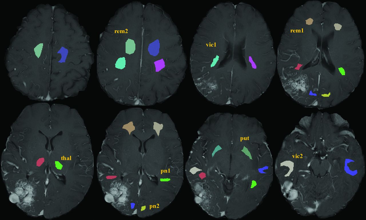
- Fig 1.
Examples of the different WM (pn1/pn2, vic1/vic2, rem1/rem2) and GM masks (put, thal) selected on the T1WI contrast-enhanced sequence. Each mask has been labeled in either of the 2 hemispheres. The set of masks was loaded on each perfusion map separately, which had previously been coregistered to the T1WI contrast-enhanced dataset. Except for some of the GM masks, almost all masks were drawn as a volume, which explains their delineation on several consecutive sections. Note the different color for the respective mask in the contralateral hemisphere, because the software did not allow the use of the same color for the respective opposite mask. pn indicates perinidal; vic, vicinity; rem, remote; thal, thalamus; put, putamen.
- Fig 2.
When superimposing the red mask representing the ipsilateral pn1 ROI previously drawn in the T1WI contrast-enhanced dataset (T1wCE; left) on the FPD-CT perfusion dataset (right), a good overlap without contamination from bAVM tissue could be observed.
- Fig 3.
Box-and-whisker plots for patients 1–4. The boxes show the median, lower, and upper quartiles. Outliers are displayed as red crosses. The mean and SD across the pixels of each ROI of patient 5 are represented in green. ref indicates reference.
- Fig 4.
Correlations between different parameters for all patients. The squares represent the ratio for a single pair of masks.


Tables
Coefficients of variation aggregating all masks, excluding the outlier (pn 1 of patient 3), across all patients
Modality Coefficient of Variation FPD-CT 21.2% ASL-CBF 11.0% ASL-arrival time 12.3% DSC-rCBF 21.3% DSC-rCBV 17.8%


