Advertisement

Main menu

  • Home
  • Content
    • Current Issue
    • Accepted Manuscripts
    • Article Preview
    • Past Issue Archive
    • Video Articles
    • AJNR Case Collection
    • Case of the Week Archive
    • Case of the Month Archive
    • Classic Case Archive
  • Special Collections
    • AJNR Awards
    • Low-Field MRI
    • Alzheimer Disease
    • ASNR Foundation Special Collection
    • Photon-Counting CT
    • View All
  • Multimedia
    • AJNR Podcasts
    • AJNR SCANtastic
    • Trainee Corner
    • MRI Safety Corner
    • Imaging Protocols
  • For Authors
    • Submit a Manuscript
    • Submit a Video Article
    • Submit an eLetter to the Editor/Response
    • Manuscript Submission Guidelines
    • Statistical Tips
    • Fast Publishing of Accepted Manuscripts
    • Graphical Abstract Preparation
    • Imaging Protocol Submission
    • Author Policies
  • About Us
    • About AJNR
    • Editorial Board
    • Editorial Board Alumni
  • More
    • Become a Reviewer/Academy of Reviewers
    • Subscribers
    • Permissions
    • Alerts
    • Feedback
    • Advertisers
    • ASNR Home

User menu

  • Alerts
  • Log in

Search

  • Advanced search
American Journal of Neuroradiology

American Journal of Neuroradiology

ASHNR American Society of Functional Neuroradiology ASHNR American Society of Pediatric Neuroradiology ASSR
  • Alerts
  • Log in

Advanced Search

  • Home
  • Content
    • Current Issue
    • Accepted Manuscripts
    • Article Preview
    • Past Issue Archive
    • Video Articles
    • AJNR Case Collection
    • Case of the Week Archive
    • Case of the Month Archive
    • Classic Case Archive
  • Special Collections
    • AJNR Awards
    • Low-Field MRI
    • Alzheimer Disease
    • ASNR Foundation Special Collection
    • Photon-Counting CT
    • View All
  • Multimedia
    • AJNR Podcasts
    • AJNR SCANtastic
    • Trainee Corner
    • MRI Safety Corner
    • Imaging Protocols
  • For Authors
    • Submit a Manuscript
    • Submit a Video Article
    • Submit an eLetter to the Editor/Response
    • Manuscript Submission Guidelines
    • Statistical Tips
    • Fast Publishing of Accepted Manuscripts
    • Graphical Abstract Preparation
    • Imaging Protocol Submission
    • Author Policies
  • About Us
    • About AJNR
    • Editorial Board
    • Editorial Board Alumni
  • More
    • Become a Reviewer/Academy of Reviewers
    • Subscribers
    • Permissions
    • Alerts
    • Feedback
    • Advertisers
    • ASNR Home
  • Follow AJNR on Twitter
  • Visit AJNR on Facebook
  • Follow AJNR on Instagram
  • Join AJNR on LinkedIn
  • RSS Feeds

AJNR Awards, New Junior Editors, and more. Read the latest AJNR updates

Research ArticleNeurointervention
Open Access

Comparison of the Diagnostic Utility of 4D-DSA with Conventional 2D- and 3D-DSA in the Diagnosis of Cerebrovascular Abnormalities

C. Sandoval-Garcia, P. Yang, T. Schubert, S. Schafer, S. Hetzel, A. Ahmed and C. Strother
American Journal of Neuroradiology April 2017, 38 (4) 729-734; DOI: https://doi.org/10.3174/ajnr.A5137
C. Sandoval-Garcia
aFrom the Departments of Neurological Surgery (C.S.-G., A.A.)
  • Find this author on Google Scholar
  • Find this author on PubMed
  • Search for this author on this site
  • ORCID record for C. Sandoval-Garcia
P. Yang
dDepartment of Neurosurgery (P.Y.), Changhai Hospital, Second Military Medical University, Shanghai, China
  • Find this author on Google Scholar
  • Find this author on PubMed
  • Search for this author on this site
  • ORCID record for P. Yang
T. Schubert
bRadiology (T.S., C.S.)
  • Find this author on Google Scholar
  • Find this author on PubMed
  • Search for this author on this site
  • ORCID record for T. Schubert
S. Schafer
eSiemens Healthineers, USA (S.S.), Hoffman Estates, Illinois.
  • Find this author on Google Scholar
  • Find this author on PubMed
  • Search for this author on this site
  • ORCID record for S. Schafer
S. Hetzel
cBiostatistics and Medical Informatics (S.H.), University of Wisconsin School of Medicine and Public Health, Madison, Wisconsin
  • Find this author on Google Scholar
  • Find this author on PubMed
  • Search for this author on this site
  • ORCID record for S. Hetzel
A. Ahmed
aFrom the Departments of Neurological Surgery (C.S.-G., A.A.)
  • Find this author on Google Scholar
  • Find this author on PubMed
  • Search for this author on this site
  • ORCID record for A. Ahmed
C. Strother
bRadiology (T.S., C.S.)
  • Find this author on Google Scholar
  • Find this author on PubMed
  • Search for this author on this site
  • ORCID record for C. Strother
  • Article
  • Figures & Data
  • Info & Metrics
  • Responses
  • References
  • PDF
Loading

Article Figures & Data

Figures

  • Tables
  • Fig 1.
    • Download figure
    • Open in new tab
    • Download powerpoint
    Fig 1.

    Screenshot of the evaluation form used in the evaluation of the 4D-DSA reconstructions.

  • Fig 2.
    • Download figure
    • Open in new tab
    • Download powerpoint
    Fig 2.

    A comparison of conventional 3D-DSA (upper row) with different timeframes from a 4D-DSA reconstruction (lower row). The projections of the 2 image types are identical. The 3D-DSA images allow viewing from any desired angle; however, in this case because of the vascular overlap, it is impossible to clearly see the relationship of the small aneurysm (yellow circle) to its parent artery. Because the 4D images allow viewing not only from any desired angle but also at any time during the passage of a contrast bolus through the vasculature, the early timeframes of the 4D images allow clear visualization of these relationships.
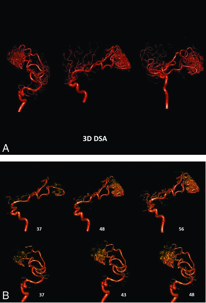
  • Fig 3.
    • Download figure
    • Open in new tab
    • Download powerpoint
    Fig 3.

    A, 3D-DSA of an AVM supplied by 2 branches of the right MCA. Despite the excellent image quality, the image is a composite of all of the 2D projections used in the reconstruction. The overlap of vascular components in the nidus precludes identification of intranidal aneurysms, the direct AVF, or venous outflow stenosis. B, Two views of early timeframes from a 4D-DSA reconstruction of the AVM shown in part A. The projections for this reconstruction are obtained at 30 frames per second. The number of each timeframe is shown beside each image. The angioarchitecture of the nidus can be clearly seen. The 4D-DSA images may be viewed from any desired angle at any time in the bolus passage.

Tables

  • Figures
    • View popup
    Table 1:

    Results of the 3 evaluators decisions regarding the presence of an abnormality on the 4D studies

    4D MethodEvaluator
    123
    No. (% Correct)No. (% Correct)No. (% Correct)
    Definitely yes17 (100)15 (100)18 (100)
    Probably yes2 (50)3 (100)1 (0)
    Unsure0 (NaN)1 (0)0 (NaN)
    Probably no2 (100)0 (NaN)0 (NaN)
    Definitely no5 (100)7 (100)7 (100)
    • Note:—NaN indicates not a number.

    • View popup
    Table 2:

    Results of the 3 evaluator's decisions when results were consolidated into responses of yes, unsure, and no

    4D MethodEvaluator
    123
    No. (% Correct)No. (% Correct)No. (% Correct)
    Yes19 (95)18 (100)19 (95)
    Unsure0 (NaN)1 (0)0 (NaN)
    No7 (100)7 (100)7 (100)
    • Note:—NaN indicates not a number.

    • View popup
    Table 3:

    Summary of correctly identifying abnormality by evaluator and consensusa

    4D MethodEvaluatorConsensus
    123
    True YesTrue NoTrue YesTrue NoTrue YesTrue NoTrue YesTrue No
    Yes181180181180
    Unsure00010000
    No07070708
    • ↵a Data are frequencies.

    • View popup
    Table 4:

    Evaluator's sensitivity, specificity, positive predictive value, negative predictive value, area under the curve, and consensus results

    MeasurementEvaluatorConsensus
    123
    Sensitivity1111
    Specificity0.8750.8750.8751
    PPV0.9470.9470.9471
    NPV1111
    AUC (95% CI)0.938 (0.815–1.000)0.938 (0.815–1.000)0.938 (0.815–1.000)1.000 (1.000–1.000)
    • Note:—PPV indicates positive predictive value; NPV, negative predictive value; AUC, area under the curve.

    • View popup
    Table 5:

    Summary of each evaluator's resulta

    4D MethodEvaluator
    123
    No.No.No.
    Aneurysm91011
    AVF322
    AVM344
    Occlusive disease–stenosis432
    • ↵a There were 9 aneurysms, 6 AVMs/AVFs, 8 normal, 3 stenosis/occlusion in the study population.

    • View popup
    Table 6:

    Summary of agreement between 4D and 2D/3D evaluations regarding type of abnormality (when there was one)a

    4D:TrueEvaluatorConsensus
    123
    AneurAVFAVMOD-SNoneAneurAVFAVMOD-SNoneAneurAVFAVMOD-SNoneAneurAVFAVMOD-SNone
    Aneur90000900109001190010
    AVF03000020000200002000
    AVM00300013000130001300
    OD-S00031000210002000020
    None00007000070000700008
    • Note:—Aneur indicates aneurysm; OD-S, occlusive disease–stenosis.

    • ↵a Data are frequencies.

PreviousNext
Back to top

In this issue

American Journal of Neuroradiology: 38 (4)
American Journal of Neuroradiology
Vol. 38, Issue 4
1 Apr 2017
  • Table of Contents
  • Index by author
  • Complete Issue (PDF)
Advertisement
Print
Download PDF
Email Article

Thank you for your interest in spreading the word on American Journal of Neuroradiology.

NOTE: We only request your email address so that the person you are recommending the page to knows that you wanted them to see it, and that it is not junk mail. We do not capture any email address.

Enter multiple addresses on separate lines or separate them with commas.
Comparison of the Diagnostic Utility of 4D-DSA with Conventional 2D- and 3D-DSA in the Diagnosis of Cerebrovascular Abnormalities
(Your Name) has sent you a message from American Journal of Neuroradiology
(Your Name) thought you would like to see the American Journal of Neuroradiology web site.
CAPTCHA
This question is for testing whether or not you are a human visitor and to prevent automated spam submissions.
Cite this article
C. Sandoval-Garcia, P. Yang, T. Schubert, S. Schafer, S. Hetzel, A. Ahmed, C. Strother
Comparison of the Diagnostic Utility of 4D-DSA with Conventional 2D- and 3D-DSA in the Diagnosis of Cerebrovascular Abnormalities
American Journal of Neuroradiology Apr 2017, 38 (4) 729-734; DOI: 10.3174/ajnr.A5137

Citation Manager Formats

  • BibTeX
  • Bookends
  • EasyBib
  • EndNote (tagged)
  • EndNote 8 (xml)
  • Medlars
  • Mendeley
  • Papers
  • RefWorks Tagged
  • Ref Manager
  • RIS
  • Zotero
0 Responses
Respond to this article
Share
Bookmark this article
Comparison of the Diagnostic Utility of 4D-DSA with Conventional 2D- and 3D-DSA in the Diagnosis of Cerebrovascular Abnormalities
C. Sandoval-Garcia, P. Yang, T. Schubert, S. Schafer, S. Hetzel, A. Ahmed, C. Strother
American Journal of Neuroradiology Apr 2017, 38 (4) 729-734; DOI: 10.3174/ajnr.A5137
del.icio.us logo Twitter logo Facebook logo Mendeley logo
  • Tweet Widget
  • Facebook Like
  • Google Plus One
Purchase

Jump to section

  • Article
    • Abstract
    • Materials and Methods
    • Results
    • Discussion
    • Conclusions
    • Footnotes
    • REFERENCES
  • Figures & Data
  • Info & Metrics
  • Responses
  • References
  • PDF

Related Articles

  • No related articles found.
  • PubMed
  • Google Scholar

Cited By...

  • Endovascular treatment in the multimodality management of brain arteriovenous malformations: report of the Society of NeuroInterventional Surgery Standards and Guidelines Committee
  • 4D-DSA: Development and Current Neurovascular Applications
  • Four-dimensional digital subtraction angiography for exploration of spinal cord vascular malformations: preliminary experience
  • Quantitative and Qualitative Comparison of 4D-DSA with 3D-DSA Using Computational Fluid Dynamics Simulations in Cerebral Aneurysms
  • Crossref
  • Google Scholar

This article has not yet been cited by articles in journals that are participating in Crossref Cited-by Linking.

More in this TOC Section

  • Rescue Reentry in Carotid Near-Occlusion
  • Contour Neurovascular System: Five Year Follow Up
  • Effect of SARS-CoV2 on Endovascular Thrombectomy
Show more Neurointervention

Similar Articles

Advertisement

Indexed Content

  • Current Issue
  • Accepted Manuscripts
  • Article Preview
  • Past Issues
  • Editorials
  • Editor's Choice
  • Fellows' Journal Club
  • Letters to the Editor
  • Video Articles

Cases

  • Case Collection
  • Archive - Case of the Week
  • Archive - Case of the Month
  • Archive - Classic Case

More from AJNR

  • Trainee Corner
  • Imaging Protocols
  • MRI Safety Corner
  • Book Reviews

Multimedia

  • AJNR Podcasts
  • AJNR Scantastics

Resources

  • Turnaround Time
  • Submit a Manuscript
  • Submit a Video Article
  • Submit an eLetter to the Editor/Response
  • Manuscript Submission Guidelines
  • Statistical Tips
  • Fast Publishing of Accepted Manuscripts
  • Graphical Abstract Preparation
  • Imaging Protocol Submission
  • Evidence-Based Medicine Level Guide
  • Publishing Checklists
  • Author Policies
  • Become a Reviewer/Academy of Reviewers
  • News and Updates

About Us

  • About AJNR
  • Editorial Board
  • Editorial Board Alumni
  • Alerts
  • Permissions
  • Not an AJNR Subscriber? Join Now
  • Advertise with Us
  • Librarian Resources
  • Feedback
  • Terms and Conditions
  • AJNR Editorial Board Alumni

American Society of Neuroradiology

  • Not an ASNR Member? Join Now

© 2025 by the American Society of Neuroradiology All rights, including for text and data mining, AI training, and similar technologies, are reserved.
Print ISSN: 0195-6108 Online ISSN: 1936-959X

Powered by HighWire