Advertisement

Main menu

  • Home
  • Content
    • Current Issue
    • Accepted Manuscripts
    • Article Preview
    • Past Issue Archive
    • Video Articles
    • AJNR Case Collection
    • Case of the Week Archive
    • Case of the Month Archive
    • Classic Case Archive
  • Special Collections
    • AJNR Awards
    • Low-Field MRI
    • Alzheimer Disease
    • ASNR Foundation Special Collection
    • Photon-Counting CT
    • View All
  • Multimedia
    • AJNR Podcasts
    • AJNR SCANtastic
    • Trainee Corner
    • MRI Safety Corner
    • Imaging Protocols
  • For Authors
    • Submit a Manuscript
    • Submit a Video Article
    • Submit an eLetter to the Editor/Response
    • Manuscript Submission Guidelines
    • Statistical Tips
    • Fast Publishing of Accepted Manuscripts
    • Graphical Abstract Preparation
    • Imaging Protocol Submission
    • Author Policies
  • About Us
    • About AJNR
    • Editorial Board
    • Editorial Board Alumni
  • More
    • Become a Reviewer/Academy of Reviewers
    • Subscribers
    • Permissions
    • Alerts
    • Feedback
    • Advertisers
    • ASNR Home

User menu

  • Alerts
  • Log in

Search

  • Advanced search
American Journal of Neuroradiology

American Journal of Neuroradiology

ASHNR American Society of Functional Neuroradiology ASHNR American Society of Pediatric Neuroradiology ASSR
  • Alerts
  • Log in

Advanced Search

  • Home
  • Content
    • Current Issue
    • Accepted Manuscripts
    • Article Preview
    • Past Issue Archive
    • Video Articles
    • AJNR Case Collection
    • Case of the Week Archive
    • Case of the Month Archive
    • Classic Case Archive
  • Special Collections
    • AJNR Awards
    • Low-Field MRI
    • Alzheimer Disease
    • ASNR Foundation Special Collection
    • Photon-Counting CT
    • View All
  • Multimedia
    • AJNR Podcasts
    • AJNR SCANtastic
    • Trainee Corner
    • MRI Safety Corner
    • Imaging Protocols
  • For Authors
    • Submit a Manuscript
    • Submit a Video Article
    • Submit an eLetter to the Editor/Response
    • Manuscript Submission Guidelines
    • Statistical Tips
    • Fast Publishing of Accepted Manuscripts
    • Graphical Abstract Preparation
    • Imaging Protocol Submission
    • Author Policies
  • About Us
    • About AJNR
    • Editorial Board
    • Editorial Board Alumni
  • More
    • Become a Reviewer/Academy of Reviewers
    • Subscribers
    • Permissions
    • Alerts
    • Feedback
    • Advertisers
    • ASNR Home
  • Follow AJNR on Twitter
  • Visit AJNR on Facebook
  • Follow AJNR on Instagram
  • Join AJNR on LinkedIn
  • RSS Feeds

AJNR Awards, New Junior Editors, and more. Read the latest AJNR updates

Research ArticleAdult Brain

Combining Diffusion Tensor Metrics and DSC Perfusion Imaging: Can It Improve the Diagnostic Accuracy in Differentiating Tumefactive Demyelination from High-Grade Glioma?

S.B. Hiremath, A. Muraleedharan, S. Kumar, C. Nagesh, C. Kesavadas, M. Abraham, T.R. Kapilamoorthy and B. Thomas
American Journal of Neuroradiology April 2017, 38 (4) 685-690; DOI: https://doi.org/10.3174/ajnr.A5089
S.B. Hiremath
aFrom the Departments of Imaging Sciences and Interventional Radiology (S.B.H., A.M., S.K., C.N., C.K., T.R.K., B.T.)
  • Find this author on Google Scholar
  • Find this author on PubMed
  • Search for this author on this site
  • ORCID record for S.B. Hiremath
A. Muraleedharan
aFrom the Departments of Imaging Sciences and Interventional Radiology (S.B.H., A.M., S.K., C.N., C.K., T.R.K., B.T.)
  • Find this author on Google Scholar
  • Find this author on PubMed
  • Search for this author on this site
  • ORCID record for A. Muraleedharan
S. Kumar
aFrom the Departments of Imaging Sciences and Interventional Radiology (S.B.H., A.M., S.K., C.N., C.K., T.R.K., B.T.)
  • Find this author on Google Scholar
  • Find this author on PubMed
  • Search for this author on this site
  • ORCID record for S. Kumar
C. Nagesh
aFrom the Departments of Imaging Sciences and Interventional Radiology (S.B.H., A.M., S.K., C.N., C.K., T.R.K., B.T.)
  • Find this author on Google Scholar
  • Find this author on PubMed
  • Search for this author on this site
  • ORCID record for C. Nagesh
C. Kesavadas
aFrom the Departments of Imaging Sciences and Interventional Radiology (S.B.H., A.M., S.K., C.N., C.K., T.R.K., B.T.)
  • Find this author on Google Scholar
  • Find this author on PubMed
  • Search for this author on this site
  • ORCID record for C. Kesavadas
M. Abraham
bNeurosurgery (M.A.), Sree Chitra Tirunal Institute for Medical Sciences and Technology, Trivandrum, Kerala, India.
  • Find this author on Google Scholar
  • Find this author on PubMed
  • Search for this author on this site
  • ORCID record for M. Abraham
T.R. Kapilamoorthy
aFrom the Departments of Imaging Sciences and Interventional Radiology (S.B.H., A.M., S.K., C.N., C.K., T.R.K., B.T.)
  • Find this author on Google Scholar
  • Find this author on PubMed
  • Search for this author on this site
  • ORCID record for T.R. Kapilamoorthy
B. Thomas
aFrom the Departments of Imaging Sciences and Interventional Radiology (S.B.H., A.M., S.K., C.N., C.K., T.R.K., B.T.)
  • Find this author on Google Scholar
  • Find this author on PubMed
  • Search for this author on this site
  • ORCID record for B. Thomas
  • Article
  • Figures & Data
  • Supplemental
  • Info & Metrics
  • Responses
  • References
  • PDF
Loading

Article Figures & Data

Figures

  • Tables
  • Fig 1.
    • Download figure
    • Open in new tab
    • Download powerpoint
    Fig 1.

    The boxplots for DTI metrics with statistical significance: p (A), q (B), L (C), and ADC (D) values of the enhancing areas and normal-appearing white matter in TDL and HGG, respectively.
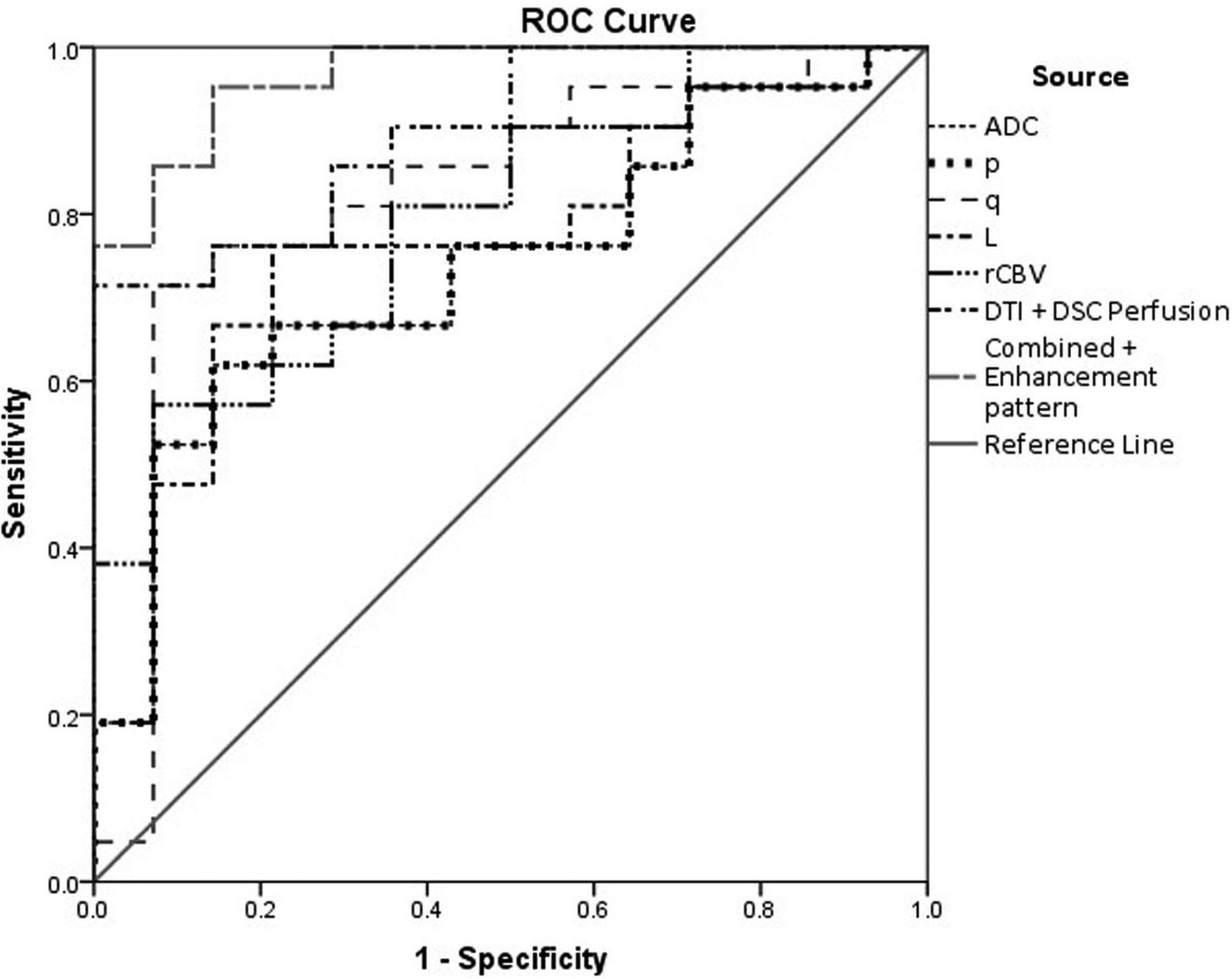
  • Fig 2.
    • Download figure
    • Open in new tab
    • Download powerpoint
    Fig 2.

    ROC curve for the diffusion tensor metrics: rCBV, combined ROC of DTI metrics and DSC perfusion, and combined ROC of enhancement pattern with DTI metrics and DSC perfusion.

Tables

  • Figures
    • View popup
    Table 1:

    Analysis of conventional imaging sequences

    Conventional AnalysisHigh-Grade GliomaNot High-Grade Glioma
    Diagnosed positive176
    Diagnosed negative48
    • View popup
    Table 2:

    Summary of results, mean ± SD

    Enhancing AreasPerilesional HyperintensityNAWMPaPb95% CI
    ADCc
        TDLs0.83 ± 0.151.56 ± 0.110.76 ± 0.06.02d.810.74–0.92
        High-grade gliomas1.01 ± 0.251.54 ± 0.20.79 ± 0.060.90–1.13
    pc
        TDLs1.44 ± 0.262.7 ± 0.191.31 ± 0.10.02d.811.29–1.59
        High-grade gliomas1.76 ± 0.442.67 ± 0.341.37 ± 0.111.56–1.96
    qc
        TDLs0.25 ± 0.070.36 ± 0.050.55 ± 0.06.004d.550.21–0.30
        High-grade gliomas0.33 ± 0.070.38 ± 0.070.54 ± 0.070.30–0.37
    Lc
        TDLs1.46 ± 0.262.7 ± 0.181.43 ± 0.11.016d.831.31–1.62
        High-grade gliomas1.79 ± 0.432.7 ± 0.341.46 ± 0.091.60–1.99
    FA
        TDLs0.21 ± 0.060.44 ± 0.330.47 ± 0.07.341.0520.17–0.25
        High-grade gliomas0.23 ± 0.060.51 ± 0.090.45 ± 0.060.20–0.27
    rCBV
        TDLs2.11 ± 1.12–.003d1.47–2.76
        High-grade gliomas3.77 ± 1.65–3.02–4.52
    • ↵a Difference in the mean values of enhancing areas of TDLs and high-grade gliomas.

    • ↵b Difference in the mean values of perilesional hyperintensity of TDLs and high-grade gliomas.

    • ↵c Values of ADC, p, q, and L are in units of 10−3 mm2/s.

    • ↵d Significant.

    • View popup
    Table 3:

    ROC curve results on sensitivity and specificity to differentiate high-grade gliomas and TDLs

    ParameterSensitivity (%)Specificity (%)AUC
    ADC valuea52.492.90.738
    p valuea61.985.70.738
    q valuea71.492.90.823
    L valuea76.278.60.765
    rCBV52.492.90.796
    DTI + DSC perfusion (combined)71.492.90.901
    Heterogeneous enhancement pattern + combined85.792.90.966
    • ↵a Values of ADC, p, q, and L are in the units of 10−3 mm2/s.

PreviousNext
Back to top

In this issue

American Journal of Neuroradiology: 38 (4)
American Journal of Neuroradiology
Vol. 38, Issue 4
1 Apr 2017
  • Table of Contents
  • Index by author
  • Complete Issue (PDF)
Advertisement
Print
Download PDF
Email Article

Thank you for your interest in spreading the word on American Journal of Neuroradiology.

NOTE: We only request your email address so that the person you are recommending the page to knows that you wanted them to see it, and that it is not junk mail. We do not capture any email address.

Enter multiple addresses on separate lines or separate them with commas.
Combining Diffusion Tensor Metrics and DSC Perfusion Imaging: Can It Improve the Diagnostic Accuracy in Differentiating Tumefactive Demyelination from High-Grade Glioma?
(Your Name) has sent you a message from American Journal of Neuroradiology
(Your Name) thought you would like to see the American Journal of Neuroradiology web site.
CAPTCHA
This question is for testing whether or not you are a human visitor and to prevent automated spam submissions.
Cite this article
S.B. Hiremath, A. Muraleedharan, S. Kumar, C. Nagesh, C. Kesavadas, M. Abraham, T.R. Kapilamoorthy, B. Thomas
Combining Diffusion Tensor Metrics and DSC Perfusion Imaging: Can It Improve the Diagnostic Accuracy in Differentiating Tumefactive Demyelination from High-Grade Glioma?
American Journal of Neuroradiology Apr 2017, 38 (4) 685-690; DOI: 10.3174/ajnr.A5089

Citation Manager Formats

  • BibTeX
  • Bookends
  • EasyBib
  • EndNote (tagged)
  • EndNote 8 (xml)
  • Medlars
  • Mendeley
  • Papers
  • RefWorks Tagged
  • Ref Manager
  • RIS
  • Zotero
0 Responses
Respond to this article
Share
Bookmark this article
Combining Diffusion Tensor Metrics and DSC Perfusion Imaging: Can It Improve the Diagnostic Accuracy in Differentiating Tumefactive Demyelination from High-Grade Glioma?
S.B. Hiremath, A. Muraleedharan, S. Kumar, C. Nagesh, C. Kesavadas, M. Abraham, T.R. Kapilamoorthy, B. Thomas
American Journal of Neuroradiology Apr 2017, 38 (4) 685-690; DOI: 10.3174/ajnr.A5089
del.icio.us logo Twitter logo Facebook logo Mendeley logo
  • Tweet Widget
  • Facebook Like
  • Google Plus One
Purchase

Jump to section

  • Article
    • Abstract
    • ABBREVIATIONS:
    • Materials and Methods
    • Results
    • Discussion
    • Conclusions
    • Footnotes
    • REFERENCES
  • Figures & Data
  • Supplemental
  • Info & Metrics
  • Responses
  • References
  • PDF

Related Articles

  • PubMed
  • Google Scholar

Cited By...

  • DSC MR Perfusion at 7T MRI: An Initial Single-Center Study for Validity and Practicability
  • Tumefactive demyelinating lesions: A literature review of recent findings
  • MRI Findings in Tumefactive Demyelinating Lesions: A Systematic Review and Meta-Analysis
  • Crossref
  • Google Scholar

This article has not yet been cited by articles in journals that are participating in Crossref Cited-by Linking.

More in this TOC Section

  • Diagnostic Neuroradiology of Monoclonal Antibodies
  • Clinical Outcomes After Chiari I Decompression
  • Segmentation of Brain Metastases with BLAST
Show more Adult Brain

Similar Articles

Advertisement

Indexed Content

  • Current Issue
  • Accepted Manuscripts
  • Article Preview
  • Past Issues
  • Editorials
  • Editor's Choice
  • Fellows' Journal Club
  • Letters to the Editor
  • Video Articles

Cases

  • Case Collection
  • Archive - Case of the Week
  • Archive - Case of the Month
  • Archive - Classic Case

More from AJNR

  • Trainee Corner
  • Imaging Protocols
  • MRI Safety Corner
  • Book Reviews

Multimedia

  • AJNR Podcasts
  • AJNR Scantastics

Resources

  • Turnaround Time
  • Submit a Manuscript
  • Submit a Video Article
  • Submit an eLetter to the Editor/Response
  • Manuscript Submission Guidelines
  • Statistical Tips
  • Fast Publishing of Accepted Manuscripts
  • Graphical Abstract Preparation
  • Imaging Protocol Submission
  • Evidence-Based Medicine Level Guide
  • Publishing Checklists
  • Author Policies
  • Become a Reviewer/Academy of Reviewers
  • News and Updates

About Us

  • About AJNR
  • Editorial Board
  • Editorial Board Alumni
  • Alerts
  • Permissions
  • Not an AJNR Subscriber? Join Now
  • Advertise with Us
  • Librarian Resources
  • Feedback
  • Terms and Conditions
  • AJNR Editorial Board Alumni

American Society of Neuroradiology

  • Not an ASNR Member? Join Now

© 2025 by the American Society of Neuroradiology All rights, including for text and data mining, AI training, and similar technologies, are reserved.
Print ISSN: 0195-6108 Online ISSN: 1936-959X

Powered by HighWire