
Article Figures & Data
Figures
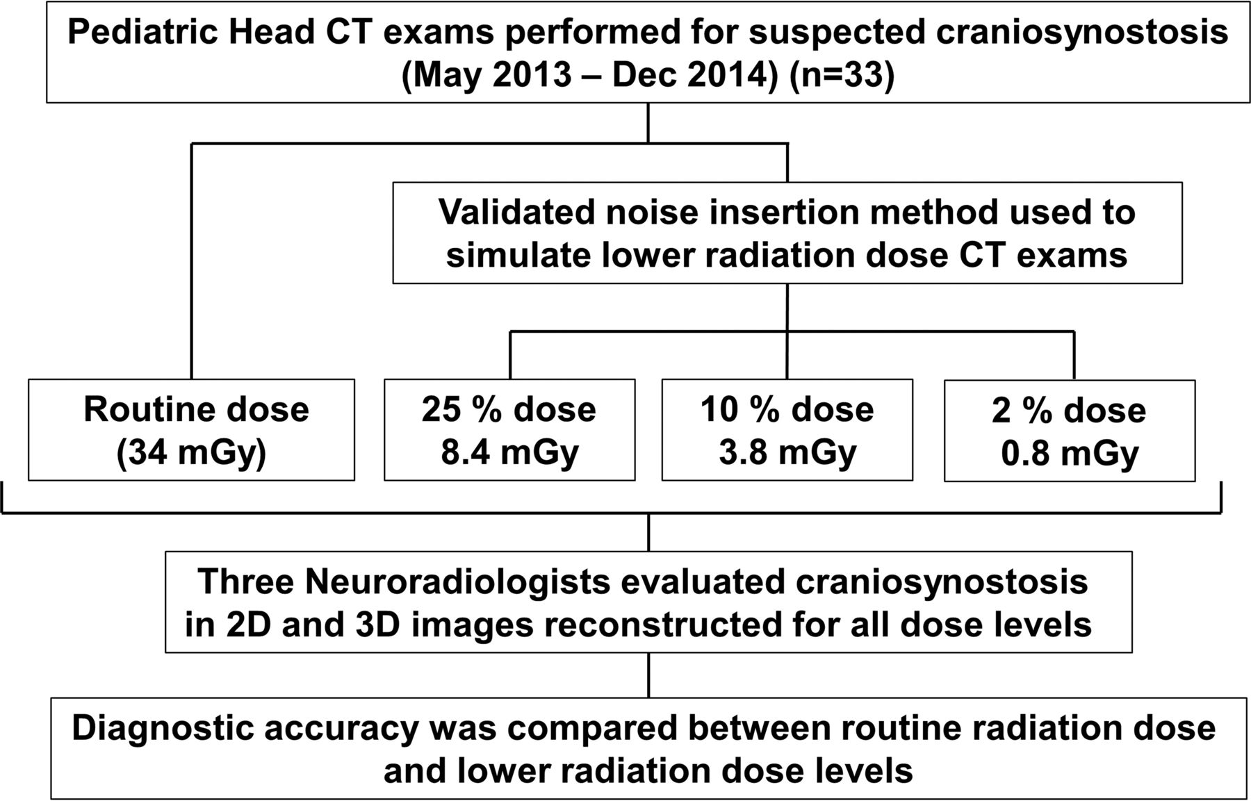
- Fig 1.
Study schema.
- Fig 2.
Automatic exposure control in the head of a pediatric patient. The quality reference tube current–time product for this study is equal to 220 mAs, corresponding to a CTDIvol equal to 34 mGy. However, because automatic exposure control was used, the mean effective tube current–time product was 90 mAs, corresponding to a CTDIvol of 13.77 mGy.
- Fig 3.
Axial and coronal images at routine dose and simulated lower radiation dose of a patient with left coronal craniosynostosis (A) and sagittal craniosynostosis (B).
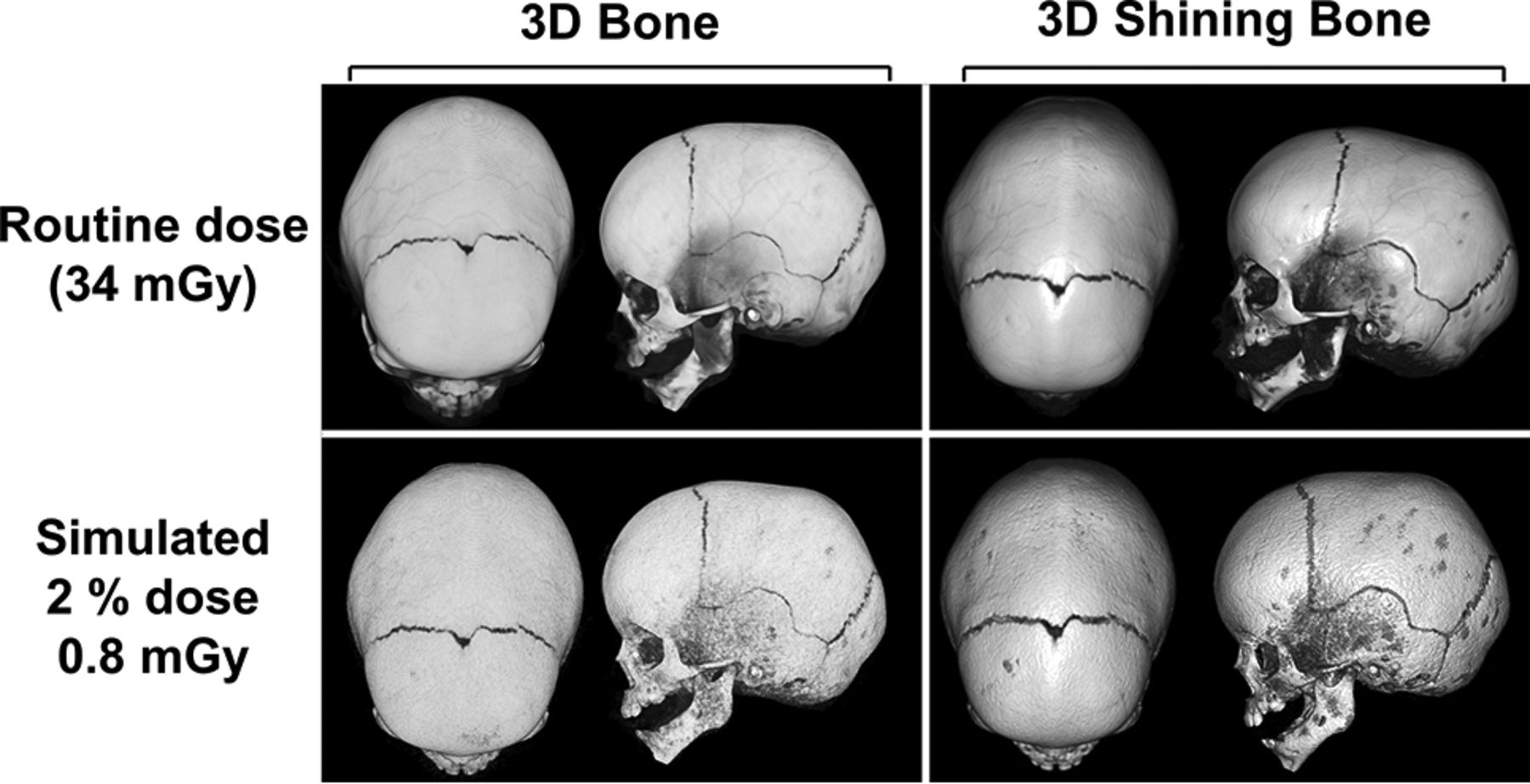
- Fig 4.
3D volume-rendered images of a patient with sagittal craniosynostosis at routine dose and 2% routine dose, created from axial images (image thickness, 0.75 mm; interval, 0.70 mm; FOV, 250 mm; J30 kernel) by using 2 different shading methods (3D bone and 3D shining bone).
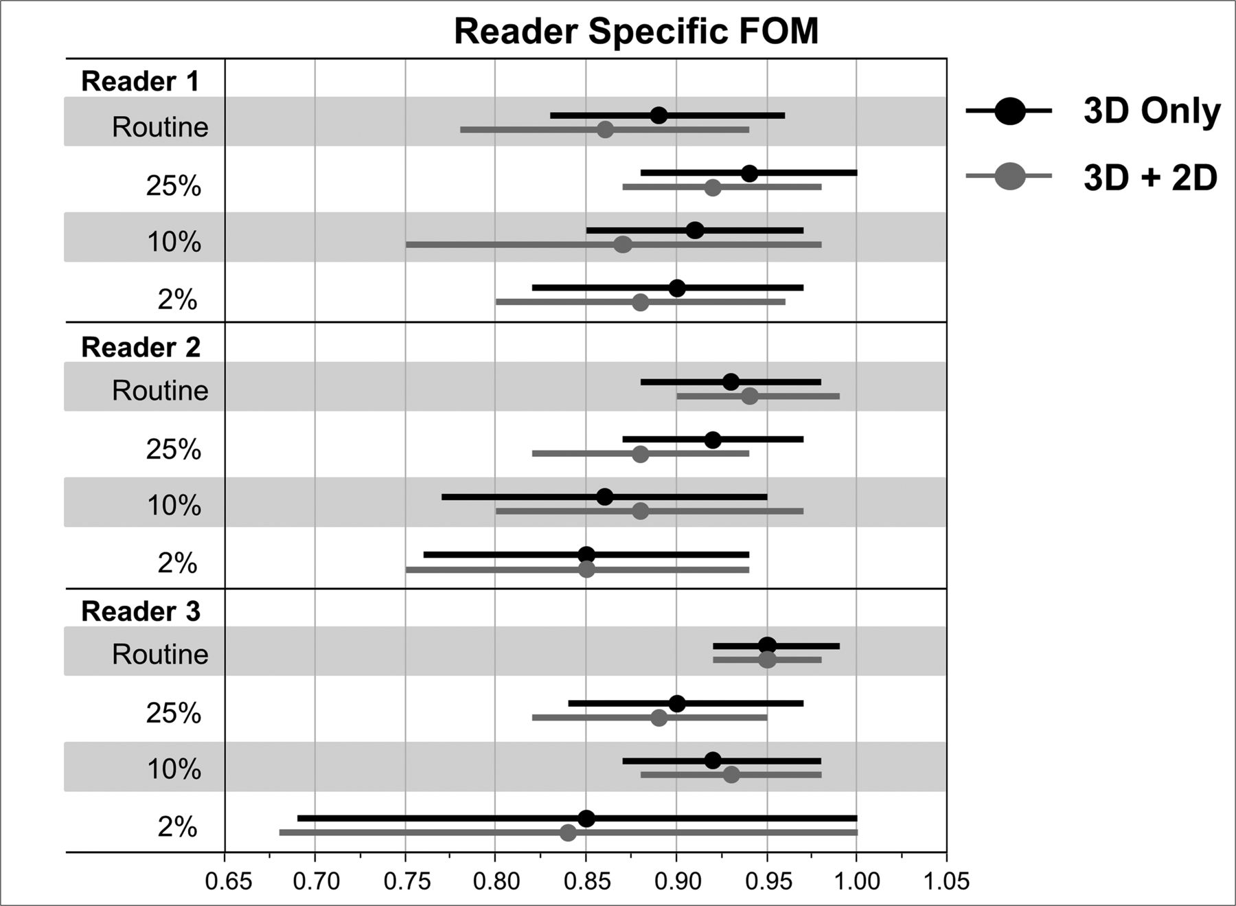
- Fig 5.
FOM and 95% CI for reader-specific evaluation of craniosynostosis by using 3D images only and 3D together with 2D images.
- Fig 6.
Average FOM and 95% CI from all readers for all radiation dose levels in 3D and 3D together with 2D evaluation.
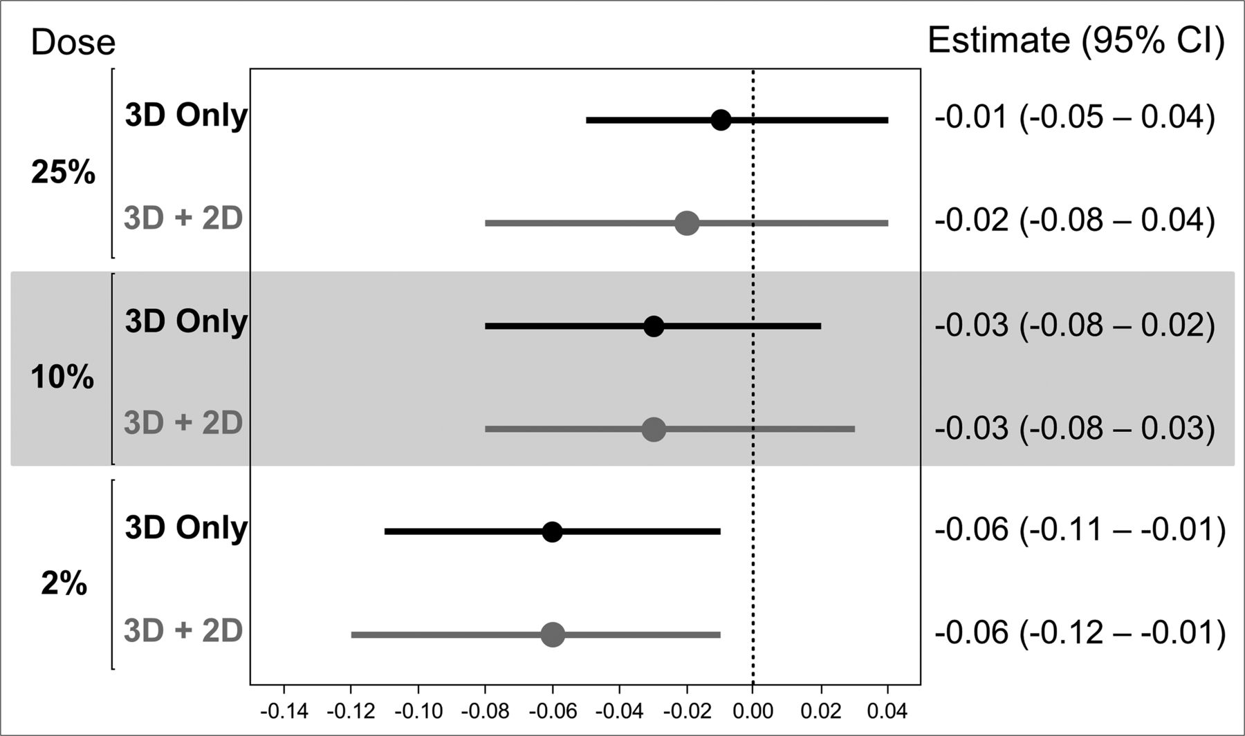
- Fig 7.
Difference in FOM (with 95% CI) between each radiation dose level and reference standard in 3D and 3D together with 2D evaluation.
- Fig 8.
Stacked percent bar plot of image quality scores for each reader and radiation dose.


Tables
Figures of merit and 95% CI for reader-specific and multireader evaluation of craniosynostosis using 3D images only and 2D images together with 3D imagesa
Routine 25% 10% 2% 3D Only 2D + 3D 3D Only 2D + 3D 3D Only 2D + 3D 3D Only 2D + 3D Reader 1 0.89 (0.83–0.96) 0.86 (0.78–0.94) 0.94 (0.88–1.00) 0.92 (0.87–0.98) 0.91 (0.85–0.97) 0.87 (0.75–0.98) 0.90 (0.82–0.97) 0.88 (0.80–0.96) Reader 2 0.93 (0.88–0.98) 0.94 (0.90–0.99) 0.92 (0.87–0.97) 0.88 (0.82–0.94) 0.86 (0.77–0.95) 0.88 (0.80–0.97) 0.85 (0.76–0.94) 0.85 (0.75–0.94) Reader 3 0.95 (0.92–0.99) 0.95 (0.92–0.98) 0.90 (0.84–0.97) 0.89 (0.82–0.95) 0.92 (0.87–0.98) 0.93 (0.88–0.98) 0.85 (0.69–1.00) 0.84 (0.68–1.00) Average 0.92 (0.90–0.95) 0.91 (0.89–0.95) 0.92 (0.88–0.97) 0.89 (0.86–0.94) 0.89 (0.84–0.95) 0.89 (0.83–0.96) 0.86 (0.79–0.94) 0.85 (0.76–0.95) ↵a Data presented as FOM (95% CI).


