
Article Figures & Data
Figures
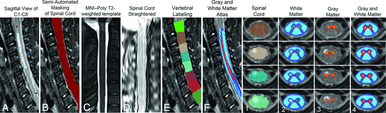
- Fig 1.
Steps for registering patient 5 (acute flaccid paralysis) to template. A, Red circles indicate manual marking of anatomic features C1 and C8. Blue lines indicate axial sections illustrated in far right grid. B, Manual masking of SC centerline was done at each axial section because of signal hyperintensity interfering with automatic reconstruction of centerline. C, MNI-Poly-AMU T2-weighted template. D, SC straightening using thin-plate spline interpolation. E, Labeling of vertebral levels after registering to template and warping back to native space. F, Sagittal view of GM and WM probabilistic atlas after registering to template and warping back to native space. Far right grid, Output of template-based atlas. Column 1, Automatic vertebral body labeling and SC space. Column 2, Probabilistic masks of WM. Column 3, Probabilistic masks of GM. Column 4, Probabilistic masks of GM and WM overlaid.
- Fig 2.
MR imaging of patient 1. A, Sagittal plane T2-weighted image centered on lesion. B, Overlay of binary thresholded image for lesion and T2-weighted sagittal image. C, Axial T2-weighted image at lesion center. D, Overlay of binary thresholded image for lesion and T2-weighted axial image.
- Fig 3.
First row, Axial T2 image from center of lesion. Second row: Probabilistic GM (orange) and WM (blue) map overlaid on thresholded axial T2 image from lesion center. Lesion Center indicates %WM and %GM weighted average metrics at axial lesion center. Lesion Segment indicates %WM and %GM weighted average metrics for lesion segment. Full Cord Atlas Volume indicates %WM and %GM weighted average metrics for full cord atlas volume. CSA Lesion Center indicates %CSA weighted average metrics at axial lesion center. CSA Lesion Segment indicates %CSA weighted average metrics at lesion segment.
- Fig 4.
Scatterplots of numeric outcome of initial strength or improvement in strength MRC score by %GM or %WM injury (weighted average metric ranking) separated by analysis types (lesion center and lesion segment).


Tables
Clinical description of 9 patients with AFM used in analysis
Patient No. Age, yr Sex Limb Weakness MRC Composite Score Enterovirus Detected MRC Score at Follow-Up Discharge Diagnosis Days to MRI 1 3 M Right UE 11 NP swab 2 Disease due to EV 9 2 7 M Bilateral LE 20 None 3 Encephalomyelitis, paraesthesia/hyperestheia 14 3 4 F Bilateral LE 9 None 1 Flaccid paralysis, unspecified 2 4 8 M Right UE 9 NP swab 0 Acute flaccid paralysis 13 5 27 M Bilateral LE 1 Serum 3 Virus-related myelitis 6 6 8 F Left UE 16 None 2 Viral meningitis 11 7 24 M Bilateral LE 24 None 3 Meningomyelitis 5 8 10 M Left UE 18 None 2 Postinfectious mycoplasma transverse myelitis 3 9 2 F Left UE 3 None 1 Hopkins syndrome 1 Note:—LE, lower extremities; NP, nasophyryngeal; UE, upper extremities.


Jump to section
Related Articles
Cited By...
- Diffusion Kurtosis Imaging of neonatal Spinal Cord in clinical routine
- Update on acute flaccid myelitis: recognition, reporting, aetiology and outcomes
- Convolutional Neural Network-Based Automated Segmentation of the Spinal Cord and Contusion Injury: Deep Learning Biomarker Correlates of Motor Impairment in Acute Spinal Cord Injury
- Test-Retest and Interreader Reproducibility of Semiautomated Atlas-Based Analysis of Diffusion Tensor Imaging Data in Acute Cervical Spine Trauma in Adult Patients