
Article Figures & Data
Figures
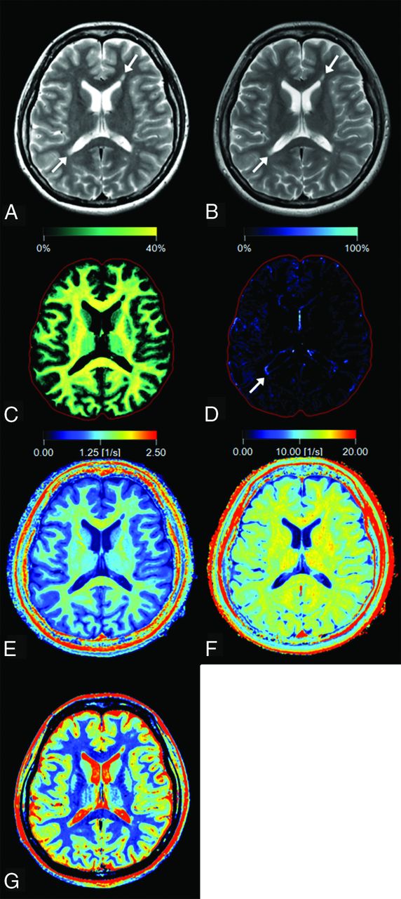
- Fig 1.
Representative images of a 27-year-old woman with multiple sclerosis. Panels show a synthetic T2-weighted image (A), a conventional T2-weighted image (B), and maps of myelin partial volume (C), excess parenchymal water partial volume (D), R1 (E), R2 (F), and PD (G). Two plaques are shown by arrows on the T2-weighted images (A and B). On the VEPW map (D), the periphery of the plaque adjacent to the trigone of the right ventricle (arrow) is visible but the one adjacent to the anterior horn of the left ventricle is not. The VEPW of this invisible plaque was very low but still higher than that of NAWM. The red intracranial outline is displayed for visual guidance in tissue images (C and D).
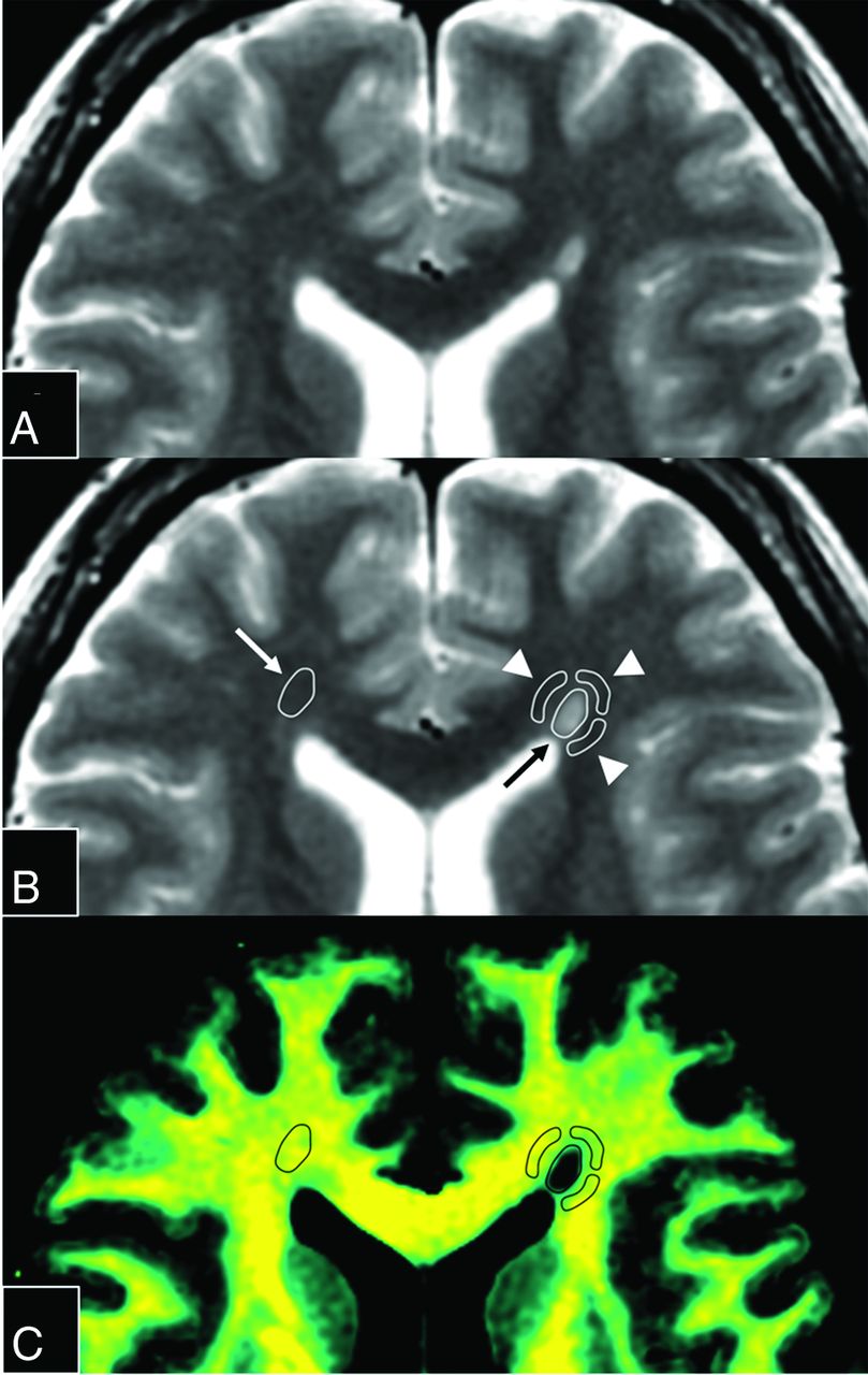
- Fig 2.
Magnified images of Fig 1A. The upper 2 panels show the same synthetic T2-weighted image without (A) or with (B) placement of ROIs. An ROI (black arrow) was drawn on a plaque adjacent to the left anterior horn, and 3 ROIs (arrowheads) were placed on periplaque white matter to encircle the plaque. The fourth ROI on PWM was discarded because it overlapped the CSF. The ROI of the plaque was copied and pasted onto the contralateral normal-appearing white matter (white arrow). These ROIs were then copied and pasted onto each quantification map. A map of the corresponding myelin partial volume (C) is shown as an example.


Tables
- Table 1:
Descriptive values of plaques, periplaque white matter, and normal-appearing white mattera
VMY (%) VEPW (%) R1 (s−1) R2 (s−1) PD (%) Plaques 12.59 ± 6.66 5.82 ± 4.75 0.90 ± 0.20 10.88 ± 1.41 78.86 ± 6.35 PWM 29.29 ± 3.73 2.31 ± 2.38 1.31 ± 0.13 13.14 ± 0.77 68.09 ± 2.49 NAWM 32.88 ± 3.12 0.92 ± 1.90 1.40 ± 0.08 13.85 ± 0.97 63.97 ± 2.07 ↵a Values are mean ± SD. P < .001 for all metrics among each tissue type.
- Table 2:
Percentage changes of VMY, VEPW, R1, R2, and PD in plaques and periplaque white matter relative to normal-appearing white mattera
VMY (%) VEPW (%) R1 (%) R2 (%) PD (%) Plaques −61.59 ± 20.28b 13.82 × 10³ ± 49.47 × 10³b −35.23 ± 13.93b −21.06 ± 11.39b 23.37 ± 10.30b PWM −10.51 ± 11.41c 51.33 × 10² ± 155.31 × 10²c −6.08 ± 8.66c −4.79 ± 6.79c 3.37 ± 4.24b ↵a Values are mean ± SD. Of the 135 ROIs, 39 were discarded after calculating the percentage change of VEPW relative to NAWM because the VEPW of these ROIs was equivalent to zero in NAWM.
↵b P < .001 in the percentage change for plaques relative to NAWM for comparison between each pair of metrics, except between R2 and PD (P = .31). Statistical analysis was performed for absolute values.
↵c P < .001 in the percentage change for PWM relative to NAWM between VEPW and other metrics, and between VMY and R2 or PD, P < .05 between VMY and R1, and P > .05 between R2 and R1 (.31) or PD (.30). Statistical analysis was performed for absolute values.


Jump to section
Related Articles
Cited By...
- Multi-Metric Quantitative MRI Identifies Spatially Distinct Age-Related Brain Differences in Female Bonnet Macaques
- Quantitative MRI in Multiple Sclerosis: From Theory to Application
- Synthetic MRI in Neurofibromatosis Type 1
- White Matter Abnormalities in Multiple Sclerosis Evaluated by Quantitative Synthetic MRI, Diffusion Tensor Imaging, and Neurite Orientation Dispersion and Density Imaging
- Effect of Gadolinium on the Estimation of Myelin and Brain Tissue Volumes Based on Quantitative Synthetic MRI
- Normal Values of Magnetic Relaxation Parameters of Spine Components with the Synthetic MRI Sequence
- Analysis of White Matter Damage in Patients with Multiple Sclerosis via a Novel In Vivo MR Method for Measuring Myelin, Axons, and G-Ratio
- Myelin Detection Using Rapid Quantitative MR Imaging Correlated to Macroscopically Registered Luxol Fast Blue-Stained Brain Specimens