Advertisement

Main menu

  • Home
  • Content
    • Current Issue
    • Accepted Manuscripts
    • Article Preview
    • Past Issue Archive
    • Video Articles
    • AJNR Case Collection
    • Case of the Week Archive
    • Case of the Month Archive
    • Classic Case Archive
  • Special Collections
    • AJNR Awards
    • Low-Field MRI
    • Alzheimer Disease
    • ASNR Foundation Special Collection
    • Photon-Counting CT
    • View All
  • Multimedia
    • AJNR Podcasts
    • AJNR SCANtastic
    • Trainee Corner
    • MRI Safety Corner
    • Imaging Protocols
  • For Authors
    • Submit a Manuscript
    • Submit a Video Article
    • Submit an eLetter to the Editor/Response
    • Manuscript Submission Guidelines
    • Statistical Tips
    • Fast Publishing of Accepted Manuscripts
    • Graphical Abstract Preparation
    • Imaging Protocol Submission
    • Author Policies
  • About Us
    • About AJNR
    • Editorial Board
    • Editorial Board Alumni
  • More
    • Become a Reviewer/Academy of Reviewers
    • Subscribers
    • Permissions
    • Alerts
    • Feedback
    • Advertisers
    • ASNR Home

User menu

  • Alerts
  • Log in

Search

  • Advanced search
American Journal of Neuroradiology

American Journal of Neuroradiology

ASHNR American Society of Functional Neuroradiology ASHNR American Society of Pediatric Neuroradiology ASSR
  • Alerts
  • Log in

Advanced Search

  • Home
  • Content
    • Current Issue
    • Accepted Manuscripts
    • Article Preview
    • Past Issue Archive
    • Video Articles
    • AJNR Case Collection
    • Case of the Week Archive
    • Case of the Month Archive
    • Classic Case Archive
  • Special Collections
    • AJNR Awards
    • Low-Field MRI
    • Alzheimer Disease
    • ASNR Foundation Special Collection
    • Photon-Counting CT
    • View All
  • Multimedia
    • AJNR Podcasts
    • AJNR SCANtastic
    • Trainee Corner
    • MRI Safety Corner
    • Imaging Protocols
  • For Authors
    • Submit a Manuscript
    • Submit a Video Article
    • Submit an eLetter to the Editor/Response
    • Manuscript Submission Guidelines
    • Statistical Tips
    • Fast Publishing of Accepted Manuscripts
    • Graphical Abstract Preparation
    • Imaging Protocol Submission
    • Author Policies
  • About Us
    • About AJNR
    • Editorial Board
    • Editorial Board Alumni
  • More
    • Become a Reviewer/Academy of Reviewers
    • Subscribers
    • Permissions
    • Alerts
    • Feedback
    • Advertisers
    • ASNR Home
  • Follow AJNR on Twitter
  • Visit AJNR on Facebook
  • Follow AJNR on Instagram
  • Join AJNR on LinkedIn
  • RSS Feeds

AJNR Awards, New Junior Editors, and more. Read the latest AJNR updates

Getting new auth cookie, if you see this message a lot, tell someone!
Research ArticleSpine Imaging and Spine Image-Guided Interventions

Advantages of 70-kV CT Angiography for the Visualization of the Adamkiewicz Artery: Comparison with 120-kV Imaging

S. Shimoyama, T. Nishii, Y. Watanabe, A.K. Kono, K. Kagawa, S. Takahashi and K. Sugimura
American Journal of Neuroradiology December 2017, 38 (12) 2399-2405; DOI: https://doi.org/10.3174/ajnr.A5372
S. Shimoyama
aFrom the Department of Radiology (S.S., T.N., Y.W., A.K.K., S.T., K.S.), Kobe University Graduate School of Medicine, Kobe, Hyogo, Japan
  • Find this author on Google Scholar
  • Find this author on PubMed
  • Search for this author on this site
  • ORCID record for S. Shimoyama
T. Nishii
aFrom the Department of Radiology (S.S., T.N., Y.W., A.K.K., S.T., K.S.), Kobe University Graduate School of Medicine, Kobe, Hyogo, Japan
  • Find this author on Google Scholar
  • Find this author on PubMed
  • Search for this author on this site
  • ORCID record for T. Nishii
Y. Watanabe
aFrom the Department of Radiology (S.S., T.N., Y.W., A.K.K., S.T., K.S.), Kobe University Graduate School of Medicine, Kobe, Hyogo, Japan
bDepartment of Radiology (Y.W., A.K.K.), National Cerebral and Cardiovascular Center, Suita, Osaka, Japan
  • Find this author on Google Scholar
  • Find this author on PubMed
  • Search for this author on this site
  • ORCID record for Y. Watanabe
A.K. Kono
aFrom the Department of Radiology (S.S., T.N., Y.W., A.K.K., S.T., K.S.), Kobe University Graduate School of Medicine, Kobe, Hyogo, Japan
bDepartment of Radiology (Y.W., A.K.K.), National Cerebral and Cardiovascular Center, Suita, Osaka, Japan
  • Find this author on Google Scholar
  • Find this author on PubMed
  • Search for this author on this site
  • ORCID record for A.K. Kono
K. Kagawa
cDivision of Radiology (K.K.), Center for Radiology and Radiation Oncology, Kobe University Hospital, Kobe, Hyogo, Japan.
  • Find this author on Google Scholar
  • Find this author on PubMed
  • Search for this author on this site
  • ORCID record for K. Kagawa
S. Takahashi
aFrom the Department of Radiology (S.S., T.N., Y.W., A.K.K., S.T., K.S.), Kobe University Graduate School of Medicine, Kobe, Hyogo, Japan
  • Find this author on Google Scholar
  • Find this author on PubMed
  • Search for this author on this site
  • ORCID record for S. Takahashi
K. Sugimura
aFrom the Department of Radiology (S.S., T.N., Y.W., A.K.K., S.T., K.S.), Kobe University Graduate School of Medicine, Kobe, Hyogo, Japan
  • Find this author on Google Scholar
  • Find this author on PubMed
  • Search for this author on this site
  • ORCID record for K. Sugimura
  • Article
  • Figures & Data
  • Info & Metrics
  • Responses
  • References
  • PDF
Loading

Article Figures & Data

Figures

  • Tables
  • Fig 1.
    • Download figure
    • Open in new tab
    • Download powerpoint
    Fig 1.

    The AKA was identified according to the following stepwise approach: 1) detect the hairpin vessel; 2) assess the continuity of the vessel to the aorta mainly around the pedicle of the vertebral arch (white arrows) as definite (a), conceivable (b), equivocal (c), or undetectable; 3) check the washout from the early-to-late arterial phase (white arrowhead), because vessels that are more prominent in the late arterial phase are likely to be veins (black arrowhead); and 4) sum the scores. We defined positive identification of the AKA as a total score of ≥3.

  • Fig 2.
    • Download figure
    • Open in new tab
    • Download powerpoint
    Fig 2.

    The ROIs placed for measuring CT numbers are shown in a coronal image of the ASA (A), a sagittal image of the ASA (B), an axial image of the spinal cord (C), and a sagittal image of the vertebral bone cortex around the vertebral foramen at the AKA (white arrow) running level (D).

  • Fig 3.
    • Download figure
    • Open in new tab
    • Download powerpoint
    Fig 3.

    Box range shows the first and third quartiles; whisker range, from the 5th to 95th percentiles. Seventy-kilovolt CTA yields significantly lower SSDE (A) and dose-length product (B) than 120-kV CTA (P < .001 for both).

  • Fig 4.
    • Download figure
    • Open in new tab
    • Download powerpoint
    Fig 4.

    Distribution of the side and level of the CSA to the AKA is shown. Two cases in which the CSA and the AKA were supplied by the axillary artery were described as “other.”

  • Fig 5.
    • Download figure
    • Open in new tab
    • Download powerpoint
    Fig 5.

    Seventy-kilovolt CTA yielded significantly higher scores than 120-kV CTA. A, Continuity score, median 3 (interquartile range, 2–3) compared with 2 (interquartile range, 1–3), respectively. B, Total score, median 5 (interquartile range, 4–5), compared with 4 (interquartile range, 3–5), respectively (P < .05 for both).
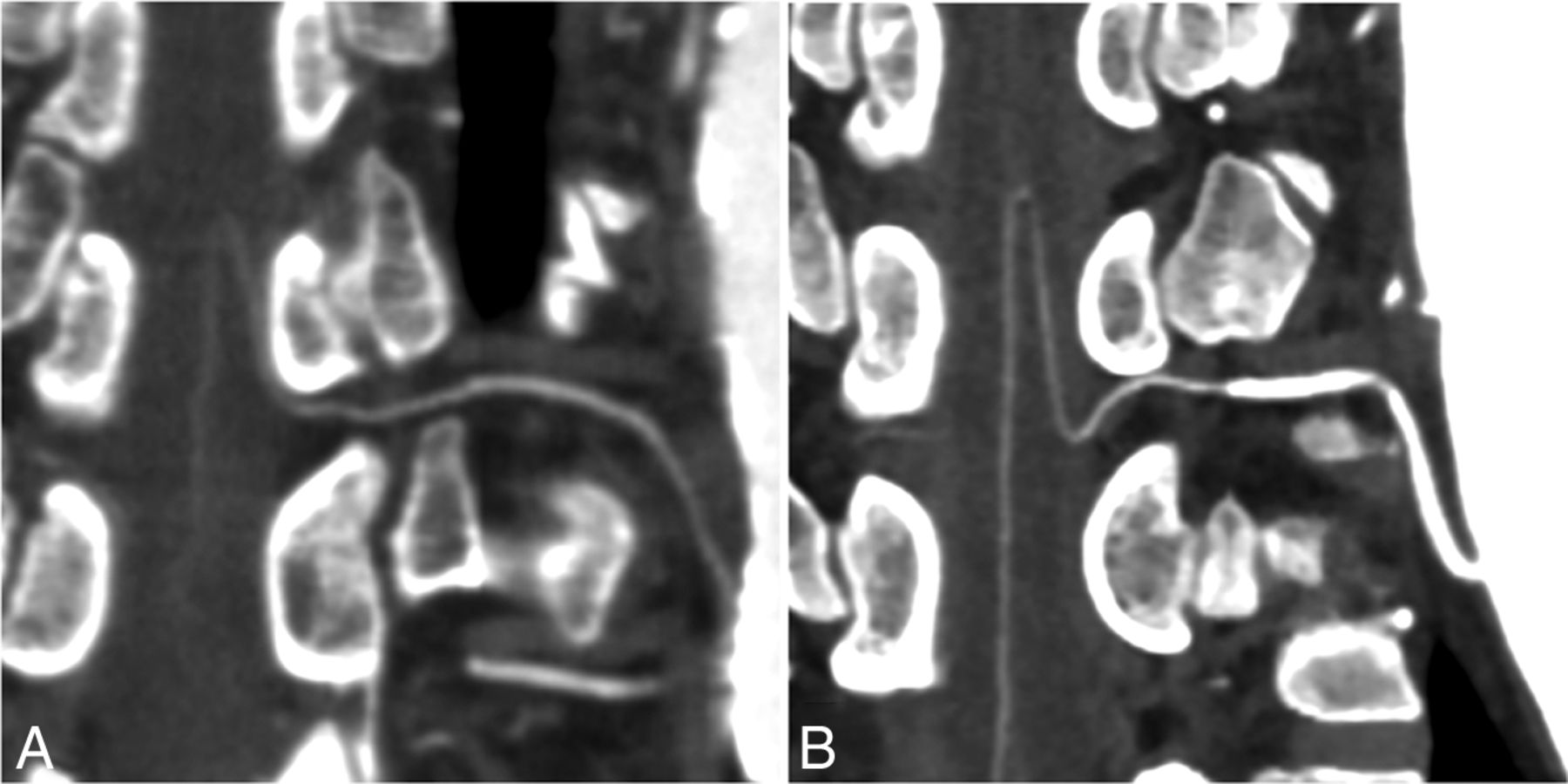
  • Fig 6.
    • Download figure
    • Open in new tab
    • Download powerpoint
    Fig 6.

    A, Curved multiplanar reformatted image of a 77-year-old woman scanned with a 120-kV protocol shows definite continuity between the AKA and the segmental artery. B, Curved multiplanar reformatted image of a 68-year-old man scanned with a 70-kV protocol shows clearer visualization of the AKA and the segmental artery from the aorta. Window center and width were set at 200 and 800.
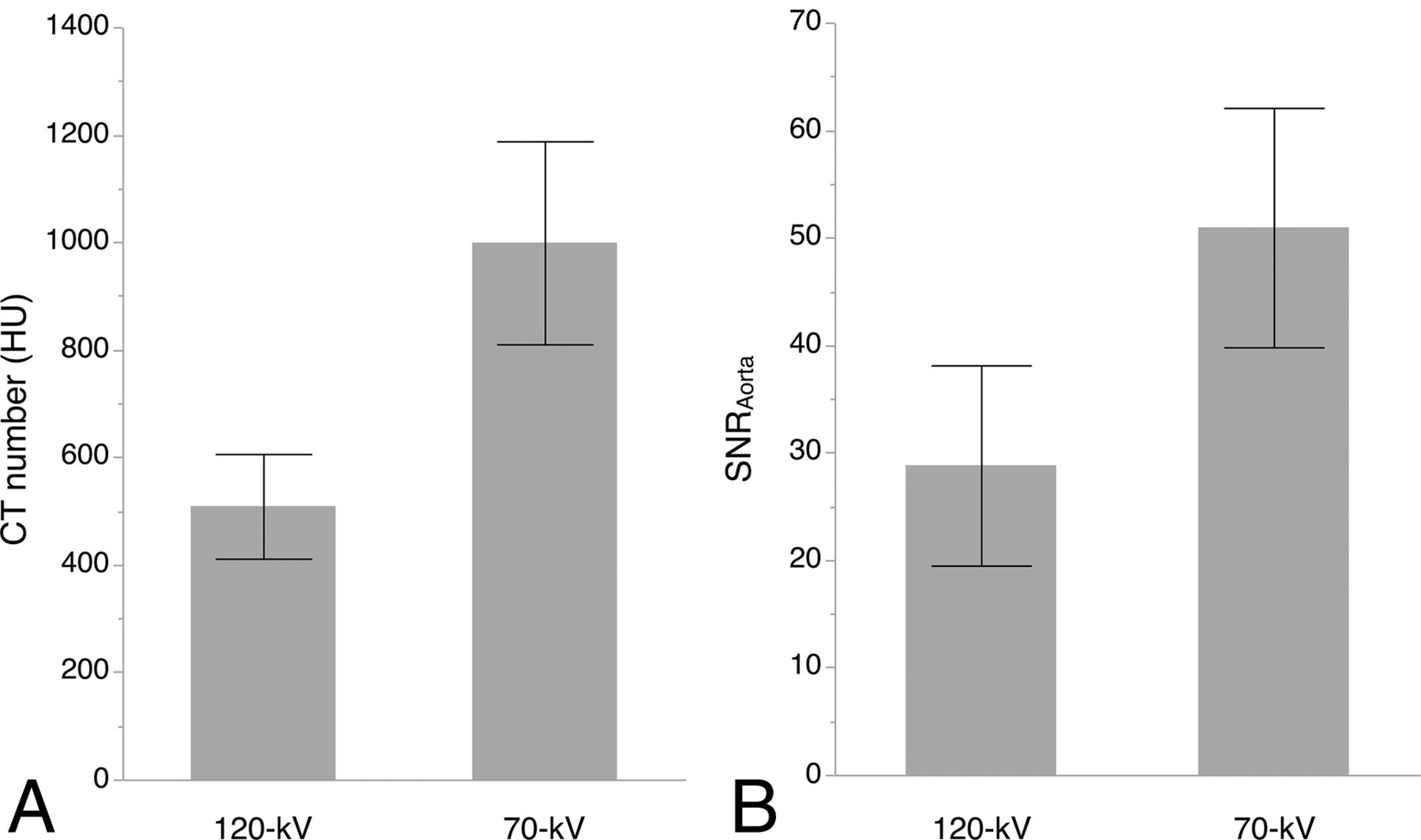
  • Fig 7.
    • Download figure
    • Open in new tab
    • Download powerpoint
    Fig 7.

    Seventy-kilovolt CTA yielded a significantly higher CT number (A) and SNRAorta (B) than 120-kV CTA (P < .001 for both analyses).

Tables

  • Figures
    • View popup
    Table 1:

    Subject demographic and clinical characteristics

    Variables120-kV (n = 60)70-kV (n = 60)P Value
    Age (yr).81
        Mean6969
        Range57.0–76.354.8–76.0
    Sex.83
        Male (No.)4445
        Female (No.)1615
    Mean body mass index (kg/m2)23.7 ± 3.323.2 ± 3.6.48
    Aortic disorder.46
        Aneurysm (No.)3531
        Dissection (No.)2529
    • View popup
    Table 2.

    Results of consensus interpretation of the critical segmental artery

    Variables120-kV70-kVP Value
    Detection of collateral pathway (No.) (%)13 (21.7)13 (21.7)1.00
    Detection of critical segmental artery (No.) (%)50 (83.3)54 (90.0).28
    • View popup
    Table 3:

    Accuracy of interpretation of the critical segmental artery

    Variables120-kV70-kVP Value
    Accuracy in all subjects
        Observer A83.395.0.035a
        Observer B83.393.3.084
    Accuracy in subjects with a collateral pathway
        Observer A61.5100.004a
        Observer B30.869.2.047a
    • ↵a Statistically significant.

    • View popup
    Table 4:

    Results of quantitative image-quality analysisa

    Variables120-kV70-kVP Value
    Spinal cord
        CT number (HU)39.4 ± 7.154.3 ± 11.6<.001b
        SD (HU)12.5 ± 3.413.7 ± 3.9.059
    Bone cortex around the vertebral foramen
        CT number (HU)657.4 ± 134.01,106.1 ± 246.2<.001b
    Anterior spinal artery
        CT number (HU)78.7 ± 13.3127.7 ± 30.0<.001b
        CNRASA-Cord3.4 ± 1.65.6 ± 2.5<.001b
        CNRASA-Bone50.3 ± 17.075.9 ± 30.5<.001b
    • Note:—CNRASA-Cord indicates contrast-to-noise ratio of the anterior spinal artery to the spinal cord; CNRASA-Bone, contrast-to-noise ratio of the anterior spinal artery to bone cortex around the vertebral foramen.

    • ↵a Data are presented as the mean ± SD.

    • ↵b Statistically significant.

PreviousNext
Back to top

In this issue

American Journal of Neuroradiology: 38 (12)
American Journal of Neuroradiology
Vol. 38, Issue 12
1 Dec 2017
  • Table of Contents
  • Index by author
  • Complete Issue (PDF)
Advertisement
Print
Download PDF
Email Article

Thank you for your interest in spreading the word on American Journal of Neuroradiology.

NOTE: We only request your email address so that the person you are recommending the page to knows that you wanted them to see it, and that it is not junk mail. We do not capture any email address.

Enter multiple addresses on separate lines or separate them with commas.
Advantages of 70-kV CT Angiography for the Visualization of the Adamkiewicz Artery: Comparison with 120-kV Imaging
(Your Name) has sent you a message from American Journal of Neuroradiology
(Your Name) thought you would like to see the American Journal of Neuroradiology web site.
CAPTCHA
This question is for testing whether or not you are a human visitor and to prevent automated spam submissions.
Cite this article
S. Shimoyama, T. Nishii, Y. Watanabe, A.K. Kono, K. Kagawa, S. Takahashi, K. Sugimura
Advantages of 70-kV CT Angiography for the Visualization of the Adamkiewicz Artery: Comparison with 120-kV Imaging
American Journal of Neuroradiology Dec 2017, 38 (12) 2399-2405; DOI: 10.3174/ajnr.A5372

Citation Manager Formats

  • BibTeX
  • Bookends
  • EasyBib
  • EndNote (tagged)
  • EndNote 8 (xml)
  • Medlars
  • Mendeley
  • Papers
  • RefWorks Tagged
  • Ref Manager
  • RIS
  • Zotero
0 Responses
Respond to this article
Share
Bookmark this article
Advantages of 70-kV CT Angiography for the Visualization of the Adamkiewicz Artery: Comparison with 120-kV Imaging
S. Shimoyama, T. Nishii, Y. Watanabe, A.K. Kono, K. Kagawa, S. Takahashi, K. Sugimura
American Journal of Neuroradiology Dec 2017, 38 (12) 2399-2405; DOI: 10.3174/ajnr.A5372
del.icio.us logo Twitter logo Facebook logo Mendeley logo
  • Tweet Widget
  • Facebook Like
  • Google Plus One
Purchase

Jump to section

  • Article
    • Abstract
    • ABBREVIATIONS:
    • Materials and Methods
    • Results
    • Discussion
    • Conclusions
    • Acknowledgments
    • Footnotes
    • References
  • Figures & Data
  • Info & Metrics
  • Responses
  • References
  • PDF

Related Articles

  • No related articles found.
  • PubMed
  • Google Scholar

Cited By...

  • Application of Spinal Subtraction and Bone Background Fusion CTA in the Accurate Diagnosis and Evaluation of Spinal Vascular Malformations
  • Assessing Vascularity of Osseous Spinal Metastases with Dual-Energy CT-DSA: A Pilot Study Compared with Catheter Angiography
  • Crossref
  • Google Scholar

This article has not yet been cited by articles in journals that are participating in Crossref Cited-by Linking.

More in this TOC Section

  • Optimization of Photon Counting CT Myelography
  • Characteristics of SIH Type I Culprit Lesions
  • Management Outcomes For VO Spine Biopsy
Show more Spine Imaging and Spine Image-Guided Interventions

Similar Articles

Advertisement

Indexed Content

  • Current Issue
  • Accepted Manuscripts
  • Article Preview
  • Past Issues
  • Editorials
  • Editor's Choice
  • Fellows' Journal Club
  • Letters to the Editor
  • Video Articles

Cases

  • Case Collection
  • Archive - Case of the Week
  • Archive - Case of the Month
  • Archive - Classic Case

More from AJNR

  • Trainee Corner
  • Imaging Protocols
  • MRI Safety Corner
  • Book Reviews

Multimedia

  • AJNR Podcasts
  • AJNR Scantastics

Resources

  • Turnaround Time
  • Submit a Manuscript
  • Submit a Video Article
  • Submit an eLetter to the Editor/Response
  • Manuscript Submission Guidelines
  • Statistical Tips
  • Fast Publishing of Accepted Manuscripts
  • Graphical Abstract Preparation
  • Imaging Protocol Submission
  • Evidence-Based Medicine Level Guide
  • Publishing Checklists
  • Author Policies
  • Become a Reviewer/Academy of Reviewers
  • News and Updates

About Us

  • About AJNR
  • Editorial Board
  • Editorial Board Alumni
  • Alerts
  • Permissions
  • Not an AJNR Subscriber? Join Now
  • Advertise with Us
  • Librarian Resources
  • Feedback
  • Terms and Conditions
  • AJNR Editorial Board Alumni

American Society of Neuroradiology

  • Not an ASNR Member? Join Now

© 2025 by the American Society of Neuroradiology All rights, including for text and data mining, AI training, and similar technologies, are reserved.
Print ISSN: 0195-6108 Online ISSN: 1936-959X

Powered by HighWire