Advertisement

Main menu

  • Home
  • Content
    • Current Issue
    • Accepted Manuscripts
    • Article Preview
    • Past Issue Archive
    • Video Articles
    • AJNR Case Collection
    • Case of the Week Archive
    • Case of the Month Archive
    • Classic Case Archive
  • Special Collections
    • AJNR Awards
    • Low-Field MRI
    • Alzheimer Disease
    • ASNR Foundation Special Collection
    • Photon-Counting CT
    • View All
  • Multimedia
    • AJNR Podcasts
    • AJNR SCANtastic
    • Trainee Corner
    • MRI Safety Corner
    • Imaging Protocols
  • For Authors
    • Submit a Manuscript
    • Submit a Video Article
    • Submit an eLetter to the Editor/Response
    • Manuscript Submission Guidelines
    • Statistical Tips
    • Fast Publishing of Accepted Manuscripts
    • Graphical Abstract Preparation
    • Imaging Protocol Submission
    • Author Policies
  • About Us
    • About AJNR
    • Editorial Board
    • Editorial Board Alumni
  • More
    • Become a Reviewer/Academy of Reviewers
    • Subscribers
    • Permissions
    • Alerts
    • Feedback
    • Advertisers
    • ASNR Home

User menu

  • Alerts
  • Log in

Search

  • Advanced search
American Journal of Neuroradiology

American Journal of Neuroradiology

ASHNR American Society of Functional Neuroradiology ASHNR American Society of Pediatric Neuroradiology ASSR
  • Alerts
  • Log in

Advanced Search

  • Home
  • Content
    • Current Issue
    • Accepted Manuscripts
    • Article Preview
    • Past Issue Archive
    • Video Articles
    • AJNR Case Collection
    • Case of the Week Archive
    • Case of the Month Archive
    • Classic Case Archive
  • Special Collections
    • AJNR Awards
    • Low-Field MRI
    • Alzheimer Disease
    • ASNR Foundation Special Collection
    • Photon-Counting CT
    • View All
  • Multimedia
    • AJNR Podcasts
    • AJNR SCANtastic
    • Trainee Corner
    • MRI Safety Corner
    • Imaging Protocols
  • For Authors
    • Submit a Manuscript
    • Submit a Video Article
    • Submit an eLetter to the Editor/Response
    • Manuscript Submission Guidelines
    • Statistical Tips
    • Fast Publishing of Accepted Manuscripts
    • Graphical Abstract Preparation
    • Imaging Protocol Submission
    • Author Policies
  • About Us
    • About AJNR
    • Editorial Board
    • Editorial Board Alumni
  • More
    • Become a Reviewer/Academy of Reviewers
    • Subscribers
    • Permissions
    • Alerts
    • Feedback
    • Advertisers
    • ASNR Home
  • Follow AJNR on Twitter
  • Visit AJNR on Facebook
  • Follow AJNR on Instagram
  • Join AJNR on LinkedIn
  • RSS Feeds

AJNR Awards, New Junior Editors, and more. Read the latest AJNR updates

Research ArticleNeurointervention

Pial Artery Supply as an Anatomic Risk Factor for Ischemic Stroke in the Treatment of Intracranial Dural Arteriovenous Fistulas

S.W. Hetts, A. Yen, D.L. Cooke, J. Nelson, P. Jolivalt, J. Banaga, M.R. Amans, C.F. Dowd, R.T. Higashida, M.T. Lawton, H. Kim and V.V. Halbach
American Journal of Neuroradiology December 2017, 38 (12) 2315-2320; DOI: https://doi.org/10.3174/ajnr.A5396
S.W. Hetts
aFrom the Department of Radiology and Biomedical Imaging (S.W.H., D.L.C., P.J., M.R.A., C.F.D., R.T.H., V.V.H.)
  • Find this author on Google Scholar
  • Find this author on PubMed
  • Search for this author on this site
  • ORCID record for S.W. Hetts
A. Yen
bSchool of Medicine (A.Y., J.B.)
  • Find this author on Google Scholar
  • Find this author on PubMed
  • Search for this author on this site
  • ORCID record for A. Yen
D.L. Cooke
aFrom the Department of Radiology and Biomedical Imaging (S.W.H., D.L.C., P.J., M.R.A., C.F.D., R.T.H., V.V.H.)
  • Find this author on Google Scholar
  • Find this author on PubMed
  • Search for this author on this site
  • ORCID record for D.L. Cooke
J. Nelson
cDepartments of Anesthesia and Perioperative Care (J.N., P.J., C.F.D., R.T.H., H.K., V.V.H.)
  • Find this author on Google Scholar
  • Find this author on PubMed
  • Search for this author on this site
  • ORCID record for J. Nelson
P. Jolivalt
aFrom the Department of Radiology and Biomedical Imaging (S.W.H., D.L.C., P.J., M.R.A., C.F.D., R.T.H., V.V.H.)
cDepartments of Anesthesia and Perioperative Care (J.N., P.J., C.F.D., R.T.H., H.K., V.V.H.)
  • Find this author on Google Scholar
  • Find this author on PubMed
  • Search for this author on this site
  • ORCID record for P. Jolivalt
J. Banaga
bSchool of Medicine (A.Y., J.B.)
  • Find this author on Google Scholar
  • Find this author on PubMed
  • Search for this author on this site
  • ORCID record for J. Banaga
M.R. Amans
aFrom the Department of Radiology and Biomedical Imaging (S.W.H., D.L.C., P.J., M.R.A., C.F.D., R.T.H., V.V.H.)
  • Find this author on Google Scholar
  • Find this author on PubMed
  • Search for this author on this site
  • ORCID record for M.R. Amans
C.F. Dowd
aFrom the Department of Radiology and Biomedical Imaging (S.W.H., D.L.C., P.J., M.R.A., C.F.D., R.T.H., V.V.H.)
cDepartments of Anesthesia and Perioperative Care (J.N., P.J., C.F.D., R.T.H., H.K., V.V.H.)
dNeurological Surgery (C.F.D., R.T.H., M.T.L., V.V.H.)
eNeurology (C.F.D., R.T.H., V.V.H.), University of California, San Francisco, San Francisco, California.
  • Find this author on Google Scholar
  • Find this author on PubMed
  • Search for this author on this site
  • ORCID record for C.F. Dowd
R.T. Higashida
aFrom the Department of Radiology and Biomedical Imaging (S.W.H., D.L.C., P.J., M.R.A., C.F.D., R.T.H., V.V.H.)
cDepartments of Anesthesia and Perioperative Care (J.N., P.J., C.F.D., R.T.H., H.K., V.V.H.)
dNeurological Surgery (C.F.D., R.T.H., M.T.L., V.V.H.)
eNeurology (C.F.D., R.T.H., V.V.H.), University of California, San Francisco, San Francisco, California.
  • Find this author on Google Scholar
  • Find this author on PubMed
  • Search for this author on this site
  • ORCID record for R.T. Higashida
M.T. Lawton
dNeurological Surgery (C.F.D., R.T.H., M.T.L., V.V.H.)
  • Find this author on Google Scholar
  • Find this author on PubMed
  • Search for this author on this site
  • ORCID record for M.T. Lawton
H. Kim
cDepartments of Anesthesia and Perioperative Care (J.N., P.J., C.F.D., R.T.H., H.K., V.V.H.)
  • Find this author on Google Scholar
  • Find this author on PubMed
  • Search for this author on this site
  • ORCID record for H. Kim
V.V. Halbach
aFrom the Department of Radiology and Biomedical Imaging (S.W.H., D.L.C., P.J., M.R.A., C.F.D., R.T.H., V.V.H.)
cDepartments of Anesthesia and Perioperative Care (J.N., P.J., C.F.D., R.T.H., H.K., V.V.H.)
dNeurological Surgery (C.F.D., R.T.H., M.T.L., V.V.H.)
eNeurology (C.F.D., R.T.H., V.V.H.), University of California, San Francisco, San Francisco, California.
  • Find this author on Google Scholar
  • Find this author on PubMed
  • Search for this author on this site
  • ORCID record for V.V. Halbach
  • Article
  • Figures & Data
  • Supplemental
  • Info & Metrics
  • Responses
  • References
  • PDF
Loading

Article Figures & Data

Figures

  • Fig 1.
    • Download figure
    • Open in new tab
    • Download powerpoint
    Fig 1.

    Postembolization infarction due to Onyx migration into a pial artery. A middle-aged woman with severe pulsatile tinnitus underwent endovascular therapy for a right transverse-sigmoid sinus junction Borden-Shucart grade I DAVF supplied principally by the middle meningeal and occipital arteries (A) and secondarily by the ipsilateral middle temporal artery (B) and tentorial branches from the contralateral middle meningeal artery (C). The fistula site is designated by a white asterisk. The fusiform gyrus branch of the middle temporal artery is indicated by a white arrow. Superselective injection of the right middle meningeal artery demonstrates the fistula site before embolization (D). Midembolization x-ray (E) demonstrates Onyx in the middle meningeal artery, fistula site, and refluxed into the pial fusiform gyrus branch of the middle temporal artery. The extent of reflux had not been evident on real-time intraprocedural blank roadmap imaging. DWI later the same day (F) demonstrates a fusiform gyrus infarction.

  • Fig 2.
    • Download figure
    • Open in new tab
    • Download powerpoint
    Fig 2.

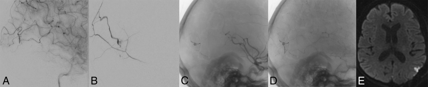
    Arterial emboli following endovascular and surgical treatments of DAVFs in 2 patients. Two middle-aged male patients undergoing posttreatment angiography were identified as having middle cerebral artery emboli (A and C, white arrows). Both patients were heparinized, and the second patient underwent superselective intra-arterial tPA treatment with minimal clot lysis. Postangiographic DWIs (B and D) demonstrate small cortical infarctions in territories associated with the MCA emboli. Although the first patient's MCA thrombus (A and B) is adjacent to the original DAVF, the second patient's is not (C and D).

  • Fig 3.
    • Download figure
    • Open in new tab
    • Download powerpoint
    Fig 3.

    Postsurgical seizure and small cortical infarction after surgical ligation of a residual DAVF. A middle-aged woman status 1 year post temporal lobe hemorrhage underwent endovascular therapy for a Borden-Shucart grade III DAVF supplied principally by the left middle meningeal artery (B) and secondarily by pial branches of the left MCA (A), with drainage directly to a cortical vein. Onyx embolization eliminated the middle meningeal artery dural supply (C), but late-phase angiographic images demonstrated persistent MCA pial supply (D). Three days following a craniotomy for successful ligation of the residual DAVF, the patient had a seizure. MR imaging at that time demonstrates a small cortical infarction (E).

  • Fig 4.
    • Download figure
    • Open in new tab
    • Download powerpoint
    Fig 4.

    Postoperative parenchymal hemorrhage and possible venous infarction following surgical ligation of a residual DAVF. A middle-aged man had intermittent speech arrest and multiple headaches for several years. Anteroposterior (A) and lateral (B) angiograms demonstrate a variant-type right transverse-sigmoid sinus junction Borden-Shucart grade III DAVF supplied by dural branches of the external carotid artery and pial branches of the MCA (C) with retrograde drainage to the vein of Labbé (D), that anastomotically drains to the vein of Trolard. Following Onyx embolization of the dural external carotid artery branches, only the pial MCA supply to the fistula continued to drain to the cortical vein as demonstrated on early (E), mid (F), and late (G) images from a lateral ICA angiogram. During surgical ligation of the residual DAVF, extensive intraoperative bleeding was noted. A CT performed immediately postoperatively (H) demonstrates right temporal intraparenchymal hemorrhage. The patient did not have a new neurologic deficit postoperatively.

PreviousNext
Back to top

In this issue

American Journal of Neuroradiology: 38 (12)
American Journal of Neuroradiology
Vol. 38, Issue 12
1 Dec 2017
  • Table of Contents
  • Index by author
  • Complete Issue (PDF)
Advertisement
Print
Download PDF
Email Article

Thank you for your interest in spreading the word on American Journal of Neuroradiology.

NOTE: We only request your email address so that the person you are recommending the page to knows that you wanted them to see it, and that it is not junk mail. We do not capture any email address.

Enter multiple addresses on separate lines or separate them with commas.
Pial Artery Supply as an Anatomic Risk Factor for Ischemic Stroke in the Treatment of Intracranial Dural Arteriovenous Fistulas
(Your Name) has sent you a message from American Journal of Neuroradiology
(Your Name) thought you would like to see the American Journal of Neuroradiology web site.
CAPTCHA
This question is for testing whether or not you are a human visitor and to prevent automated spam submissions.
Cite this article
S.W. Hetts, A. Yen, D.L. Cooke, J. Nelson, P. Jolivalt, J. Banaga, M.R. Amans, C.F. Dowd, R.T. Higashida, M.T. Lawton, H. Kim, V.V. Halbach
Pial Artery Supply as an Anatomic Risk Factor for Ischemic Stroke in the Treatment of Intracranial Dural Arteriovenous Fistulas
American Journal of Neuroradiology Dec 2017, 38 (12) 2315-2320; DOI: 10.3174/ajnr.A5396

Citation Manager Formats

  • BibTeX
  • Bookends
  • EasyBib
  • EndNote (tagged)
  • EndNote 8 (xml)
  • Medlars
  • Mendeley
  • Papers
  • RefWorks Tagged
  • Ref Manager
  • RIS
  • Zotero
0 Responses
Respond to this article
Share
Bookmark this article
Pial Artery Supply as an Anatomic Risk Factor for Ischemic Stroke in the Treatment of Intracranial Dural Arteriovenous Fistulas
S.W. Hetts, A. Yen, D.L. Cooke, J. Nelson, P. Jolivalt, J. Banaga, M.R. Amans, C.F. Dowd, R.T. Higashida, M.T. Lawton, H. Kim, V.V. Halbach
American Journal of Neuroradiology Dec 2017, 38 (12) 2315-2320; DOI: 10.3174/ajnr.A5396
del.icio.us logo Twitter logo Facebook logo Mendeley logo
  • Tweet Widget
  • Facebook Like
  • Google Plus One
Purchase

Jump to section

  • Article
    • Abstract
    • ABBREVIATION:
    • Materials and Methods
    • Results
    • Discussion
    • Conclusions
    • Footnotes
    • References
  • Figures & Data
  • Supplemental
  • Info & Metrics
  • Responses
  • References
  • PDF

Related Articles

  • PubMed
  • Google Scholar

Cited By...

  • Safety and Efficacy of Combined Venous Sinus Balloon Protection Technique in Transarterial Embolization of Low- and Intermediate-Grade Transverse-Sigmoid Sinus Dural Arteriovenous Fistulas: A Cohort of 161 Patients
  • Endovascular Treatment for Tentorial Dural Arteriovenous Fistulas: A Retrospective Single-Center Study
  • Dural arteriovenous fistula research and management in China (DREAM-INI): initial characterization and patient cohort outcomes
  • Pia-FLOW: Deciphering hemodynamic maps of the pial vascular connectome and its response to arterial occlusion
  • Imaging of the pial arterial vasculature of the human brain in vivo using high-resolution 7T time-of-flight angiography
  • Crossref
  • Google Scholar

This article has not yet been cited by articles in journals that are participating in Crossref Cited-by Linking.

More in this TOC Section

  • Rescue Reentry in Carotid Near-Occlusion
  • Contour Neurovascular System: Five Year Follow Up
  • Effect of SARS-CoV2 on Endovascular Thrombectomy
Show more Neurointervention

Similar Articles

Advertisement

Indexed Content

  • Current Issue
  • Accepted Manuscripts
  • Article Preview
  • Past Issues
  • Editorials
  • Editor's Choice
  • Fellows' Journal Club
  • Letters to the Editor
  • Video Articles

Cases

  • Case Collection
  • Archive - Case of the Week
  • Archive - Case of the Month
  • Archive - Classic Case

More from AJNR

  • Trainee Corner
  • Imaging Protocols
  • MRI Safety Corner
  • Book Reviews

Multimedia

  • AJNR Podcasts
  • AJNR Scantastics

Resources

  • Turnaround Time
  • Submit a Manuscript
  • Submit a Video Article
  • Submit an eLetter to the Editor/Response
  • Manuscript Submission Guidelines
  • Statistical Tips
  • Fast Publishing of Accepted Manuscripts
  • Graphical Abstract Preparation
  • Imaging Protocol Submission
  • Evidence-Based Medicine Level Guide
  • Publishing Checklists
  • Author Policies
  • Become a Reviewer/Academy of Reviewers
  • News and Updates

About Us

  • About AJNR
  • Editorial Board
  • Editorial Board Alumni
  • Alerts
  • Permissions
  • Not an AJNR Subscriber? Join Now
  • Advertise with Us
  • Librarian Resources
  • Feedback
  • Terms and Conditions
  • AJNR Editorial Board Alumni

American Society of Neuroradiology

  • Not an ASNR Member? Join Now

© 2025 by the American Society of Neuroradiology All rights, including for text and data mining, AI training, and similar technologies, are reserved.
Print ISSN: 0195-6108 Online ISSN: 1936-959X

Powered by HighWire