Advertisement

Main menu

  • Home
  • Content
    • Current Issue
    • Accepted Manuscripts
    • Article Preview
    • Past Issue Archive
    • Video Articles
    • AJNR Case Collection
    • Case of the Week Archive
    • Case of the Month Archive
    • Classic Case Archive
  • Special Collections
    • AJNR Awards
    • Low-Field MRI
    • Alzheimer Disease
    • ASNR Foundation Special Collection
    • Photon-Counting CT
    • View All
  • Multimedia
    • AJNR Podcasts
    • AJNR SCANtastic
    • Trainee Corner
    • MRI Safety Corner
    • Imaging Protocols
  • For Authors
    • Submit a Manuscript
    • Submit a Video Article
    • Submit an eLetter to the Editor/Response
    • Manuscript Submission Guidelines
    • Statistical Tips
    • Fast Publishing of Accepted Manuscripts
    • Graphical Abstract Preparation
    • Imaging Protocol Submission
    • Author Policies
  • About Us
    • About AJNR
    • Editorial Board
    • Editorial Board Alumni
  • More
    • Become a Reviewer/Academy of Reviewers
    • Subscribers
    • Permissions
    • Alerts
    • Feedback
    • Advertisers
    • ASNR Home

User menu

  • Alerts
  • Log in

Search

  • Advanced search
American Journal of Neuroradiology

American Journal of Neuroradiology

ASHNR American Society of Functional Neuroradiology ASHNR American Society of Pediatric Neuroradiology ASSR
  • Alerts
  • Log in

Advanced Search

  • Home
  • Content
    • Current Issue
    • Accepted Manuscripts
    • Article Preview
    • Past Issue Archive
    • Video Articles
    • AJNR Case Collection
    • Case of the Week Archive
    • Case of the Month Archive
    • Classic Case Archive
  • Special Collections
    • AJNR Awards
    • Low-Field MRI
    • Alzheimer Disease
    • ASNR Foundation Special Collection
    • Photon-Counting CT
    • View All
  • Multimedia
    • AJNR Podcasts
    • AJNR SCANtastic
    • Trainee Corner
    • MRI Safety Corner
    • Imaging Protocols
  • For Authors
    • Submit a Manuscript
    • Submit a Video Article
    • Submit an eLetter to the Editor/Response
    • Manuscript Submission Guidelines
    • Statistical Tips
    • Fast Publishing of Accepted Manuscripts
    • Graphical Abstract Preparation
    • Imaging Protocol Submission
    • Author Policies
  • About Us
    • About AJNR
    • Editorial Board
    • Editorial Board Alumni
  • More
    • Become a Reviewer/Academy of Reviewers
    • Subscribers
    • Permissions
    • Alerts
    • Feedback
    • Advertisers
    • ASNR Home
  • Follow AJNR on Twitter
  • Visit AJNR on Facebook
  • Follow AJNR on Instagram
  • Join AJNR on LinkedIn
  • RSS Feeds

AJNR Awards, New Junior Editors, and more. Read the latest AJNR updates

Research ArticleNeurointervention
Open Access

Endovascular Therapy of M2 Occlusion in IMS III: Role of M2 Segment Definition and Location on Clinical and Revascularization Outcomes

T.A. Tomsick, J. Carrozzella, L. Foster, M.D. Hill, R. von Kummer, M. Goyal, A.M. Demchuk, P. Khatri, Y. Palesch, J.P. Broderick, S.D. Yeatts, D.S. Liebeskind and for the IMS III Investigators
American Journal of Neuroradiology January 2017, 38 (1) 84-89; DOI: https://doi.org/10.3174/ajnr.A4979
T.A. Tomsick
aFrom the Department of Radiology (T.A.T., J.C.), University of Cincinnati Academic Health Center, University Hospital, Cincinnati, Ohio
  • Find this author on Google Scholar
  • Find this author on PubMed
  • Search for this author on this site
  • ORCID record for T.A. Tomsick
J. Carrozzella
aFrom the Department of Radiology (T.A.T., J.C.), University of Cincinnati Academic Health Center, University Hospital, Cincinnati, Ohio
  • Find this author on Google Scholar
  • Find this author on PubMed
  • Search for this author on this site
  • ORCID record for J. Carrozzella
L. Foster
bDepartment of Biostatistics, Bioinformatics, and Epidemiology (L.F., Y.P., S.D.Y.), Medical University of South Carolina, Charleston, South Carolina
  • Find this author on Google Scholar
  • Find this author on PubMed
  • Search for this author on this site
  • ORCID record for L. Foster
M.D. Hill
cCalgary Stroke Program (M.D.H., A.M.D.), Department of Clinical Neurosciences, Medicine, Community Health Sciences, Hotchkiss Brain Institute, University of Calgary, Foothills Hospital, Calgary, Alberta, Canada
  • Find this author on Google Scholar
  • Find this author on PubMed
  • Search for this author on this site
  • ORCID record for M.D. Hill
R. von Kummer
dDepartment of Neuroradiology (R.v.K.), Dresden University Stroke Center, Universitätsklinikum Carl Gustav Carusan deTechnischen Universität Dresden, Dresden, Germany
  • Find this author on Google Scholar
  • Find this author on PubMed
  • Search for this author on this site
  • ORCID record for R. von Kummer
M. Goyal
eDepartment of Radiology and Clinical Neurosciences (M.G.), University of Calgary, Calgary, Alberta, Canada
  • Find this author on Google Scholar
  • Find this author on PubMed
  • Search for this author on this site
  • ORCID record for M. Goyal
A.M. Demchuk
cCalgary Stroke Program (M.D.H., A.M.D.), Department of Clinical Neurosciences, Medicine, Community Health Sciences, Hotchkiss Brain Institute, University of Calgary, Foothills Hospital, Calgary, Alberta, Canada
  • Find this author on Google Scholar
  • Find this author on PubMed
  • Search for this author on this site
  • ORCID record for A.M. Demchuk
P. Khatri
fDepartment of Neurology (P.K., J.P.B.), University of Cincinnati Academic Health Center, Cincinnati, Ohio
  • Find this author on Google Scholar
  • Find this author on PubMed
  • Search for this author on this site
  • ORCID record for P. Khatri
Y. Palesch
bDepartment of Biostatistics, Bioinformatics, and Epidemiology (L.F., Y.P., S.D.Y.), Medical University of South Carolina, Charleston, South Carolina
  • Find this author on Google Scholar
  • Find this author on PubMed
  • Search for this author on this site
  • ORCID record for Y. Palesch
J.P. Broderick
fDepartment of Neurology (P.K., J.P.B.), University of Cincinnati Academic Health Center, Cincinnati, Ohio
  • Find this author on Google Scholar
  • Find this author on PubMed
  • Search for this author on this site
  • ORCID record for J.P. Broderick
S.D. Yeatts
bDepartment of Biostatistics, Bioinformatics, and Epidemiology (L.F., Y.P., S.D.Y.), Medical University of South Carolina, Charleston, South Carolina
  • Find this author on Google Scholar
  • Find this author on PubMed
  • Search for this author on this site
  • ORCID record for S.D. Yeatts
D.S. Liebeskind
gUniversity of California Los Angeles Stroke Center (D.S.L.), Los Angeles, California.
  • Find this author on Google Scholar
  • Find this author on PubMed
  • Search for this author on this site
  • ORCID record for D.S. Liebeskind
  • Article
  • Figures & Data
  • Supplemental
  • Info & Metrics
  • Responses
  • References
  • PDF
Loading

Article Figures & Data

Figures

  • Tables
  • Fig 1.
    • Download figure
    • Open in new tab
    • Download powerpoint
    Fig 1.

    A, Right M1 trunk gives rise to the ATA with the posterior temporal branch filling on microcatheter injection. B, Lateral view baseline common carotid arteriogram confirms mid- and posterior temporal lobe cortical supply from the patent posterior temporal artery.

  • Fig 2.
    • Download figure
    • Open in new tab
    • Download powerpoint
    Fig 2.

    A, Anteroposterior: short M1 trunk with no ATA arising is shown. An isolated M2 holotemporal branch originates, simulating and giving origin to the ATA. It then exits the insular cistern, with multiple middle and posterior temporal arteries draping over and supplying the remainder of the temporal lobe (B). B, Lateral view common carotid arteriogram confirms filling of the holotemporal branch, with no other MCA branches filling.
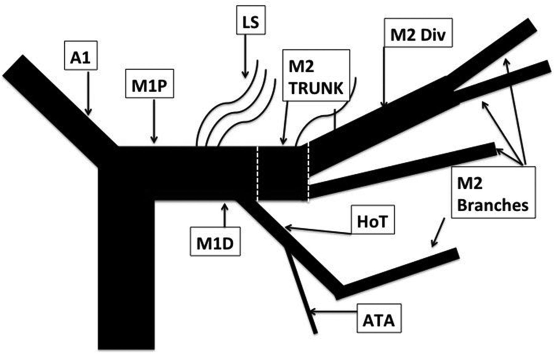
  • Fig 3.
    • Download figure
    • Open in new tab
    • Download powerpoint
    Fig 3.

    Composite diagram of M1-M2 trunk anatomy based on IMS III post hoc analysis. The M1 trunk proximal to the lenticulostriate arteries (LS) is termed “M1P.” The anterior temporal artery arises from the holotemporal M2 branch (HoT). The M2 trunk is a continuation of the distal M1 trunk, beyond a holotemporal (HoT) or posterior temporal M2 branch. The M2 trunk divides into M2 divisions (M2 Div) or branches. M2 divisions divide further into M2 branches.

Tables

  • Figures
  • mRS 0–2 outcomes for 79 evaluable M2 trunk, division, and branch occlusion grouped according to mTICI 0–1, 2a, 2b–3 reperfusion gradea

    mTICI GradeM2 Trunk (n = 9)M2 Division (n = 42)M2 Branch (n = 28)
    N% mRS 0–2N% mRS 0–2N% mRS 0–2
    0–120601136.4
    2a101866.7922.2
    2b–36b5018b44.4837.5
    • ↵a This table uses only the evaluable sample.

    • ↵b mTICI 3: n = 1.

PreviousNext
Back to top

In this issue

American Journal of Neuroradiology: 38 (1)
American Journal of Neuroradiology
Vol. 38, Issue 1
1 Jan 2017
  • Table of Contents
  • Index by author
  • Complete Issue (PDF)
Advertisement
Print
Download PDF
Email Article

Thank you for your interest in spreading the word on American Journal of Neuroradiology.

NOTE: We only request your email address so that the person you are recommending the page to knows that you wanted them to see it, and that it is not junk mail. We do not capture any email address.

Enter multiple addresses on separate lines or separate them with commas.
Endovascular Therapy of M2 Occlusion in IMS III: Role of M2 Segment Definition and Location on Clinical and Revascularization Outcomes
(Your Name) has sent you a message from American Journal of Neuroradiology
(Your Name) thought you would like to see the American Journal of Neuroradiology web site.
CAPTCHA
This question is for testing whether or not you are a human visitor and to prevent automated spam submissions.
Cite this article
T.A. Tomsick, J. Carrozzella, L. Foster, M.D. Hill, R. von Kummer, M. Goyal, A.M. Demchuk, P. Khatri, Y. Palesch, J.P. Broderick, S.D. Yeatts, D.S. Liebeskind, for the IMS III Investigators
Endovascular Therapy of M2 Occlusion in IMS III: Role of M2 Segment Definition and Location on Clinical and Revascularization Outcomes
American Journal of Neuroradiology Jan 2017, 38 (1) 84-89; DOI: 10.3174/ajnr.A4979

Citation Manager Formats

  • BibTeX
  • Bookends
  • EasyBib
  • EndNote (tagged)
  • EndNote 8 (xml)
  • Medlars
  • Mendeley
  • Papers
  • RefWorks Tagged
  • Ref Manager
  • RIS
  • Zotero
0 Responses
Respond to this article
Share
Bookmark this article
Endovascular Therapy of M2 Occlusion in IMS III: Role of M2 Segment Definition and Location on Clinical and Revascularization Outcomes
T.A. Tomsick, J. Carrozzella, L. Foster, M.D. Hill, R. von Kummer, M. Goyal, A.M. Demchuk, P. Khatri, Y. Palesch, J.P. Broderick, S.D. Yeatts, D.S. Liebeskind, for the IMS III Investigators
American Journal of Neuroradiology Jan 2017, 38 (1) 84-89; DOI: 10.3174/ajnr.A4979
del.icio.us logo Twitter logo Facebook logo Mendeley logo
  • Tweet Widget
  • Facebook Like
  • Google Plus One
Purchase

Jump to section

  • Article
    • Abstract
    • ABBREVIATIONS:
    • Materials and Methods
    • Results
    • Discussion
    • Conclusions
    • Footnotes
    • References
  • Figures & Data
  • Supplemental
  • Info & Metrics
  • Responses
  • References
  • PDF

Related Articles

  • PubMed
  • Google Scholar

Cited By...

  • Anatomical distribution and clinical significance of middle cerebral artery M2 segment vessel occlusions and its cortical branches in acute ischaemic stroke patients
  • Predictors and Impact of Sulcal SAH after Mechanical Thrombectomy in Patients with Isolated M2 Occlusion
  • Cost-effectiveness of endovascular thrombectomy in patients with acute stroke and M2 occlusion
  • Multiphase CT Angiography: A Useful Technique in Acute Stroke Imaging--Collaterals and Beyond
  • eTICI reperfusion: defining success in endovascular stroke therapy
  • Frontline ADAPT therapy to treat patients with symptomatic M2 and M3 occlusions in acute ischemic stroke: initial experience with the Penumbra ACE and 3MAX reperfusion system
  • Caution; Confusion Ahead...
  • Crossref
  • Google Scholar

This article has not yet been cited by articles in journals that are participating in Crossref Cited-by Linking.

More in this TOC Section

  • Rescue Reentry in Carotid Near-Occlusion
  • Contour Neurovascular System: Five Year Follow Up
  • Effect of SARS-CoV2 on Endovascular Thrombectomy
Show more Neurointervention

Similar Articles

Advertisement

Indexed Content

  • Current Issue
  • Accepted Manuscripts
  • Article Preview
  • Past Issues
  • Editorials
  • Editor's Choice
  • Fellows' Journal Club
  • Letters to the Editor
  • Video Articles

Cases

  • Case Collection
  • Archive - Case of the Week
  • Archive - Case of the Month
  • Archive - Classic Case

More from AJNR

  • Trainee Corner
  • Imaging Protocols
  • MRI Safety Corner
  • Book Reviews

Multimedia

  • AJNR Podcasts
  • AJNR Scantastics

Resources

  • Turnaround Time
  • Submit a Manuscript
  • Submit a Video Article
  • Submit an eLetter to the Editor/Response
  • Manuscript Submission Guidelines
  • Statistical Tips
  • Fast Publishing of Accepted Manuscripts
  • Graphical Abstract Preparation
  • Imaging Protocol Submission
  • Evidence-Based Medicine Level Guide
  • Publishing Checklists
  • Author Policies
  • Become a Reviewer/Academy of Reviewers
  • News and Updates

About Us

  • About AJNR
  • Editorial Board
  • Editorial Board Alumni
  • Alerts
  • Permissions
  • Not an AJNR Subscriber? Join Now
  • Advertise with Us
  • Librarian Resources
  • Feedback
  • Terms and Conditions
  • AJNR Editorial Board Alumni

American Society of Neuroradiology

  • Not an ASNR Member? Join Now

© 2025 by the American Society of Neuroradiology All rights, including for text and data mining, AI training, and similar technologies, are reserved.
Print ISSN: 0195-6108 Online ISSN: 1936-959X

Powered by HighWire