Advertisement

Main menu

  • Home
  • Content
    • Current Issue
    • Accepted Manuscripts
    • Article Preview
    • Past Issue Archive
    • Video Articles
    • AJNR Case Collection
    • Case of the Week Archive
    • Case of the Month Archive
    • Classic Case Archive
  • Special Collections
    • AJNR Awards
    • Low-Field MRI
    • Alzheimer Disease
    • ASNR Foundation Special Collection
    • Photon-Counting CT
    • View All
  • Multimedia
    • AJNR Podcasts
    • AJNR SCANtastic
    • Trainee Corner
    • MRI Safety Corner
    • Imaging Protocols
  • For Authors
    • Submit a Manuscript
    • Submit a Video Article
    • Submit an eLetter to the Editor/Response
    • Manuscript Submission Guidelines
    • Statistical Tips
    • Fast Publishing of Accepted Manuscripts
    • Graphical Abstract Preparation
    • Imaging Protocol Submission
    • Author Policies
  • About Us
    • About AJNR
    • Editorial Board
    • Editorial Board Alumni
  • More
    • Become a Reviewer/Academy of Reviewers
    • Subscribers
    • Permissions
    • Alerts
    • Feedback
    • Advertisers
    • ASNR Home

User menu

  • Alerts
  • Log in

Search

  • Advanced search
American Journal of Neuroradiology

American Journal of Neuroradiology

ASHNR American Society of Functional Neuroradiology ASHNR American Society of Pediatric Neuroradiology ASSR
  • Alerts
  • Log in

Advanced Search

  • Home
  • Content
    • Current Issue
    • Accepted Manuscripts
    • Article Preview
    • Past Issue Archive
    • Video Articles
    • AJNR Case Collection
    • Case of the Week Archive
    • Case of the Month Archive
    • Classic Case Archive
  • Special Collections
    • AJNR Awards
    • Low-Field MRI
    • Alzheimer Disease
    • ASNR Foundation Special Collection
    • Photon-Counting CT
    • View All
  • Multimedia
    • AJNR Podcasts
    • AJNR SCANtastic
    • Trainee Corner
    • MRI Safety Corner
    • Imaging Protocols
  • For Authors
    • Submit a Manuscript
    • Submit a Video Article
    • Submit an eLetter to the Editor/Response
    • Manuscript Submission Guidelines
    • Statistical Tips
    • Fast Publishing of Accepted Manuscripts
    • Graphical Abstract Preparation
    • Imaging Protocol Submission
    • Author Policies
  • About Us
    • About AJNR
    • Editorial Board
    • Editorial Board Alumni
  • More
    • Become a Reviewer/Academy of Reviewers
    • Subscribers
    • Permissions
    • Alerts
    • Feedback
    • Advertisers
    • ASNR Home
  • Follow AJNR on Twitter
  • Visit AJNR on Facebook
  • Follow AJNR on Instagram
  • Join AJNR on LinkedIn
  • RSS Feeds

AJNR Awards, New Junior Editors, and more. Read the latest AJNR updates

Getting new auth cookie, if you see this message a lot, tell someone!
Research ArticleNeurointervention

Quantifying the Cerebral Hemodynamics of Dural Arteriovenous Fistula in Transverse Sigmoid Sinus Complicated by Sinus Stenosis: A Retrospective Cohort Study

W.-Y. Guo, C.-C.J. Lee, C.-J. Lin, H.-C. Yang, H.-M. Wu, C.-C. Wu, W.-Y. Chung and K.-D. Liu
American Journal of Neuroradiology January 2017, 38 (1) 132-138; DOI: https://doi.org/10.3174/ajnr.A4960
W.-Y. Guo
aFrom the Department of Radiology (W.-Y.G., C.-J.L., H.-M.W., C.-C.W.)
cSchool of Medicine (W.-Y.G., C.-C.J.L., C.-J.L., H.-C.Y., H.-M.W., C.-C.W., W.-Y.C., K.-D.L.), National Yang-Ming University, Taipei, Taiwan.
  • Find this author on Google Scholar
  • Find this author on PubMed
  • Search for this author on this site
  • ORCID record for W.-Y. Guo
C.-C.J. Lee
bDepartment of Neurosurgery (C.-C.J.L., H.-C.Y., W.-Y.C., K.-D.L.), Neurological Institute, Taipei Veterans General Hospital, Taipei, Taiwan
cSchool of Medicine (W.-Y.G., C.-C.J.L., C.-J.L., H.-C.Y., H.-M.W., C.-C.W., W.-Y.C., K.-D.L.), National Yang-Ming University, Taipei, Taiwan.
  • Find this author on Google Scholar
  • Find this author on PubMed
  • Search for this author on this site
  • ORCID record for C.-C.J. Lee
C.-J. Lin
aFrom the Department of Radiology (W.-Y.G., C.-J.L., H.-M.W., C.-C.W.)
cSchool of Medicine (W.-Y.G., C.-C.J.L., C.-J.L., H.-C.Y., H.-M.W., C.-C.W., W.-Y.C., K.-D.L.), National Yang-Ming University, Taipei, Taiwan.
  • Find this author on Google Scholar
  • Find this author on PubMed
  • Search for this author on this site
  • ORCID record for C.-J. Lin
H.-C. Yang
bDepartment of Neurosurgery (C.-C.J.L., H.-C.Y., W.-Y.C., K.-D.L.), Neurological Institute, Taipei Veterans General Hospital, Taipei, Taiwan
cSchool of Medicine (W.-Y.G., C.-C.J.L., C.-J.L., H.-C.Y., H.-M.W., C.-C.W., W.-Y.C., K.-D.L.), National Yang-Ming University, Taipei, Taiwan.
  • Find this author on Google Scholar
  • Find this author on PubMed
  • Search for this author on this site
  • ORCID record for H.-C. Yang
H.-M. Wu
aFrom the Department of Radiology (W.-Y.G., C.-J.L., H.-M.W., C.-C.W.)
cSchool of Medicine (W.-Y.G., C.-C.J.L., C.-J.L., H.-C.Y., H.-M.W., C.-C.W., W.-Y.C., K.-D.L.), National Yang-Ming University, Taipei, Taiwan.
  • Find this author on Google Scholar
  • Find this author on PubMed
  • Search for this author on this site
  • ORCID record for H.-M. Wu
C.-C. Wu
aFrom the Department of Radiology (W.-Y.G., C.-J.L., H.-M.W., C.-C.W.)
cSchool of Medicine (W.-Y.G., C.-C.J.L., C.-J.L., H.-C.Y., H.-M.W., C.-C.W., W.-Y.C., K.-D.L.), National Yang-Ming University, Taipei, Taiwan.
  • Find this author on Google Scholar
  • Find this author on PubMed
  • Search for this author on this site
  • ORCID record for C.-C. Wu
W.-Y. Chung
bDepartment of Neurosurgery (C.-C.J.L., H.-C.Y., W.-Y.C., K.-D.L.), Neurological Institute, Taipei Veterans General Hospital, Taipei, Taiwan
cSchool of Medicine (W.-Y.G., C.-C.J.L., C.-J.L., H.-C.Y., H.-M.W., C.-C.W., W.-Y.C., K.-D.L.), National Yang-Ming University, Taipei, Taiwan.
  • Find this author on Google Scholar
  • Find this author on PubMed
  • Search for this author on this site
  • ORCID record for W.-Y. Chung
K.-D. Liu
bDepartment of Neurosurgery (C.-C.J.L., H.-C.Y., W.-Y.C., K.-D.L.), Neurological Institute, Taipei Veterans General Hospital, Taipei, Taiwan
cSchool of Medicine (W.-Y.G., C.-C.J.L., C.-J.L., H.-C.Y., H.-M.W., C.-C.W., W.-Y.C., K.-D.L.), National Yang-Ming University, Taipei, Taiwan.
  • Find this author on Google Scholar
  • Find this author on PubMed
  • Search for this author on this site
  • ORCID record for K.-D. Liu
  • Article
  • Figures & Data
  • Info & Metrics
  • Responses
  • References
  • PDF
Loading

Article Figures & Data

Figures

  • Tables
  • Fig 1.
    • Download figure
    • Open in new tab
    • Download powerpoint
    Fig 1.

    Quantitative color-coded digital subtraction angiography of the anteroposterior (A) and lateral (B) views of a Cognard type I DAVF. ROI1: internal carotid artery; ROI2: ipsilateral normal transverse sinus; ROI3: internal jugular vein; ROI4: parietal vein; ROI5: vein of Labbé; ROI6: prestenotic segment; ROI7: poststenotic segment. The Arrow indicates the stenotic sinus segment.
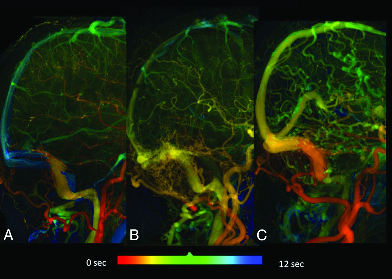
  • Fig 2.
    • Download figure
    • Open in new tab
    • Download powerpoint
    Fig 2.

    A, Quantitative digital subtraction angiography of a healthy subject. B, One single peak appears at 9.87 seconds (venous phase) in the internal jugular vein. C, Quantitative digital subtraction angiography of a Cognard type I DAVF in the left transverse sinus in a 50-year-old woman. D, Time-density curve of the internal jugular vein demonstrates 2 peaks. The first peak comes from arterial flow from the DAVF shunt; the second peak comes from blood flow from normal brain parenchyma. E, Quantitative digital subtraction of a case of Cognard type IIa+b in the left transverse sigmoid sinus in a 73-year-old man. F, Only a single peak can be depicted in a time-density curve of the ipsilateral jugular vein, indicating that it lacks the drainage function of the normal brain.

  • Fig 3.
    • Download figure
    • Open in new tab
    • Download powerpoint
    Fig 3.

    The process of case selection from the angiosuite logbook for our study cohort.

  • Fig 4.
    • Download figure
    • Open in new tab
    • Download powerpoint
    Fig 4.

    Kaplan-Meier analysis of complete regression (A) and favorable outcomes (B) among types I, IIa, and IIa+b or higher.

  • Fig 5.
    • Download figure
    • Open in new tab
    • Download powerpoint
    Fig 5.

    Quantitative DSA of cases of Cognard type I (A), Cognard type IIa (B), and Cognard type IIa+b (C). Severe sinus stenosis is more commonly encountered in more aggressive DAVF types. The TFT (TTP of the internal jugular vein) was largest in type IIa+b (green), followed by types IIa (yellow-green) and I (yellow). Progressive shortening of the TTP in the superior sagittal sinuses in type IIa and type IIa+b is also depicted.

Tables

  • Figures
    • View popup
    Table 1:

    Comparison of patient characteristics in 3 different groups: type I, type IIa, and types with CVDa

    Type IType IIaTypes with CVD
    No.22813
    Age (yr)58 (52.8–64.2)54 (35.5–73.7)52 (41.3–63.8)
    Headaches4 (18.2; 2.1–34.3)3 (37.5; 6.2–79.5)4 (30.8; 5.7–55.9)
    Hemorrhage/neurologic deficits008 (61.5; 35.1–88.0)b
    Venous stenosis8 (36.4; 16.3–56.5)b8 (100%)11 (84.6; 65–100)
    Loss of double peak7 (31.8; 12.4–51.3)c8 (100%)c9 (69.2; 44.1–94.3)c
    • ↵a The numbers inside the parentheses for age indicate the 95% confidence intervals. The numbers inside the parentheses for headaches, hemorrhage/neurologic deficits, venous stenosis, and loss of double peak indicate the percentage of the observed variable in the group with 95% confidence intervals.

    • ↵b Significant difference compared with the other 2 groups.

    • ↵c Significant difference across the 3 groups.

    • View popup
    Table 2:

    Intra- and interobserver variability in different time parametersa

    Reader AReader BInterobserver
    TFT0.98 (0.96–0.99)0.97 (0.96–0.98)0.94 (0.90–0.97)
    TST0.97 (0.95–0.99)0.96 (0.94–0.99)0.93 (0.90–0.96)
    TTPPV0.98 (0.96–0.99)0.95 (0.90–0.97)0.91 (0.86–0.94)
    TTPVL0.98 (0.96–0.99)0.92 (0.89–0.96)0.92 (0.87–0.94)
    TTPTS0.95 (0.97–0.91)0.96 (0.93–0.99)0.94 (0.90–0.98)
    • Note:—TTPTS indicates TTP of the ipsilateral normal transverse sinus.

    • ↵a Data are 95% CI.

    • View popup
    Table 3:

    Comparison of different time parameters among the 3 groupsa

    Type IType IIaTypes with CVD
    TFT1.04 (0.80–1.00)0.42 (0.27–0.4)2.09 (1.06–2.26)b
    TST0.03 (0–0)0.34 (0.3–0.53)b0.67 (−0.54–0.8)b
    TTPPV4.57 (4.14–5.60)4.25 (3.20–4.94)5.6 (4.13–6.26)
    TTPVL4.20 (4.26–5.20)4.40 (3.50–6.00)1.4 (0.93–3.46)b
    TTPTS6.01 (4.93–7.47)b1.17 (1.06–3.6)1.1 (1.06–1.86)
    • Note:—TTPTS indicates TTP of the ipsilateral normal transverse sinus.

    • ↵a Data are 95% CI.

    • ↵b Significant difference compared with the other 2 groups.

    • View popup
    Table 4:

    Clinical characteristics, treatment strategy, and response in 13 patients with DAVF types with CVD

    Case No.SexAge (yr)Cognard Type before SRSTreatmentCognard Type after Venoplasty/StentTreatment after SRSFollow-Up Duration (mo)Response
    1F41IIa+bVenoplasty/stentIIa–46PR
    2M63IIa+bVenoplastyIIa–38CR
    3M56IIa+bVenoplasty/stentIIa–19PRa
    4M45IIa+b–NA–15CR
    5F63IIa+bVenoplasty/stentIIa+b–10PRa
    6M73IIa+b–NA–10PR
    7M17IIa+bVenoplasty/stentIIa+b–6PR
    8M55IIb–NA–5.6PR
    9M19III–NATAE twice17PR
    10M27IIIVenoplastyIIa–27CRb
    11M12III––13CR
    12M27IV––27.6PR
    13M55IV––NANA
    • Note:—TAE indicates transarterial embolization; CR, complete regression; PR, partial regression; –, no adjunct treatment was performed; NA, not available.

    • ↵a Restenosis of sinus after venoplasty and stenting.

    • ↵b Asymptomatic intracranial hemorrhage on MR imaging.

PreviousNext
Back to top

In this issue

American Journal of Neuroradiology: 38 (1)
American Journal of Neuroradiology
Vol. 38, Issue 1
1 Jan 2017
  • Table of Contents
  • Index by author
  • Complete Issue (PDF)
Advertisement
Print
Download PDF
Email Article

Thank you for your interest in spreading the word on American Journal of Neuroradiology.

NOTE: We only request your email address so that the person you are recommending the page to knows that you wanted them to see it, and that it is not junk mail. We do not capture any email address.

Enter multiple addresses on separate lines or separate them with commas.
Quantifying the Cerebral Hemodynamics of Dural Arteriovenous Fistula in Transverse Sigmoid Sinus Complicated by Sinus Stenosis: A Retrospective Cohort Study
(Your Name) has sent you a message from American Journal of Neuroradiology
(Your Name) thought you would like to see the American Journal of Neuroradiology web site.
CAPTCHA
This question is for testing whether or not you are a human visitor and to prevent automated spam submissions.
Cite this article
W.-Y. Guo, C.-C.J. Lee, C.-J. Lin, H.-C. Yang, H.-M. Wu, C.-C. Wu, W.-Y. Chung, K.-D. Liu
Quantifying the Cerebral Hemodynamics of Dural Arteriovenous Fistula in Transverse Sigmoid Sinus Complicated by Sinus Stenosis: A Retrospective Cohort Study
American Journal of Neuroradiology Jan 2017, 38 (1) 132-138; DOI: 10.3174/ajnr.A4960

Citation Manager Formats

  • BibTeX
  • Bookends
  • EasyBib
  • EndNote (tagged)
  • EndNote 8 (xml)
  • Medlars
  • Mendeley
  • Papers
  • RefWorks Tagged
  • Ref Manager
  • RIS
  • Zotero
0 Responses
Respond to this article
Share
Bookmark this article
Quantifying the Cerebral Hemodynamics of Dural Arteriovenous Fistula in Transverse Sigmoid Sinus Complicated by Sinus Stenosis: A Retrospective Cohort Study
W.-Y. Guo, C.-C.J. Lee, C.-J. Lin, H.-C. Yang, H.-M. Wu, C.-C. Wu, W.-Y. Chung, K.-D. Liu
American Journal of Neuroradiology Jan 2017, 38 (1) 132-138; DOI: 10.3174/ajnr.A4960
del.icio.us logo Twitter logo Facebook logo Mendeley logo
  • Tweet Widget
  • Facebook Like
  • Google Plus One
Purchase

Jump to section

  • Article
    • Abstract
    • ABBREVIATIONS:
    • Materials and Methods
    • Results
    • Discussion
    • Conclusions
    • Acknowledgments
    • Footnotes
    • References
  • Figures & Data
  • Info & Metrics
  • Responses
  • References
  • PDF

Related Articles

  • No related articles found.
  • PubMed
  • Google Scholar

Cited By...

  • No citing articles found.
  • Crossref
  • Google Scholar

This article has not yet been cited by articles in journals that are participating in Crossref Cited-by Linking.

More in this TOC Section

  • Rescue Reentry in Carotid Near-Occlusion
  • Contour Neurovascular System: Five Year Follow Up
  • Effect of SARS-CoV2 on Endovascular Thrombectomy
Show more Neurointervention

Similar Articles

Advertisement

Indexed Content

  • Current Issue
  • Accepted Manuscripts
  • Article Preview
  • Past Issues
  • Editorials
  • Editor's Choice
  • Fellows' Journal Club
  • Letters to the Editor
  • Video Articles

Cases

  • Case Collection
  • Archive - Case of the Week
  • Archive - Case of the Month
  • Archive - Classic Case

More from AJNR

  • Trainee Corner
  • Imaging Protocols
  • MRI Safety Corner
  • Book Reviews

Multimedia

  • AJNR Podcasts
  • AJNR Scantastics

Resources

  • Turnaround Time
  • Submit a Manuscript
  • Submit a Video Article
  • Submit an eLetter to the Editor/Response
  • Manuscript Submission Guidelines
  • Statistical Tips
  • Fast Publishing of Accepted Manuscripts
  • Graphical Abstract Preparation
  • Imaging Protocol Submission
  • Evidence-Based Medicine Level Guide
  • Publishing Checklists
  • Author Policies
  • Become a Reviewer/Academy of Reviewers
  • News and Updates

About Us

  • About AJNR
  • Editorial Board
  • Editorial Board Alumni
  • Alerts
  • Permissions
  • Not an AJNR Subscriber? Join Now
  • Advertise with Us
  • Librarian Resources
  • Feedback
  • Terms and Conditions
  • AJNR Editorial Board Alumni

American Society of Neuroradiology

  • Not an ASNR Member? Join Now

© 2025 by the American Society of Neuroradiology All rights, including for text and data mining, AI training, and similar technologies, are reserved.
Print ISSN: 0195-6108 Online ISSN: 1936-959X

Powered by HighWire