Advertisement

Main menu

  • Home
  • Content
    • Current Issue
    • Accepted Manuscripts
    • Article Preview
    • Past Issue Archive
    • Video Articles
    • AJNR Case Collection
    • Case of the Week Archive
    • Case of the Month Archive
    • Classic Case Archive
  • Special Collections
    • AJNR Awards
    • Low-Field MRI
    • Alzheimer Disease
    • ASNR Foundation Special Collection
    • Photon-Counting CT
    • View All
  • Multimedia
    • AJNR Podcasts
    • AJNR SCANtastic
    • Trainee Corner
    • MRI Safety Corner
    • Imaging Protocols
  • For Authors
    • Submit a Manuscript
    • Submit a Video Article
    • Submit an eLetter to the Editor/Response
    • Manuscript Submission Guidelines
    • Statistical Tips
    • Fast Publishing of Accepted Manuscripts
    • Graphical Abstract Preparation
    • Imaging Protocol Submission
    • Author Policies
  • About Us
    • About AJNR
    • Editorial Board
    • Editorial Board Alumni
  • More
    • Become a Reviewer/Academy of Reviewers
    • Subscribers
    • Permissions
    • Alerts
    • Feedback
    • Advertisers
    • ASNR Home

User menu

  • Alerts
  • Log in

Search

  • Advanced search
American Journal of Neuroradiology

American Journal of Neuroradiology

ASHNR American Society of Functional Neuroradiology ASHNR American Society of Pediatric Neuroradiology ASSR
  • Alerts
  • Log in

Advanced Search

  • Home
  • Content
    • Current Issue
    • Accepted Manuscripts
    • Article Preview
    • Past Issue Archive
    • Video Articles
    • AJNR Case Collection
    • Case of the Week Archive
    • Case of the Month Archive
    • Classic Case Archive
  • Special Collections
    • AJNR Awards
    • Low-Field MRI
    • Alzheimer Disease
    • ASNR Foundation Special Collection
    • Photon-Counting CT
    • View All
  • Multimedia
    • AJNR Podcasts
    • AJNR SCANtastic
    • Trainee Corner
    • MRI Safety Corner
    • Imaging Protocols
  • For Authors
    • Submit a Manuscript
    • Submit a Video Article
    • Submit an eLetter to the Editor/Response
    • Manuscript Submission Guidelines
    • Statistical Tips
    • Fast Publishing of Accepted Manuscripts
    • Graphical Abstract Preparation
    • Imaging Protocol Submission
    • Author Policies
  • About Us
    • About AJNR
    • Editorial Board
    • Editorial Board Alumni
  • More
    • Become a Reviewer/Academy of Reviewers
    • Subscribers
    • Permissions
    • Alerts
    • Feedback
    • Advertisers
    • ASNR Home
  • Follow AJNR on Twitter
  • Visit AJNR on Facebook
  • Follow AJNR on Instagram
  • Join AJNR on LinkedIn
  • RSS Feeds

AJNR Awards, New Junior Editors, and more. Read the latest AJNR updates

Research ArticleNeurointervention

Hemodynamic and Anatomic Variations Require an Adaptable Approach during Intra-Arterial Chemotherapy for Intraocular Retinoblastoma: Alternative Routes, Strategies, and Follow-Up

E. Bertelli, S. Leonini, D. Galimberti, S. Moretti, R. Tinturini, T. Hadjistilianou, S. De Francesco, D.G. Romano, I.M. Vallone, S. Cioni, P. Gennari, P. Galluzzi, I. Grazzini, S. Rossi and S. Bracco
American Journal of Neuroradiology July 2016, 37 (7) 1289-1295; DOI: https://doi.org/10.3174/ajnr.A4741
E. Bertelli
aFrom the Department of Molecular and Developmental Medicine (E.B., S.R.), University of Siena, Siena, Italy
  • Find this author on Google Scholar
  • Find this author on PubMed
  • Search for this author on this site
  • ORCID record for E. Bertelli
S. Leonini
bUnits of Neuroimaging and Neurointervention (S.L., D.G.R., I.M.V., S.C., P. Gennari, P. Galluzzi, I.G., S.B.)
  • Find this author on Google Scholar
  • Find this author on PubMed
  • Search for this author on this site
  • ORCID record for S. Leonini
D. Galimberti
cPediatrics (D.G., S.M.)
  • Find this author on Google Scholar
  • Find this author on PubMed
  • Search for this author on this site
  • ORCID record for D. Galimberti
S. Moretti
cPediatrics (D.G., S.M.)
  • Find this author on Google Scholar
  • Find this author on PubMed
  • Search for this author on this site
  • ORCID record for S. Moretti
R. Tinturini
dNeurosurgical Intensive Care (R.T.), Azienda Ospedaliera Universitaria Senese, University of Siena, Policlinico “Santa Maria alle Scotte,” Siena, Italy
  • Find this author on Google Scholar
  • Find this author on PubMed
  • Search for this author on this site
  • ORCID record for R. Tinturini
T. Hadjistilianou
eDepartment of Medicine, Surgery and Neuroscience (T.H., S.D.F.), Unit of Ophthalmology, University of Siena, Policlinico “Santa Maria alle Scotte,” Siena, Italy.
  • Find this author on Google Scholar
  • Find this author on PubMed
  • Search for this author on this site
  • ORCID record for T. Hadjistilianou
S. De Francesco
eDepartment of Medicine, Surgery and Neuroscience (T.H., S.D.F.), Unit of Ophthalmology, University of Siena, Policlinico “Santa Maria alle Scotte,” Siena, Italy.
  • Find this author on Google Scholar
  • Find this author on PubMed
  • Search for this author on this site
  • ORCID record for S. De Francesco
D.G. Romano
bUnits of Neuroimaging and Neurointervention (S.L., D.G.R., I.M.V., S.C., P. Gennari, P. Galluzzi, I.G., S.B.)
  • Find this author on Google Scholar
  • Find this author on PubMed
  • Search for this author on this site
  • ORCID record for D.G. Romano
I.M. Vallone
bUnits of Neuroimaging and Neurointervention (S.L., D.G.R., I.M.V., S.C., P. Gennari, P. Galluzzi, I.G., S.B.)
  • Find this author on Google Scholar
  • Find this author on PubMed
  • Search for this author on this site
  • ORCID record for I.M. Vallone
S. Cioni
bUnits of Neuroimaging and Neurointervention (S.L., D.G.R., I.M.V., S.C., P. Gennari, P. Galluzzi, I.G., S.B.)
  • Find this author on Google Scholar
  • Find this author on PubMed
  • Search for this author on this site
  • ORCID record for S. Cioni
P. Gennari
bUnits of Neuroimaging and Neurointervention (S.L., D.G.R., I.M.V., S.C., P. Gennari, P. Galluzzi, I.G., S.B.)
  • Find this author on Google Scholar
  • Find this author on PubMed
  • Search for this author on this site
  • ORCID record for P. Gennari
P. Galluzzi
bUnits of Neuroimaging and Neurointervention (S.L., D.G.R., I.M.V., S.C., P. Gennari, P. Galluzzi, I.G., S.B.)
  • Find this author on Google Scholar
  • Find this author on PubMed
  • Search for this author on this site
  • ORCID record for P. Galluzzi
I. Grazzini
bUnits of Neuroimaging and Neurointervention (S.L., D.G.R., I.M.V., S.C., P. Gennari, P. Galluzzi, I.G., S.B.)
  • Find this author on Google Scholar
  • Find this author on PubMed
  • Search for this author on this site
  • ORCID record for I. Grazzini
S. Rossi
aFrom the Department of Molecular and Developmental Medicine (E.B., S.R.), University of Siena, Siena, Italy
  • Find this author on Google Scholar
  • Find this author on PubMed
  • Search for this author on this site
  • ORCID record for S. Rossi
S. Bracco
bUnits of Neuroimaging and Neurointervention (S.L., D.G.R., I.M.V., S.C., P. Gennari, P. Galluzzi, I.G., S.B.)
  • Find this author on Google Scholar
  • Find this author on PubMed
  • Search for this author on this site
  • ORCID record for S. Bracco
  • Article
  • Figures & Data
  • Supplemental
  • Info & Metrics
  • Responses
  • References
  • PDF
Loading

Article Figures & Data

Figures

  • Tables
  • Fig 1.
    • Download figure
    • Open in new tab
    • Download powerpoint
    Fig 1.

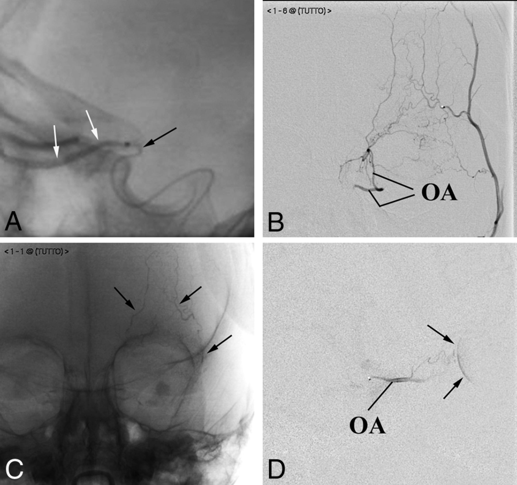
    An exemplary case of adaptable approaches to the IAC. The patient has bilateral retinoblastoma previously enucleated on 1 side (the prosthesis is visible). The remaining eye is a case of VP of drug delivery. A, The OA is successfully catheterized and used in 2 sessions of IAC because the choroidal blush (arrows) is regularly achieved. B–E, Third session of IAC. Selective angiography of the ICA does not show any visible OA (B). Nevertheless catheterization of the OA is successful (C), though the contrast medium flows back into the ICA (On-line Video). Superselective angiography of the MMA shows a good anastomotic pathway to the OA (D), which allows achieving the choroidal blush (arrows in E). F and G, Fourth session of IAC. This time the anastomosis between the MMA and the OA does not guarantee the choroidal blush. An alternative route for drug delivery through the ECA is sought and found between the frontal branch of the superficial temporal artery (arrows point to the microcatheter within the artery) and the supratrochlear artery (F). A later angiographic phase shows that this pathway guarantees the choroidal blush (arrows) (G). H–J, Fifth session of IAC. The anastomosis between the MMA and the OA is working again. However, the contrast medium flows back even into a large branch of the MMA (arrows in H). To reduce the volume of distribution, we glued the meningeal branch (arrows point to the cast in I), and the following injection of contrast medium achieves the choroidal blush (arrows in J).

  • Fig 2.
    • Download figure
    • Open in new tab
    • Download powerpoint
    Fig 2.

    Particular angiographic procedures. A, Customization of the microcatheter. The tip of the microcatheter (black arrow), manually bent to fit the angioanatomy of the patient, has been firmly placed at the entrance of the OA to release the contrast medium (white arrows). B–D, Flow anterograde redirection within the OA. Anteroposterior projections. Contrast medium injection into the superficial temporal artery shows a rich network of small vessels connecting the superficial temporal artery with the OA (B). Embolization of the arterial network. The cast of glue outlines the embolized frontal vessels (arrows in C). The flow in the OA, redirected anterogradely (D), allows achieving the choroidal blush (arrows in D).

Tables

  • Figures
    • View popup
    Table 1:

    Outcome of the eyes according to the pattern of IAC drug delivery

    Tumor RemissionRecurrence/Enucleation
    FPO4627a
    FPEC72
    VP89
    • ↵a One patient died. After all conservative treatments were tried, the disease still progressed. Nevertheless, the parents refused enucleation of the eye.

    • View popup
    Table 2:

    Ophthalmologic response according to the pattern of IAC drug delivery

    Low ResponseMedium ResponseHigh Response
    FPO4366
    FPEC207
    VP1115
    • View popup
    Table 3:

    Local adverse events according to the pattern of IAC drug delivery

    Adverse Events (No.) (%)FPOFPECVP
    Transient
        Palpebral edema/hyperemia, 55 (55.6%)4069
        Frontal edema/hyperemia, 14 (14.1%)1013
        Retinal bleeding, 12 (12.1%)1002
        Anterior ischemia of the optic nerve, 2 (2%)101
        Madarosis, 2 (2%)200
        Frontal alopecia, 2 (2%)101
        Orbital cellulitis, 1 (1%)100
        Glaucoma, 1 (1%)001
        Roth spots, 1 (1%)100
        Iridocyclitis, 1 (1%)100
    Permanent
        Chorioretinic atrophy, 16 (16.2%)1321
        Exotropia, 4 (4%)220
        Ptosis, 1 (1%)100
        Anisocoria, 1 (1%)001
        Cutaneous scar necrosis, 1 (1%)010
        Iris heterochromia, 1 (1%)001
PreviousNext
Back to top

In this issue

American Journal of Neuroradiology: 37 (7)
American Journal of Neuroradiology
Vol. 37, Issue 7
1 Jul 2016
  • Table of Contents
  • Index by author
  • Complete Issue (PDF)
Advertisement
Print
Download PDF
Email Article

Thank you for your interest in spreading the word on American Journal of Neuroradiology.

NOTE: We only request your email address so that the person you are recommending the page to knows that you wanted them to see it, and that it is not junk mail. We do not capture any email address.

Enter multiple addresses on separate lines or separate them with commas.
Hemodynamic and Anatomic Variations Require an Adaptable Approach during Intra-Arterial Chemotherapy for Intraocular Retinoblastoma: Alternative Routes, Strategies, and Follow-Up
(Your Name) has sent you a message from American Journal of Neuroradiology
(Your Name) thought you would like to see the American Journal of Neuroradiology web site.
CAPTCHA
This question is for testing whether or not you are a human visitor and to prevent automated spam submissions.
Cite this article
E. Bertelli, S. Leonini, D. Galimberti, S. Moretti, R. Tinturini, T. Hadjistilianou, S. De Francesco, D.G. Romano, I.M. Vallone, S. Cioni, P. Gennari, P. Galluzzi, I. Grazzini, S. Rossi, S. Bracco
Hemodynamic and Anatomic Variations Require an Adaptable Approach during Intra-Arterial Chemotherapy for Intraocular Retinoblastoma: Alternative Routes, Strategies, and Follow-Up
American Journal of Neuroradiology Jul 2016, 37 (7) 1289-1295; DOI: 10.3174/ajnr.A4741

Citation Manager Formats

  • BibTeX
  • Bookends
  • EasyBib
  • EndNote (tagged)
  • EndNote 8 (xml)
  • Medlars
  • Mendeley
  • Papers
  • RefWorks Tagged
  • Ref Manager
  • RIS
  • Zotero
0 Responses
Respond to this article
Share
Bookmark this article
Hemodynamic and Anatomic Variations Require an Adaptable Approach during Intra-Arterial Chemotherapy for Intraocular Retinoblastoma: Alternative Routes, Strategies, and Follow-Up
E. Bertelli, S. Leonini, D. Galimberti, S. Moretti, R. Tinturini, T. Hadjistilianou, S. De Francesco, D.G. Romano, I.M. Vallone, S. Cioni, P. Gennari, P. Galluzzi, I. Grazzini, S. Rossi, S. Bracco
American Journal of Neuroradiology Jul 2016, 37 (7) 1289-1295; DOI: 10.3174/ajnr.A4741
del.icio.us logo Twitter logo Facebook logo Mendeley logo
  • Tweet Widget
  • Facebook Like
  • Google Plus One
Purchase

Jump to section

  • Article
    • Abstract
    • ABBREVIATIONS:
    • Materials and Methods
    • Results
    • Discussion
    • Conclusions
    • Footnotes
    • REFERENCES
  • Figures & Data
  • Supplemental
  • Info & Metrics
  • Responses
  • References
  • PDF

Related Articles

  • PubMed
  • Google Scholar

Cited By...

  • Absorbable gelatin compressed sponge (Gelfoam) embolization of distal external carotid artery branches in intra-arterial chemotherapy for retinoblastoma
  • Risk factors for cataract in retinoblastoma management
  • Absorbable gelatin compressed sponge (Gelfoam) embolization of distal external carotid artery branches in intra-arterial chemotherapy for retinoblastoma
  • Risk factors for cataract in retinoblastoma management
  • Angiographic investigation of orbital vascular variations in the rabbit and implications for endovascular intra-arterial chemotherapy models
  • Correlation of Technical and Adjunctive Factors with Quantitative Tumor Reduction in Children Undergoing Selective Ophthalmic Artery Infusion Chemotherapy for Retinoblastoma
  • Initial experience with the Scepter Mini dual-lumen balloon for transophthalmic artery embolization of anterior cranial fossa dural arteriovenous fistulae
  • Ophthalmic artery catheterization for retinoblastoma treatment: does reflux affect tumor response?
  • Orbital infarction syndrome after mechanical thrombectomy in acute ischaemic stroke
  • Retrograde Approach through the Posterior Communicating Artery and Anterior Communicating Artery to the Ophthalmic Artery
  • Reply:
  • Embryologic Anatomic Variations: Challenges in Intra-Arterial Chemotherapy for Intraocular Retinoblastoma
  • Crossref
  • Google Scholar

This article has not yet been cited by articles in journals that are participating in Crossref Cited-by Linking.

More in this TOC Section

Neurointervention

  • Rescue Reentry in Carotid Near-Occlusion
  • Contour Neurovascular System: Five Year Follow Up
  • Effect of SARS-CoV2 on Endovascular Thrombectomy
Show more Neurointervention

Pediatric Neuroimaging

  • Frontal Paraventricular Cysts
  • Sodium MRI in Pediatric Brain Tumors
  • FRACTURE MR in Congenital Vertebral Anomalies
Show more Pediatric Neuroimaging

Extracranial Vascular

  • Frontal Paraventricular Cysts
  • Sodium MRI in Pediatric Brain Tumors
  • FRACTURE MR in Congenital Vertebral Anomalies
Show more Extracranial Vascular

Similar Articles

Advertisement

Indexed Content

  • Current Issue
  • Accepted Manuscripts
  • Article Preview
  • Past Issues
  • Editorials
  • Editor's Choice
  • Fellows' Journal Club
  • Letters to the Editor
  • Video Articles

Cases

  • Case Collection
  • Archive - Case of the Week
  • Archive - Case of the Month
  • Archive - Classic Case

More from AJNR

  • Trainee Corner
  • Imaging Protocols
  • MRI Safety Corner
  • Book Reviews

Multimedia

  • AJNR Podcasts
  • AJNR Scantastics

Resources

  • Turnaround Time
  • Submit a Manuscript
  • Submit a Video Article
  • Submit an eLetter to the Editor/Response
  • Manuscript Submission Guidelines
  • Statistical Tips
  • Fast Publishing of Accepted Manuscripts
  • Graphical Abstract Preparation
  • Imaging Protocol Submission
  • Evidence-Based Medicine Level Guide
  • Publishing Checklists
  • Author Policies
  • Become a Reviewer/Academy of Reviewers
  • News and Updates

About Us

  • About AJNR
  • Editorial Board
  • Editorial Board Alumni
  • Alerts
  • Permissions
  • Not an AJNR Subscriber? Join Now
  • Advertise with Us
  • Librarian Resources
  • Feedback
  • Terms and Conditions
  • AJNR Editorial Board Alumni

American Society of Neuroradiology

  • Not an ASNR Member? Join Now

© 2025 by the American Society of Neuroradiology All rights, including for text and data mining, AI training, and similar technologies, are reserved.
Print ISSN: 0195-6108 Online ISSN: 1936-959X

Powered by HighWire