
Article Figures & Data
Figures
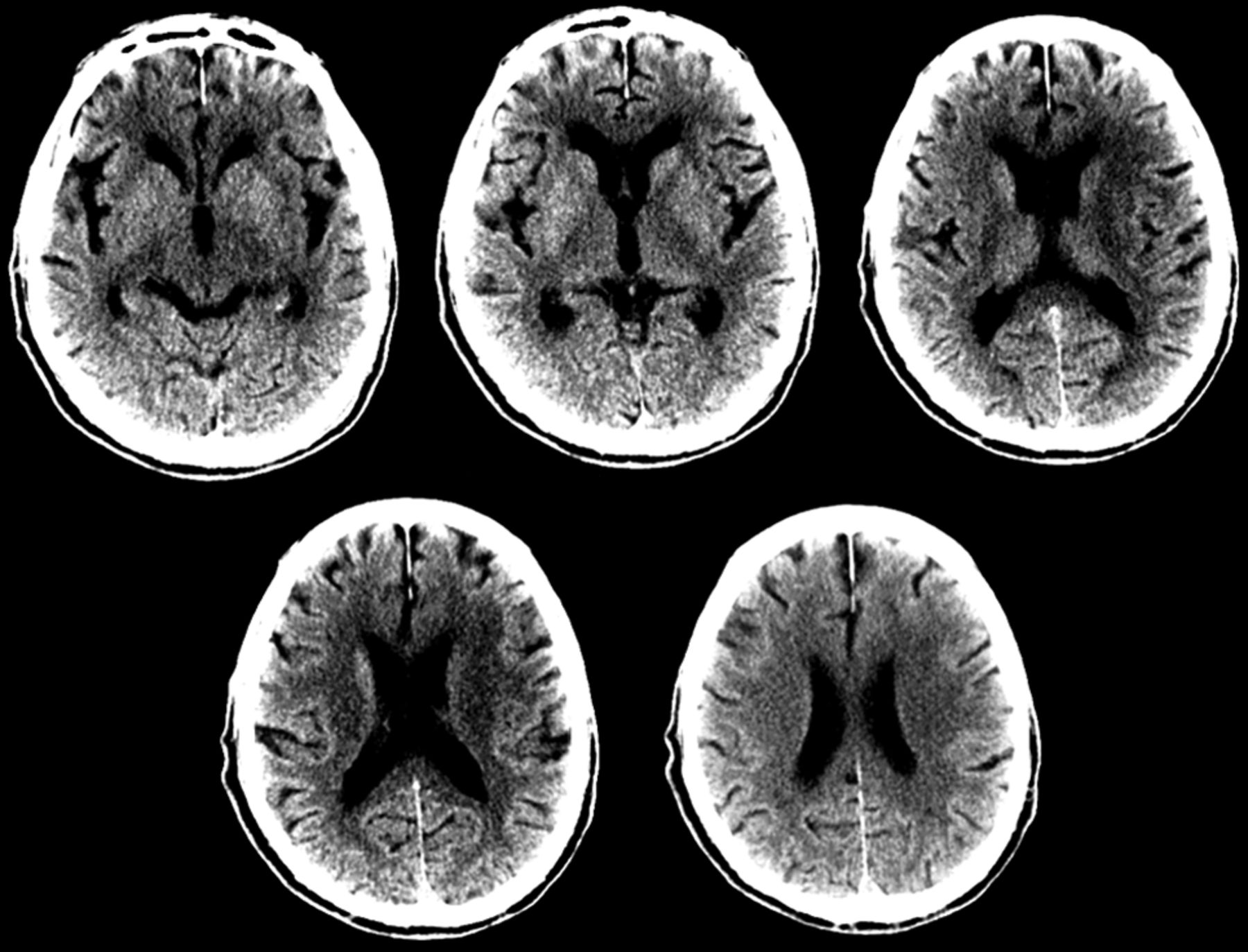
- Fig 1.
The unenhanced CT ASPECTS score is 7, with 1 point subtracted for early ischemic changes in the caudate nucleus, lentiform nucleus, and insula on the left.
- Fig 2.
A, Axial multiphase CT angiography (3 phases) maximum-intensity-projection images (arterial, phase 1 [P1]; midvenous, phase 2 [P2]; and late venous, phase 3 [P3] from top to bottom) show left middle cerebral artery segment 1 occlusion. In addition, during phase 1, there is a delay in contrast opacification of the left middle cerebral artery branches compared with the contralateral normal side; however, during phase 2, the contrast opacification appears to be symmetric. This is labeled as “one phase delay.” There is also symmetric “extent” of collaterals (red oval) compared with the contralateral normal side (yellow oval). Overall, this gives an impression of fairly good collaterals. B, Magnified axial MIP image in the second delayed phase (P3) shows the possible clot length (arrow) calculated as the distance between the site of abrupt vessel cutoff (proximal end) and the site of distal vessel opacification, either through slow anterograde or retrograde collateral filling (distal end). This can help in preplanning the length of the stent retriever to be used.
- Fig 3.
Measuring the extent of revascularization. Comparison between the TICI scoring before (TICI 0, A) and after (TICI 2b, B) mechanical thrombectomy in the late arterial phase. The blue outline depicts the normal extent of the MCA territory. Note, the region shown by the red arrow suggesting non-/slow filling of a few distal cortical branches.


Jump to section
Related Articles
Cited By...
- Recent developments in pre-hospital and in-hospital triage for endovascular stroke treatment
- Workflow patterns and potential for optimization in endovascular stroke treatment across the world: results from a multinational survey
- Dynamic CTA-Derived Perfusion Maps Predict Final Infarct Volume: The Simple Perfusion Reconstruction Algorithm
- Workflow and factors associated with delay in the delivery of intra-arterial treatment for acute ischemic stroke in the MR CLEAN trial
- Wide Variability in Prethrombectomy Workflow Practices in the United States: A Multicenter Survey
- Improved Detection of Anterior Circulation Occlusions: The "Delayed Vessel Sign" on Multiphase CT Angiography
- Multiphase CT angiography increases detection of anterior circulation intracranial occlusion