Advertisement

Main menu

  • Home
  • Content
    • Current Issue
    • Accepted Manuscripts
    • Article Preview
    • Past Issue Archive
    • Video Articles
    • AJNR Case Collection
    • Case of the Week Archive
    • Case of the Month Archive
    • Classic Case Archive
  • Special Collections
    • AJNR Awards
    • Low-Field MRI
    • Alzheimer Disease
    • ASNR Foundation Special Collection
    • Photon-Counting CT
    • View All
  • Multimedia
    • AJNR Podcasts
    • AJNR SCANtastic
    • Trainee Corner
    • MRI Safety Corner
    • Imaging Protocols
  • For Authors
    • Submit a Manuscript
    • Submit a Video Article
    • Submit an eLetter to the Editor/Response
    • Manuscript Submission Guidelines
    • Statistical Tips
    • Fast Publishing of Accepted Manuscripts
    • Graphical Abstract Preparation
    • Imaging Protocol Submission
    • Author Policies
  • About Us
    • About AJNR
    • Editorial Board
    • Editorial Board Alumni
  • More
    • Become a Reviewer/Academy of Reviewers
    • Subscribers
    • Permissions
    • Alerts
    • Feedback
    • Advertisers
    • ASNR Home

User menu

  • Alerts
  • Log in

Search

  • Advanced search
American Journal of Neuroradiology

American Journal of Neuroradiology

ASHNR American Society of Functional Neuroradiology ASHNR American Society of Pediatric Neuroradiology ASSR
  • Alerts
  • Log in

Advanced Search

  • Home
  • Content
    • Current Issue
    • Accepted Manuscripts
    • Article Preview
    • Past Issue Archive
    • Video Articles
    • AJNR Case Collection
    • Case of the Week Archive
    • Case of the Month Archive
    • Classic Case Archive
  • Special Collections
    • AJNR Awards
    • Low-Field MRI
    • Alzheimer Disease
    • ASNR Foundation Special Collection
    • Photon-Counting CT
    • View All
  • Multimedia
    • AJNR Podcasts
    • AJNR SCANtastic
    • Trainee Corner
    • MRI Safety Corner
    • Imaging Protocols
  • For Authors
    • Submit a Manuscript
    • Submit a Video Article
    • Submit an eLetter to the Editor/Response
    • Manuscript Submission Guidelines
    • Statistical Tips
    • Fast Publishing of Accepted Manuscripts
    • Graphical Abstract Preparation
    • Imaging Protocol Submission
    • Author Policies
  • About Us
    • About AJNR
    • Editorial Board
    • Editorial Board Alumni
  • More
    • Become a Reviewer/Academy of Reviewers
    • Subscribers
    • Permissions
    • Alerts
    • Feedback
    • Advertisers
    • ASNR Home
  • Follow AJNR on Twitter
  • Visit AJNR on Facebook
  • Follow AJNR on Instagram
  • Join AJNR on LinkedIn
  • RSS Feeds

AJNR Awards, New Junior Editors, and more. Read the latest AJNR updates

Research Article

Nonmotor Functions of the Cerebellum: An Introduction

A.P. Klein, J.L. Ulmer, S.A. Quinet, V. Mathews and L.P. Mark
American Journal of Neuroradiology June 2016, 37 (6) 1005-1009; DOI: https://doi.org/10.3174/ajnr.A4720
A.P. Klein
aFrom the Medical College of Wisconsin, Department of Radiology, Neuroradiology Section, Froedtert Hospital, Milwaukee, Wisconsin.
  • Find this author on Google Scholar
  • Find this author on PubMed
  • Search for this author on this site
  • ORCID record for A.P. Klein
J.L. Ulmer
aFrom the Medical College of Wisconsin, Department of Radiology, Neuroradiology Section, Froedtert Hospital, Milwaukee, Wisconsin.
  • Find this author on Google Scholar
  • Find this author on PubMed
  • Search for this author on this site
  • ORCID record for J.L. Ulmer
S.A. Quinet
aFrom the Medical College of Wisconsin, Department of Radiology, Neuroradiology Section, Froedtert Hospital, Milwaukee, Wisconsin.
  • Find this author on Google Scholar
  • Find this author on PubMed
  • Search for this author on this site
  • ORCID record for S.A. Quinet
V. Mathews
aFrom the Medical College of Wisconsin, Department of Radiology, Neuroradiology Section, Froedtert Hospital, Milwaukee, Wisconsin.
  • Find this author on Google Scholar
  • Find this author on PubMed
  • Search for this author on this site
  • ORCID record for V. Mathews
L.P. Mark
aFrom the Medical College of Wisconsin, Department of Radiology, Neuroradiology Section, Froedtert Hospital, Milwaukee, Wisconsin.
  • Find this author on Google Scholar
  • Find this author on PubMed
  • Search for this author on this site
  • ORCID record for L.P. Mark
  • Article
  • Figures & Data
  • Info & Metrics
  • Responses
  • References
  • PDF
Loading

Article Figures & Data

Figures

  • Fig 1.
    • Download figure
    • Open in new tab
    • Download powerpoint
    Fig 1.

    Unfolded view of the cerebellar cortex showing the lobes, lobules (by name on the right and number on the left), and main fissures (blue font). The hemispheric lobules are designated with the prefix H followed by the Roman numeral indicating their corresponding vermian lobules. Adapted from Haines DE. Fundamental Neuroscience for Basic and Clinical Applications. 4th ed. Philadelphia: Elsevier/Saunders; 2013 with permission of Elsevier.30

  • Fig 2.
    • Download figure
    • Open in new tab
    • Download powerpoint
    Fig 2.

    The cerebrocerebellum is the phylogenetically newest and largest portion of the cerebellum. It primarily receives input indirectly from many cerebral cortical areas. The spinocerebellum occupies the median and paramedian zone of the cerebellum and receives input directly from the spinal cord. The vestibulocerebellum is the phylogenetically oldest part of the cerebellum, and it receives input from the vestibular nuclei of the brain stem. Adapted with permission from Purves et al.11
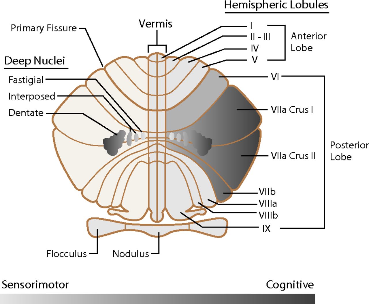
  • Fig 3.
    • Download figure
    • Open in new tab
    • Download powerpoint
    Fig 3.

    Unfolded view of the cerebellum illustrating sensorimotor-to-cognitive functions distributed in a gradient-like fashion from medial to lateral. The sensorimotor functions are distributed more toward the midline, while the cognitive functions are located more laterally in the cerebellar hemispheres. The same medial-to-lateral organization is seen in the corresponding cerebellar nuclei. Adapted with permission from Fatemi et al.12
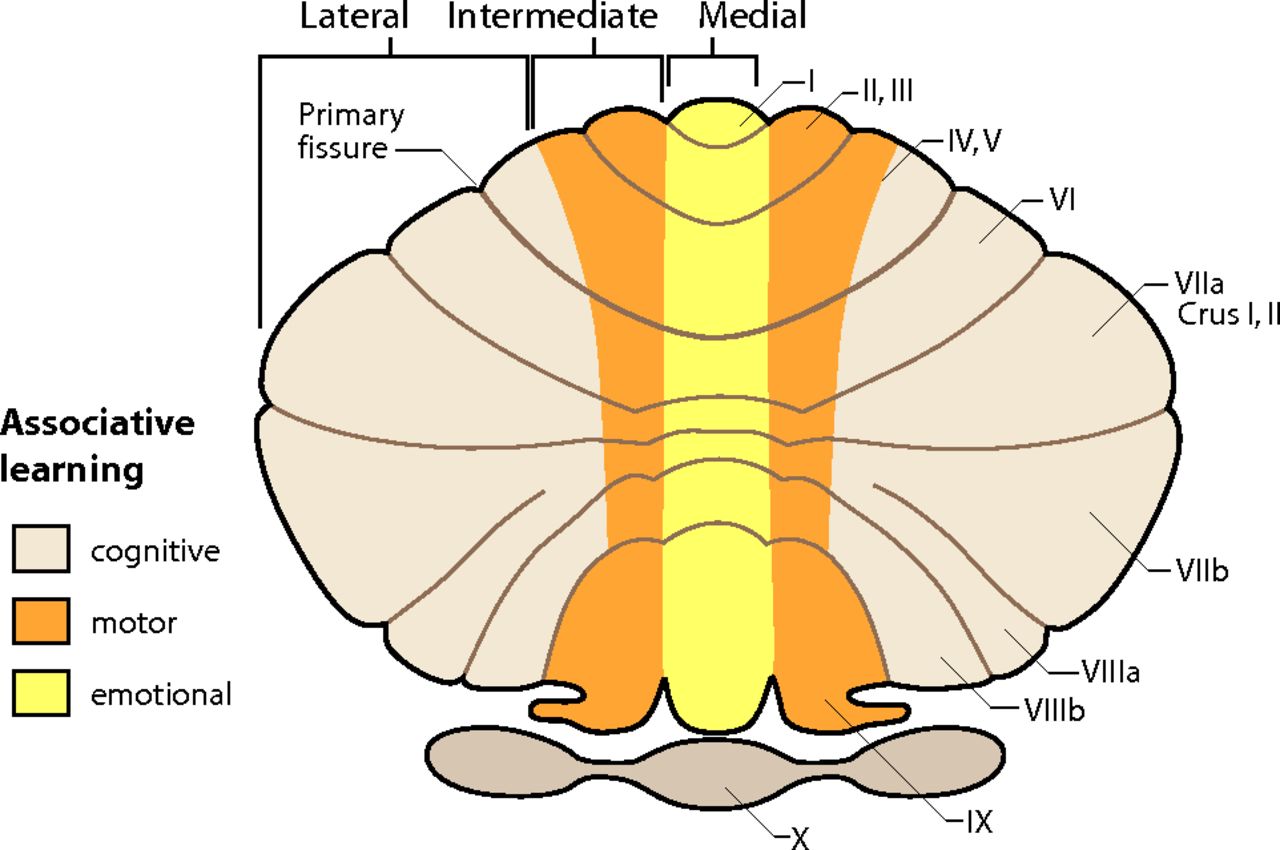
  • Fig 4.
    • Download figure
    • Open in new tab
    • Download powerpoint
    Fig 4.

    Unfolded view of the cerebellum demonstrating associative learning with emotional tasks located in the medial zone (yellow). Motor tasks locate to the intermediate zone (orange), and cognitive tasks occupy most of the cerebellar hemisphere in the lateral zone (beige). Adapted from Timmann D, Drepper J, Frings M, et al. The human cerebellum contributes to motor, emotional and cognitive associative learning: a review. Cortex 2010;46:845–57 with permission of Elsevier.16

  • Fig 5.
    • Download figure
    • Open in new tab
    • Download powerpoint
    Fig 5.

    Unfolded view of the cerebellum showing the asymmetric distribution of some cerebellar functions. The right cerebellar hemisphere is associated with language, and the left cerebellar hemisphere, with visuospatial functions. Executive functions, including verbal working memory, are related to both hemispheres. Attention is also a neocerebellar function. The vermis or “limbic cerebellum” is involved in modulating affective behavior. Adapted from Konczak J, Timmann D. The effect of damage to the cerebellum on sensorimotor and cognitive function in children and adolescents. Neurosci Biobehav Rev 2007;31:1101–13 with permission of Elsevier.17

  • Fig 6.
    • Download figure
    • Open in new tab
    • Download powerpoint
    Fig 6.

    A, Schematic demonstration of the cerebral and cerebellar functional locations of the foot (green), hand (red), and face (blue) in the monkey. B, Cerebellar locations of the foot (green), hand (red), and tongue (blue) in humans measured by fMRI. “fcMRI” refers to results based on functional connectivity studies. “Task” refers to results from task-based fMRI studies. C, Cerebellar locations of foot (F, green), hand (H, red), and tongue (T, blue) representations in humans from fcMRI studies displayed on a parasagittal image. Note the mirror image representation of the somatomotor functions with the primary or dominant location in the anterior lobe of the cerebellum. Adapted from Buckner RL. The cerebellum and cognitive function: 25 years of insight from anatomy and neuroimaging. Neuron 2013;80:807–15 with permission of Elsevier.31

  • Fig 7.
    • Download figure
    • Open in new tab
    • Download powerpoint
    Fig 7.

    Topographic map of various cerebellar functions displayed on a parasagittal view of the left cerebellar hemisphere. The primary cerebellar distribution of cerebral functions is an orderly map of the somatomotor functions of the foot, hand, and tongue (blue) in the anterior lobe followed by a hierarchy of association cortices labeled 1–4 (magenta, green, orange, red) on the superior surface of the cerebellum. An inverted secondary representation in reverse order is seen on the inferior cerebellar surface. A small tertiary representative map is hypothesized to be present in the posterior lobe as well. Adapted from Buckner RL. The cerebellum and cognitive function: 25 years of insight from anatomy and neuroimaging. Neuron 2013;80:807–15 with permission of Elsevier.31

  • Fig 8.
    • Download figure
    • Open in new tab
    • Download powerpoint
    Fig 8.

    The 3 images on the left represent multiple coronal sections of the cerebellum with colors representing different cortical functions. The right-sided image is the cerebrum with the colors representing the different functional areas. The somatomotor cortex is blue. This cortex is represented at the more medial aspect of the cerebellum. Most of the human cerebellum, however, is linked to cerebral association networks, including executive networks (orange) and the default network (red). These association networks have multiple cerebellar representations. Adapted from Buckner RL. The cerebellum and cognitive function: 25 years of insight from anatomy and neuroimaging. Neuron 2013;80:807–15 with permission of Elsevier.31

PreviousNext
Back to top

In this issue

American Journal of Neuroradiology: 37 (6)
American Journal of Neuroradiology
Vol. 37, Issue 6
1 Jun 2016
  • Table of Contents
  • Index by author
  • Complete Issue (PDF)
Advertisement
Print
Download PDF
Email Article

Thank you for your interest in spreading the word on American Journal of Neuroradiology.

NOTE: We only request your email address so that the person you are recommending the page to knows that you wanted them to see it, and that it is not junk mail. We do not capture any email address.

Enter multiple addresses on separate lines or separate them with commas.
Nonmotor Functions of the Cerebellum: An Introduction
(Your Name) has sent you a message from American Journal of Neuroradiology
(Your Name) thought you would like to see the American Journal of Neuroradiology web site.
CAPTCHA
This question is for testing whether or not you are a human visitor and to prevent automated spam submissions.
Cite this article
A.P. Klein, J.L. Ulmer, S.A. Quinet, V. Mathews, L.P. Mark
Nonmotor Functions of the Cerebellum: An Introduction
American Journal of Neuroradiology Jun 2016, 37 (6) 1005-1009; DOI: 10.3174/ajnr.A4720

Citation Manager Formats

  • BibTeX
  • Bookends
  • EasyBib
  • EndNote (tagged)
  • EndNote 8 (xml)
  • Medlars
  • Mendeley
  • Papers
  • RefWorks Tagged
  • Ref Manager
  • RIS
  • Zotero
0 Responses
Respond to this article
Share
Bookmark this article
Nonmotor Functions of the Cerebellum: An Introduction
A.P. Klein, J.L. Ulmer, S.A. Quinet, V. Mathews, L.P. Mark
American Journal of Neuroradiology Jun 2016, 37 (6) 1005-1009; DOI: 10.3174/ajnr.A4720
del.icio.us logo Twitter logo Facebook logo Mendeley logo
  • Tweet Widget
  • Facebook Like
  • Google Plus One
Purchase

Jump to section

  • Article
    • Footnotes
    • References
  • Figures & Data
  • Info & Metrics
  • Responses
  • References
  • PDF

Related Articles

  • No related articles found.
  • PubMed
  • Google Scholar

Cited By...

  • Lateralized cerebellar connectivity differentiates auditory pathways in echolocating and non-echolocating whales
  • Human-specific features of cerebellar astrocytes and Purkinje cells: an anatomical comparison with mice and macaques
  • Affection of Motor Network Regions by Tau Pathology Across the Alzheimer's Disease Spectrum
  • Large Data on the Small Brain: Population-wide Cerebellar Growth Models of Children and Adolescents
  • "Upregulation of TLR4/MyD88 pathway in alcohol-induced Wernickes encephalopathy: findings in preclinical models and in a postmortem human case"
  • Deleting Mecp2 from the entire cerebellum rather than its neuronal subtypes causes a delay in motor learning in mice
  • 18F-FDG-PET Hyperactivity in Alzheimers Disease Cerebellum and Primary Olfactory Cortex
  • Biometry of the Cerebellar Vermis and Brain Stem in Children: MR Imaging Reference Data from Measurements in 718 Children
  • Crossref
  • Google Scholar

This article has not yet been cited by articles in journals that are participating in Crossref Cited-by Linking.

Similar Articles

Advertisement

Indexed Content

  • Current Issue
  • Accepted Manuscripts
  • Article Preview
  • Past Issues
  • Editorials
  • Editor's Choice
  • Fellows' Journal Club
  • Letters to the Editor
  • Video Articles

Cases

  • Case Collection
  • Archive - Case of the Week
  • Archive - Case of the Month
  • Archive - Classic Case

More from AJNR

  • Trainee Corner
  • Imaging Protocols
  • MRI Safety Corner
  • Book Reviews

Multimedia

  • AJNR Podcasts
  • AJNR Scantastics

Resources

  • Turnaround Time
  • Submit a Manuscript
  • Submit a Video Article
  • Submit an eLetter to the Editor/Response
  • Manuscript Submission Guidelines
  • Statistical Tips
  • Fast Publishing of Accepted Manuscripts
  • Graphical Abstract Preparation
  • Imaging Protocol Submission
  • Evidence-Based Medicine Level Guide
  • Publishing Checklists
  • Author Policies
  • Become a Reviewer/Academy of Reviewers
  • News and Updates

About Us

  • About AJNR
  • Editorial Board
  • Editorial Board Alumni
  • Alerts
  • Permissions
  • Not an AJNR Subscriber? Join Now
  • Advertise with Us
  • Librarian Resources
  • Feedback
  • Terms and Conditions
  • AJNR Editorial Board Alumni

American Society of Neuroradiology

  • Not an ASNR Member? Join Now

© 2025 by the American Society of Neuroradiology All rights, including for text and data mining, AI training, and similar technologies, are reserved.
Print ISSN: 0195-6108 Online ISSN: 1936-959X

Powered by HighWire