
Article Figures & Data
Figures
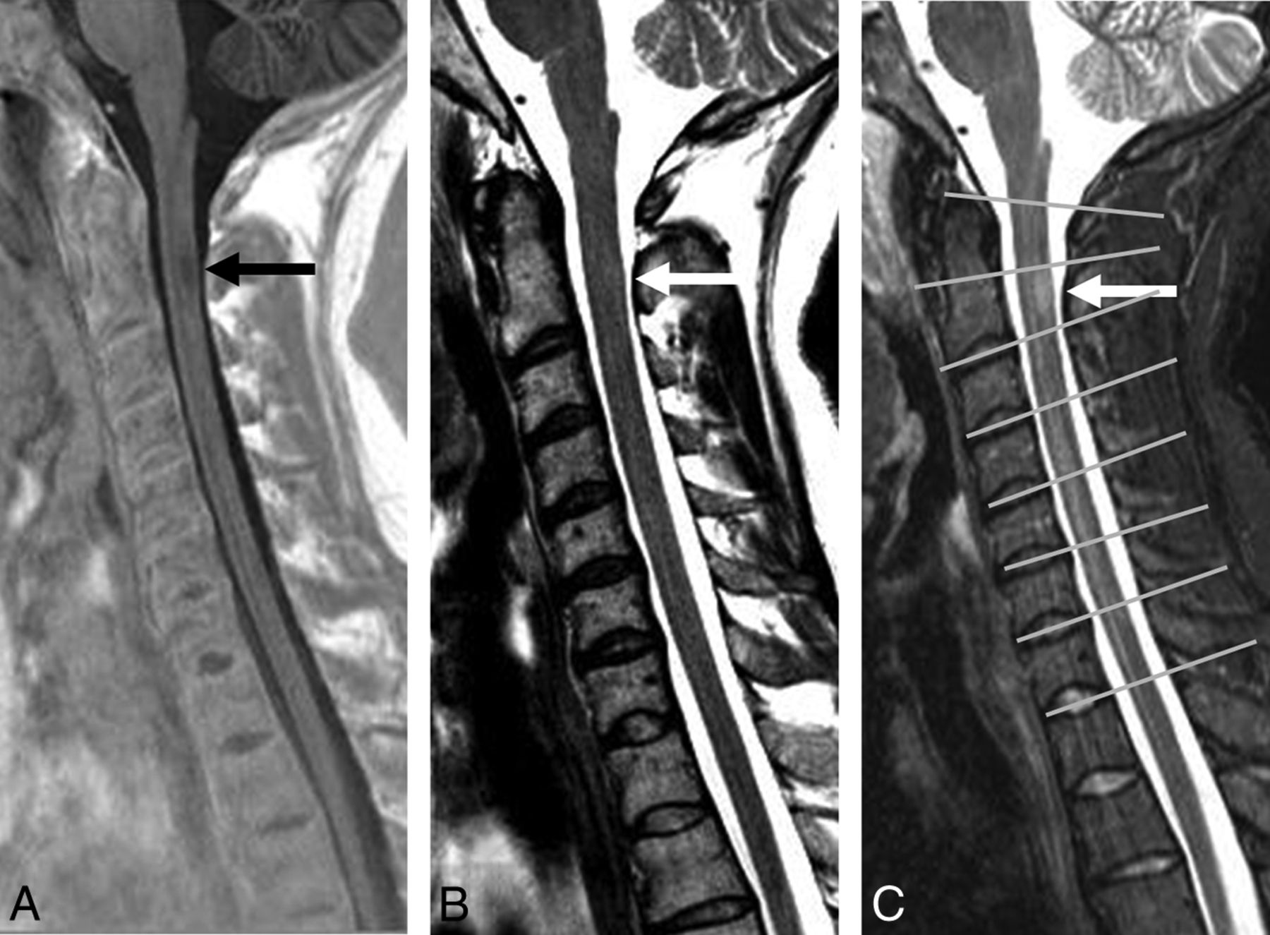
- Fig 1.
Sagittal PSIR (A), FSE T2 (B), and STIR (C) sequences in a patient with MS. A demyelinating lesion at the C2 level (arrows) is adequately visualized on PSIR (A) and STIR (C) but not on FSE T2 (B). C, An example of reference lines through the center on the disc spaces used to separate the vertebral levels (gray lines).
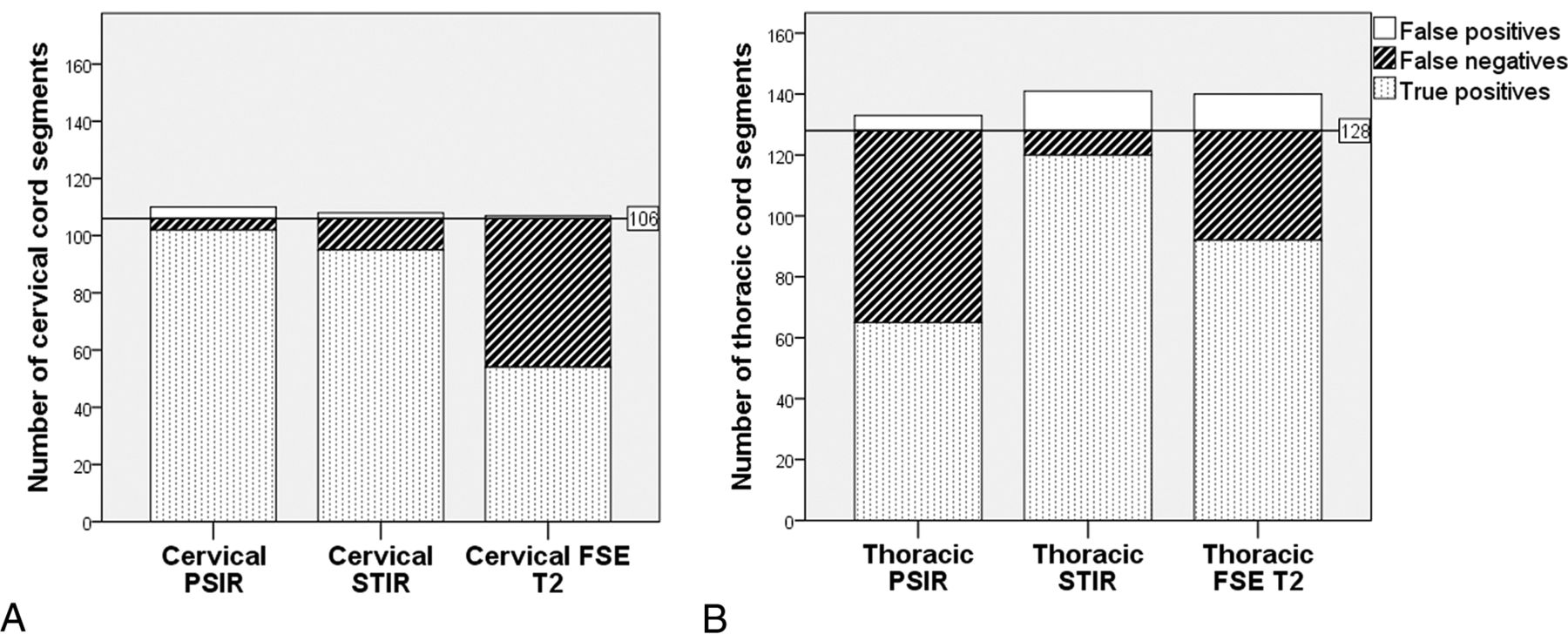
- Fig 2.
Stacked bar charts show the total number of true-positive, false-negative, and false-positive spinal cord segments in the cervical (A) and thoracic (B) regions on the 3 different sagittal sequences evaluated. The horizontal lines represent the total number of segments truly affected by lesions as per criterion standard evaluation.
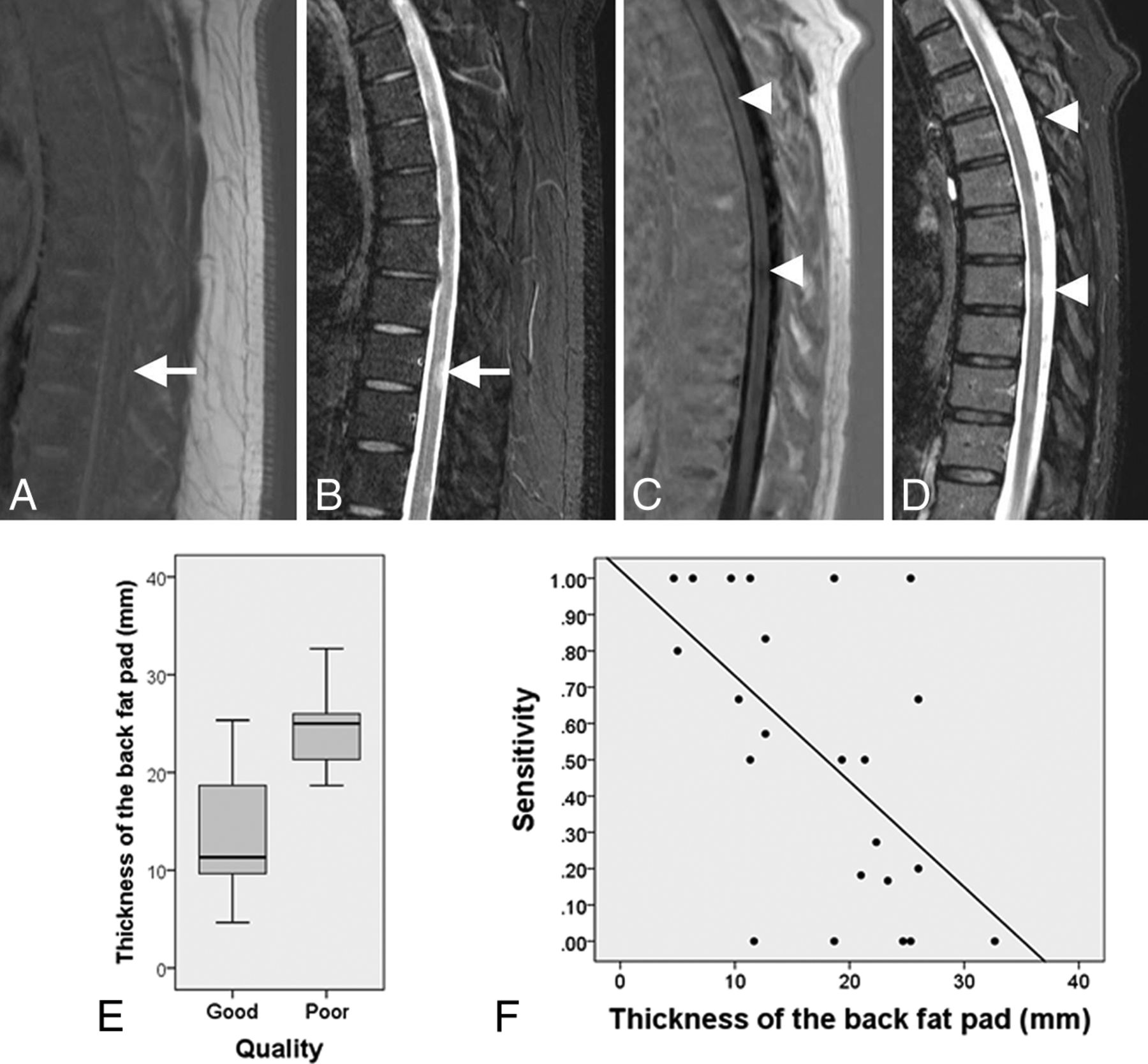
- Fig 3.
A, Thoracic sagittal PSIR image of a patient with MS with a thick back fat pad shows increased noise and failure of CSF suppression. A demyelinating lesion (white arrow) is clearly identified on STIR (B) but not seen on PSIR (A). Sagittal PSIR (C) and STIR (D) sequences of a slim patient with MS show adequate contrast between the lesions (arrow caps) and the cord and adequate CSF signal suppression on PSIR. E, The boxplot shows the distribution of the variable thickness of the back fat pad in the group of patients with good- and poor-quality thoracic PSIR images. The bottom and the top of the boxes represent the 25th and 75th percentiles, respectively; the median is represented by the horizontal line in the box; and the range corresponds to the whiskers that extend from the box. Patients with poor-quality PSIR sequences of the thoracic spine have a median back fat pad size significantly thicker than that in patients with good-quality images. F, The scatterplot shows the negative correlation between the sensitivity of the PSIR sequence of the thoracic segment and the thickness of the back fat pad (r = −0.53).


Tables
Acquisition parameters of MR imaging sequences
Segment Sequence Plane FOV (mm) Acquisition Matrix Thickness (mm) TR (ms) TE (ms) TI (ms) No. of Signals Acquired Acquisition Time (min:sec) Parallel Imaging Cervical PSIR Sagittal 220 320 × 224 3 2400 9.4 400 2 4:45 GRAPPA 2 Cervical STIR Sagittal 300 448 × 336 3 400 51 200 1 5:46 GRAPPA 2 Cervical FSE PD Sagittal 220 320 × 288 3 2500 23 1 2:02 None Cervical FSE T2 Sagittal 220 384 × 307 3 3500 106 2 3:35 None Cervical GE T2 Transverse 180 256 × 218 3 740 17 2 4:25 GRAPPA 2 Thoracic PSIR Sagittal 330 384 × 269 3 2400 9.5 400 2 4:45 GRAPPA 2 Thoracic STIR Sagittal 330 448 × 336 3 4000 50 200 1 5:46 GRAPPA 2 Thoracic FSE PD Sagittal 330 320 × 288 3 2500 21 1 4:02 None Thoracic FSE T2 Sagittal 330 448 × 336 3 4000 96 1 4:26 None Thoracic FSE T2 Transverse 180 256 × 176 5 6780 107 2 4:46 None Note:—GE indicates gradient-echo; PD, proton density; GRAPPA, generalized autocalibrating partially parallel acquisition.


