Advertisement

Main menu

  • Home
  • Content
    • Current Issue
    • Accepted Manuscripts
    • Article Preview
    • Past Issue Archive
    • Video Articles
    • AJNR Case Collection
    • Case of the Week Archive
    • Case of the Month Archive
    • Classic Case Archive
  • Special Collections
    • AJNR Awards
    • Low-Field MRI
    • Alzheimer Disease
    • ASNR Foundation Special Collection
    • Photon-Counting CT
    • View All
  • Multimedia
    • AJNR Podcasts
    • AJNR SCANtastic
    • Trainee Corner
    • MRI Safety Corner
    • Imaging Protocols
  • For Authors
    • Submit a Manuscript
    • Submit a Video Article
    • Submit an eLetter to the Editor/Response
    • Manuscript Submission Guidelines
    • Statistical Tips
    • Fast Publishing of Accepted Manuscripts
    • Graphical Abstract Preparation
    • Imaging Protocol Submission
    • Author Policies
  • About Us
    • About AJNR
    • Editorial Board
    • Editorial Board Alumni
  • More
    • Become a Reviewer/Academy of Reviewers
    • Subscribers
    • Permissions
    • Alerts
    • Feedback
    • Advertisers
    • ASNR Home

User menu

  • Alerts
  • Log in

Search

  • Advanced search
American Journal of Neuroradiology

American Journal of Neuroradiology

ASHNR American Society of Functional Neuroradiology ASHNR American Society of Pediatric Neuroradiology ASSR
  • Alerts
  • Log in

Advanced Search

  • Home
  • Content
    • Current Issue
    • Accepted Manuscripts
    • Article Preview
    • Past Issue Archive
    • Video Articles
    • AJNR Case Collection
    • Case of the Week Archive
    • Case of the Month Archive
    • Classic Case Archive
  • Special Collections
    • AJNR Awards
    • Low-Field MRI
    • Alzheimer Disease
    • ASNR Foundation Special Collection
    • Photon-Counting CT
    • View All
  • Multimedia
    • AJNR Podcasts
    • AJNR SCANtastic
    • Trainee Corner
    • MRI Safety Corner
    • Imaging Protocols
  • For Authors
    • Submit a Manuscript
    • Submit a Video Article
    • Submit an eLetter to the Editor/Response
    • Manuscript Submission Guidelines
    • Statistical Tips
    • Fast Publishing of Accepted Manuscripts
    • Graphical Abstract Preparation
    • Imaging Protocol Submission
    • Author Policies
  • About Us
    • About AJNR
    • Editorial Board
    • Editorial Board Alumni
  • More
    • Become a Reviewer/Academy of Reviewers
    • Subscribers
    • Permissions
    • Alerts
    • Feedback
    • Advertisers
    • ASNR Home
  • Follow AJNR on Twitter
  • Visit AJNR on Facebook
  • Follow AJNR on Instagram
  • Join AJNR on LinkedIn
  • RSS Feeds

AJNR Awards, New Junior Editors, and more. Read the latest AJNR updates

Research ArticlePediatric Neuroimaging

Quantitative Assessment of Neovascularization after Indirect Bypass Surgery: Color-Coded Digital Subtraction Angiography in Pediatric Moyamoya Disease

H.-H. Cho, J.-E. Cheon, S.-K. Kim, Y.H. Choi, I.-O. Kim, W.S. Kim, S.-M. Lee, S.K. You and S.-M. Shin
American Journal of Neuroradiology May 2016, 37 (5) 932-938; DOI: https://doi.org/10.3174/ajnr.A4614
H.-H. Cho
aFrom the Department of Radiology (H.-H.C., J.-E.C., Y.H.C., I.-O.K., W.S.K., S.-M.L., S.K.Y.), Seoul National University Hospital, Seoul, Korea
  • Find this author on Google Scholar
  • Find this author on PubMed
  • Search for this author on this site
  • ORCID record for H.-H. Cho
J.-E. Cheon
aFrom the Department of Radiology (H.-H.C., J.-E.C., Y.H.C., I.-O.K., W.S.K., S.-M.L., S.K.Y.), Seoul National University Hospital, Seoul, Korea
bDepartment of Radiology (J.-E.C., Y.H.C., I.-O.K., W.S.K.), Seoul National University College of Medicine, Seoul, Korea
cInstitute of Radiation Medicine (J.-E.C., I.-O.K., W.S.K.), Seoul National University Medical Research Center, Seoul, Korea
  • Find this author on Google Scholar
  • Find this author on PubMed
  • Search for this author on this site
  • ORCID record for J.-E. Cheon
S.-K. Kim
dDivision of Pediatric Neurosurgery (S.-K.K.), Seoul National University Children's Hospital, Seoul, Korea
  • Find this author on Google Scholar
  • Find this author on PubMed
  • Search for this author on this site
  • ORCID record for S.-K. Kim
Y.H. Choi
aFrom the Department of Radiology (H.-H.C., J.-E.C., Y.H.C., I.-O.K., W.S.K., S.-M.L., S.K.Y.), Seoul National University Hospital, Seoul, Korea
bDepartment of Radiology (J.-E.C., Y.H.C., I.-O.K., W.S.K.), Seoul National University College of Medicine, Seoul, Korea
  • Find this author on Google Scholar
  • Find this author on PubMed
  • Search for this author on this site
  • ORCID record for Y.H. Choi
I.-O. Kim
aFrom the Department of Radiology (H.-H.C., J.-E.C., Y.H.C., I.-O.K., W.S.K., S.-M.L., S.K.Y.), Seoul National University Hospital, Seoul, Korea
bDepartment of Radiology (J.-E.C., Y.H.C., I.-O.K., W.S.K.), Seoul National University College of Medicine, Seoul, Korea
cInstitute of Radiation Medicine (J.-E.C., I.-O.K., W.S.K.), Seoul National University Medical Research Center, Seoul, Korea
  • Find this author on Google Scholar
  • Find this author on PubMed
  • Search for this author on this site
  • ORCID record for I.-O. Kim
W.S. Kim
aFrom the Department of Radiology (H.-H.C., J.-E.C., Y.H.C., I.-O.K., W.S.K., S.-M.L., S.K.Y.), Seoul National University Hospital, Seoul, Korea
bDepartment of Radiology (J.-E.C., Y.H.C., I.-O.K., W.S.K.), Seoul National University College of Medicine, Seoul, Korea
cInstitute of Radiation Medicine (J.-E.C., I.-O.K., W.S.K.), Seoul National University Medical Research Center, Seoul, Korea
  • Find this author on Google Scholar
  • Find this author on PubMed
  • Search for this author on this site
  • ORCID record for W.S. Kim
S.-M. Lee
aFrom the Department of Radiology (H.-H.C., J.-E.C., Y.H.C., I.-O.K., W.S.K., S.-M.L., S.K.Y.), Seoul National University Hospital, Seoul, Korea
  • Find this author on Google Scholar
  • Find this author on PubMed
  • Search for this author on this site
  • ORCID record for S.-M. Lee
S.K. You
aFrom the Department of Radiology (H.-H.C., J.-E.C., Y.H.C., I.-O.K., W.S.K., S.-M.L., S.K.Y.), Seoul National University Hospital, Seoul, Korea
  • Find this author on Google Scholar
  • Find this author on PubMed
  • Search for this author on this site
  • ORCID record for S.K. You
S.-M. Shin
eDepartment of Radiology (S.-M.S.), Seoul Metropolitan Government–Seoul National University, Boramae Medical Center, Seoul, Korea.
  • Find this author on Google Scholar
  • Find this author on PubMed
  • Search for this author on this site
  • ORCID record for S.-M. Shin
  • Article
  • Figures & Data
  • Info & Metrics
  • Responses
  • References
  • PDF
Loading

Article Figures & Data

Figures

  • Tables
  • Fig 1.
    • Download figure
    • Open in new tab
    • Download powerpoint
    Fig 1.

    Postprocessing cascade of the DSA image. A, A color-coded single image is obtained from DSA images of CCA angiography; then, we draw selected ROIs along the estimated EDAS flap site. A reference ROI is drawn within the proximal input artery. B, The time-attenuation intensity curve is obtained automatically. The superior green line represents the time-attenuation intensity curve within the selected ROI, and the inferior red line represents that of the reference ROI. C, Using the time-attenuation intensity curve, we calculated the TTP and AUC within the selected ROI, adjusted by the reference ROI.

  • Fig 2.
    • Download figure
    • Open in new tab
    • Download powerpoint
    Fig 2.

    A 4-year-old boy with excellent clinical outcome after EDAS neovascularization. Comparing pre- (A) and post- (E) operative ECA angiograms shows good neovascularization after EDAS. Postprocessed pre- (B) and post- (F) operative CCA angiograms show shortening of TTP of approximately 2.3 seconds and an increase of the percentage of AUC difference of about 51.0% after the operation. Approximately a 1080.5% increment of the percentage of AUC difference after surgery is noted when comparing pre- (C) and post- (G) operative ECA angiograms, and an approximate 44.0% of AUC difference decrease is noted when comparing pre- (D) and post- (H) operative ICA angiograms.
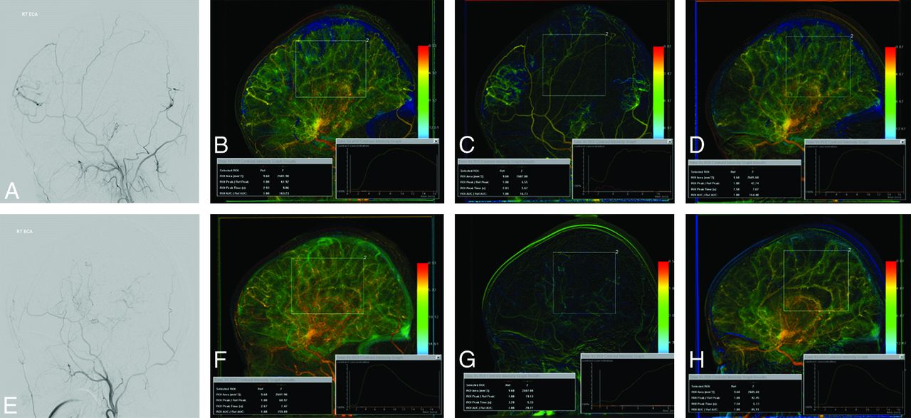
  • Fig 3.
    • Download figure
    • Open in new tab
    • Download powerpoint
    Fig 3.

    A 5-year-old boy with poor clinical outcome after EDAS neovascularization. Comparing pre- (A) and post- (E) operative ECA angiograms shows poor neovascularization after EDAS. Postprocessed pre- (B) and post- (F) operative CCA angiograms show no definite shortening of the TTP (0.0 seconds), with a decrease of the percentage of AUC difference of approximately 2.0% after the operation. About a 41.2% increase of the percentage of AUC difference after the operation is noted when comparing pre- (C) and post- (G) operative ECA angiograms, and about a 13.7% of AUC difference increase is noted when comparing pre- (D) and post- (H) operative ICA angiograms.

Tables

  • Figures
    • View popup
    Table 1:

    Relationships between DSA parameters and clinical outcomes

    GroupExcellent (n = 16)Good (n = 4)Fair (n = 8)Poor (n = 4)P Value
    ΔTTP in CCA (sec)2.021.00.80−0.13.003
    ΔAUC ratio in CCA (%)68.131.59.1−11.3.000
    ΔAUC ratio in ECA (%)1119.8768.2279.5248.7.000
    ΔAUC ratio in ICA (%)−41.0−35.6−44.2−22.3.418
    • View popup
    Table 2:

    Relationships between DSA parameters and angiographic outcomes

    GroupGood (n = 16)Fair (n = 7)Poor (n = 9)P Value
    ΔTTP in CCA (sec)2.010.970.49.005
    ΔAUC ratio in CCA (%)64.529.80.4.000
    ΔAUC ratio in ECA (%)1065.9753.1210.5.000
    ΔAUC ratio in ICA (%)−41.0−42.4−32.2.424
    • View popup
    Table 3:

    Cutoff values between favorable and unfavorable groups

    ParameterCriterionSensitivity (%)Specificity (%)
    ΔTTP in CCA (sec)>1.2781.491.7
    ΔAUC ratio in CCA (%)>33.581100
    ΔAUC ratio in ECA (%)>50485.392
    ΔAUC ratio in ICA (%)≤20.285.933.4
PreviousNext
Back to top

In this issue

American Journal of Neuroradiology: 37 (5)
American Journal of Neuroradiology
Vol. 37, Issue 5
1 May 2016
  • Table of Contents
  • Index by author
  • Complete Issue (PDF)
Advertisement
Print
Download PDF
Email Article

Thank you for your interest in spreading the word on American Journal of Neuroradiology.

NOTE: We only request your email address so that the person you are recommending the page to knows that you wanted them to see it, and that it is not junk mail. We do not capture any email address.

Enter multiple addresses on separate lines or separate them with commas.
Quantitative Assessment of Neovascularization after Indirect Bypass Surgery: Color-Coded Digital Subtraction Angiography in Pediatric Moyamoya Disease
(Your Name) has sent you a message from American Journal of Neuroradiology
(Your Name) thought you would like to see the American Journal of Neuroradiology web site.
CAPTCHA
This question is for testing whether or not you are a human visitor and to prevent automated spam submissions.
Cite this article
H.-H. Cho, J.-E. Cheon, S.-K. Kim, Y.H. Choi, I.-O. Kim, W.S. Kim, S.-M. Lee, S.K. You, S.-M. Shin
Quantitative Assessment of Neovascularization after Indirect Bypass Surgery: Color-Coded Digital Subtraction Angiography in Pediatric Moyamoya Disease
American Journal of Neuroradiology May 2016, 37 (5) 932-938; DOI: 10.3174/ajnr.A4614

Citation Manager Formats

  • BibTeX
  • Bookends
  • EasyBib
  • EndNote (tagged)
  • EndNote 8 (xml)
  • Medlars
  • Mendeley
  • Papers
  • RefWorks Tagged
  • Ref Manager
  • RIS
  • Zotero
0 Responses
Respond to this article
Share
Bookmark this article
Quantitative Assessment of Neovascularization after Indirect Bypass Surgery: Color-Coded Digital Subtraction Angiography in Pediatric Moyamoya Disease
H.-H. Cho, J.-E. Cheon, S.-K. Kim, Y.H. Choi, I.-O. Kim, W.S. Kim, S.-M. Lee, S.K. You, S.-M. Shin
American Journal of Neuroradiology May 2016, 37 (5) 932-938; DOI: 10.3174/ajnr.A4614
del.icio.us logo Twitter logo Facebook logo Mendeley logo
  • Tweet Widget
  • Facebook Like
  • Google Plus One
Purchase

Jump to section

  • Article
    • Abstract
    • ABBREVIATIONS:
    • Materials and Methods
    • Results
    • Discussion
    • Conclusions
    • Acknowledgments
    • References
  • Figures & Data
  • Info & Metrics
  • Responses
  • References
  • PDF

Related Articles

  • No related articles found.
  • PubMed
  • Google Scholar

Cited By...

  • Quantitative angiographic haemodynamic evaluation of bypasses for complex aneurysms: a preliminary study
  • Association of intracranial arteriovenous malformation embolization with more rapid rate of perfusion in the peri-nidal region on color-coded quantitative digital subtraction angiography
  • Crossref
  • Google Scholar

This article has not yet been cited by articles in journals that are participating in Crossref Cited-by Linking.

More in this TOC Section

Pediatric Neuroimaging

  • Frontal Paraventricular Cysts
  • Sodium MRI in Pediatric Brain Tumors
  • FRACTURE MR in Congenital Vertebral Anomalies
Show more Pediatric Neuroimaging

Neurointervention

  • Rescue Reentry in Carotid Near-Occlusion
  • Contour Neurovascular System: Five Year Follow Up
  • Effect of SARS-CoV2 on Endovascular Thrombectomy
Show more Neurointervention

Similar Articles

Advertisement

Indexed Content

  • Current Issue
  • Accepted Manuscripts
  • Article Preview
  • Past Issues
  • Editorials
  • Editor's Choice
  • Fellows' Journal Club
  • Letters to the Editor
  • Video Articles

Cases

  • Case Collection
  • Archive - Case of the Week
  • Archive - Case of the Month
  • Archive - Classic Case

More from AJNR

  • Trainee Corner
  • Imaging Protocols
  • MRI Safety Corner
  • Book Reviews

Multimedia

  • AJNR Podcasts
  • AJNR Scantastics

Resources

  • Turnaround Time
  • Submit a Manuscript
  • Submit a Video Article
  • Submit an eLetter to the Editor/Response
  • Manuscript Submission Guidelines
  • Statistical Tips
  • Fast Publishing of Accepted Manuscripts
  • Graphical Abstract Preparation
  • Imaging Protocol Submission
  • Evidence-Based Medicine Level Guide
  • Publishing Checklists
  • Author Policies
  • Become a Reviewer/Academy of Reviewers
  • News and Updates

About Us

  • About AJNR
  • Editorial Board
  • Editorial Board Alumni
  • Alerts
  • Permissions
  • Not an AJNR Subscriber? Join Now
  • Advertise with Us
  • Librarian Resources
  • Feedback
  • Terms and Conditions
  • AJNR Editorial Board Alumni

American Society of Neuroradiology

  • Not an ASNR Member? Join Now

© 2025 by the American Society of Neuroradiology All rights, including for text and data mining, AI training, and similar technologies, are reserved.
Print ISSN: 0195-6108 Online ISSN: 1936-959X

Powered by HighWire