Advertisement

Main menu

  • Home
  • Content
    • Current Issue
    • Accepted Manuscripts
    • Article Preview
    • Past Issue Archive
    • Video Articles
    • AJNR Case Collection
    • Case of the Week Archive
    • Case of the Month Archive
    • Classic Case Archive
  • Special Collections
    • AJNR Awards
    • Low-Field MRI
    • Alzheimer Disease
    • ASNR Foundation Special Collection
    • Photon-Counting CT
    • View All
  • Multimedia
    • AJNR Podcasts
    • AJNR SCANtastic
    • Trainee Corner
    • MRI Safety Corner
    • Imaging Protocols
  • For Authors
    • Submit a Manuscript
    • Submit a Video Article
    • Submit an eLetter to the Editor/Response
    • Manuscript Submission Guidelines
    • Statistical Tips
    • Fast Publishing of Accepted Manuscripts
    • Graphical Abstract Preparation
    • Imaging Protocol Submission
    • Author Policies
  • About Us
    • About AJNR
    • Editorial Board
    • Editorial Board Alumni
  • More
    • Become a Reviewer/Academy of Reviewers
    • Subscribers
    • Permissions
    • Alerts
    • Feedback
    • Advertisers
    • ASNR Home

User menu

  • Alerts
  • Log in

Search

  • Advanced search
American Journal of Neuroradiology

American Journal of Neuroradiology

ASHNR American Society of Functional Neuroradiology ASHNR American Society of Pediatric Neuroradiology ASSR
  • Alerts
  • Log in

Advanced Search

  • Home
  • Content
    • Current Issue
    • Accepted Manuscripts
    • Article Preview
    • Past Issue Archive
    • Video Articles
    • AJNR Case Collection
    • Case of the Week Archive
    • Case of the Month Archive
    • Classic Case Archive
  • Special Collections
    • AJNR Awards
    • Low-Field MRI
    • Alzheimer Disease
    • ASNR Foundation Special Collection
    • Photon-Counting CT
    • View All
  • Multimedia
    • AJNR Podcasts
    • AJNR SCANtastic
    • Trainee Corner
    • MRI Safety Corner
    • Imaging Protocols
  • For Authors
    • Submit a Manuscript
    • Submit a Video Article
    • Submit an eLetter to the Editor/Response
    • Manuscript Submission Guidelines
    • Statistical Tips
    • Fast Publishing of Accepted Manuscripts
    • Graphical Abstract Preparation
    • Imaging Protocol Submission
    • Author Policies
  • About Us
    • About AJNR
    • Editorial Board
    • Editorial Board Alumni
  • More
    • Become a Reviewer/Academy of Reviewers
    • Subscribers
    • Permissions
    • Alerts
    • Feedback
    • Advertisers
    • ASNR Home
  • Follow AJNR on Twitter
  • Visit AJNR on Facebook
  • Follow AJNR on Instagram
  • Join AJNR on LinkedIn
  • RSS Feeds

AJNR Awards, New Junior Editors, and more. Read the latest AJNR updates

Research ArticlePediatric Neuroimaging

Asymptomatic Interhypothalamic Adhesions in Children

F.N. Ahmed, N.V. Stence and D.M. Mirsky
American Journal of Neuroradiology April 2016, 37 (4) 726-729; DOI: https://doi.org/10.3174/ajnr.A4602
F.N. Ahmed
aFrom the Department of Radiology (F.N.A.), University of Colorado Anschutz Medical Campus, Aurora, Colorado
  • Find this author on Google Scholar
  • Find this author on PubMed
  • Search for this author on this site
  • ORCID record for F.N. Ahmed
N.V. Stence
bDepartment of Radiology (N.V.S., D.M.M.), Children's Hospital Colorado, Aurora, Colorado.
  • Find this author on Google Scholar
  • Find this author on PubMed
  • Search for this author on this site
  • ORCID record for N.V. Stence
D.M. Mirsky
bDepartment of Radiology (N.V.S., D.M.M.), Children's Hospital Colorado, Aurora, Colorado.
  • Find this author on Google Scholar
  • Find this author on PubMed
  • Search for this author on this site
  • ORCID record for D.M. Mirsky
  • Article
  • Figures & Data
  • Info & Metrics
  • Responses
  • References
  • PDF
Loading

Article Figures & Data

Figures

  • Tables
  • Fig 1.
    • Download figure
    • Open in new tab
    • Download powerpoint
    Fig 1.

    A band of tissue spanning the anterior recess of the third ventricle that appeared linear in the axial and coronal planes and nodular in the sagittal plane as seen on 3T T1WI. A and B, A flat band of tissue behind the lamina terminalis. C and D, A flat band of tissue above the tuber cinereum. E and F, A nodular band of tissue contacting both lamina terminalis and tuber cinereum.
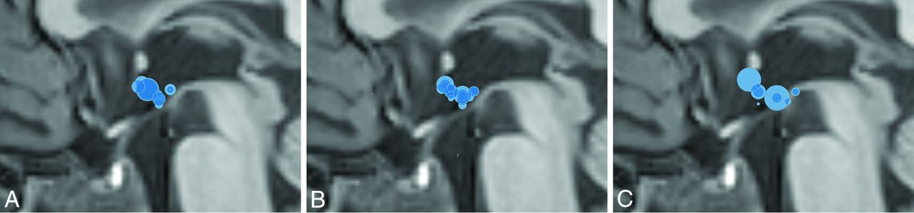
  • Fig 2.
    • Download figure
    • Open in new tab
    • Download powerpoint
    Fig 2.

    Position maps of the interhypothalamic adhesions. A, All IHAs in our study. B, Asymptomatic IHAs. C, Symptomatic IHAs.

Tables

  • Figures
    • View popup
    Table 1:

    IHA demographics and imaging indications of the cohort

    No Referable SymptomsSymptomaticP Value
    Total4017
    Demographics
        Male207.54
        Female2010.54
    Average age (yr)6.110.2.30
    Age range8 Days to 20 years11 Months to 17 years
    Imaging indication
        Seizures95.58
        Developmental delays83.84
        Headaches/migraines40.18
        Vision problems41.62
        Hearing problems20.35
        Trauma20.35
        Chiari diagnosed in utero20.35
        Short stature12.15
        Fetal alcohol syndrome11.53
        Other8a5b
    • ↵a Tongue hemihypertrophy, myelomeningocele, scalp mass, Horner syndrome, multiple sclerosis, lower extremity pain, and spasms.

    • ↵b Craniosynostosis, hydrocephalus on fetal ultrasound, panhypopituitarism, and hypothyroidism.

    • View popup
    Table 2:

    Imaging characteristics of the cohort: structural features by imaging

    No Referable SymptomsSymptomaticP Value
    IHA volume (mm3)48.878.04
    Otherwise normal144.39
    Gray matter heterotopias176.61
    Septo-optic dysplasia04.001
    Chiari malformation30.25
    Periventricular leukomalacia21.89
    Neoplasm20.35
    Polymicrogyria01.12
    Other8a6b
    • ↵a Dilated perivascular spaces, multiple sclerosis with a velum interpositum, right cochlear nerve hypoplasia, right cerebral closed lip schizencephaly, vermian hypoplasia, sinus pericranii, and 2 isolated cases of cavum septum pellucidum.

    • ↵b Abnormal sulcation, transmantle cortical dysplasia, frontonasal dysplasia, inferior vermian hypoplasia with absent septum pellucidum, cavum septum pellucidum (with septo-optic dysplasia), and velum interpositum.

    • View popup
    Table 3:

    Clinical symptoms of the cohort

    No Referable SymptomsSymptomaticP Value
    Healthy18a0<.01
    Seizures9b5.58
    Endocrine problems03<.01
    Mild developmental delays86.22
    Severe developmental delays08<.01
    Heart anomalies30.25
    Sensory hearing loss20.35
    Premature birthb52.94
    • ↵a Four of these individuals had initially presented with headaches, and 1, with vertigo, all considered to be isolated issues without ongoing symptoms at the time of this review. All 3 individuals with Chiari malformations were included in the group lacking referable symptoms. Two additional patients had presented with trauma and were healthy after their other musculoskeletal injuries healed. Two more of these patients were evaluated for unilateral sensory hearing loss but were considered clinically healthy otherwise. An individual with a low-grade glial neoplasm had originally presented with a seizure but had no other symptoms after resection. One individual with multiple sclerosis had 2 episodes of lower extremity weakness and tingling that had resolved. The remaining patients lacking symptoms referable to the IHA had presenting symptoms that resolved with treatment or conservative measures (tongue hemihypertrophy with an arteriovascular malformation, scalp mass [sinus pericranii], Horner syndrome, lower extremity pain, and spasms).

    • ↵b Born at 26–34 weeks' gestation.

PreviousNext
Back to top

In this issue

American Journal of Neuroradiology: 37 (4)
American Journal of Neuroradiology
Vol. 37, Issue 4
1 Apr 2016
  • Table of Contents
  • Index by author
  • Complete Issue (PDF)
Advertisement
Print
Download PDF
Email Article

Thank you for your interest in spreading the word on American Journal of Neuroradiology.

NOTE: We only request your email address so that the person you are recommending the page to knows that you wanted them to see it, and that it is not junk mail. We do not capture any email address.

Enter multiple addresses on separate lines or separate them with commas.
Asymptomatic Interhypothalamic Adhesions in Children
(Your Name) has sent you a message from American Journal of Neuroradiology
(Your Name) thought you would like to see the American Journal of Neuroradiology web site.
CAPTCHA
This question is for testing whether or not you are a human visitor and to prevent automated spam submissions.
Cite this article
F.N. Ahmed, N.V. Stence, D.M. Mirsky
Asymptomatic Interhypothalamic Adhesions in Children
American Journal of Neuroradiology Apr 2016, 37 (4) 726-729; DOI: 10.3174/ajnr.A4602

Citation Manager Formats

  • BibTeX
  • Bookends
  • EasyBib
  • EndNote (tagged)
  • EndNote 8 (xml)
  • Medlars
  • Mendeley
  • Papers
  • RefWorks Tagged
  • Ref Manager
  • RIS
  • Zotero
0 Responses
Respond to this article
Share
Bookmark this article
Asymptomatic Interhypothalamic Adhesions in Children
F.N. Ahmed, N.V. Stence, D.M. Mirsky
American Journal of Neuroradiology Apr 2016, 37 (4) 726-729; DOI: 10.3174/ajnr.A4602
del.icio.us logo Twitter logo Facebook logo Mendeley logo
  • Tweet Widget
  • Facebook Like
  • Google Plus One
Purchase

Jump to section

  • Article
    • Abstract
    • ABBREVIATIONS:
    • Materials and Methods
    • Results
    • Discussion
    • Conclusions
    • Footnotes
    • REFERENCES
  • Figures & Data
  • Info & Metrics
  • Responses
  • References
  • PDF

Related Articles

  • No related articles found.
  • PubMed
  • Google Scholar

Cited By...

  • CDK5RAP2 primary microcephaly is associated with hypothalamic, retinal and cochlear developmental defects
  • Thalamic Massa Intermedia in Children with and without Midline Brain Malformations
  • Crossref
  • Google Scholar

This article has not yet been cited by articles in journals that are participating in Crossref Cited-by Linking.

More in this TOC Section

  • Frontal Paraventricular Cysts
  • Sodium MRI in Pediatric Brain Tumors
  • FRACTURE MR in Congenital Vertebral Anomalies
Show more Pediatric Neuroimaging

Similar Articles

Advertisement

Indexed Content

  • Current Issue
  • Accepted Manuscripts
  • Article Preview
  • Past Issues
  • Editorials
  • Editor's Choice
  • Fellows' Journal Club
  • Letters to the Editor
  • Video Articles

Cases

  • Case Collection
  • Archive - Case of the Week
  • Archive - Case of the Month
  • Archive - Classic Case

More from AJNR

  • Trainee Corner
  • Imaging Protocols
  • MRI Safety Corner
  • Book Reviews

Multimedia

  • AJNR Podcasts
  • AJNR Scantastics

Resources

  • Turnaround Time
  • Submit a Manuscript
  • Submit a Video Article
  • Submit an eLetter to the Editor/Response
  • Manuscript Submission Guidelines
  • Statistical Tips
  • Fast Publishing of Accepted Manuscripts
  • Graphical Abstract Preparation
  • Imaging Protocol Submission
  • Evidence-Based Medicine Level Guide
  • Publishing Checklists
  • Author Policies
  • Become a Reviewer/Academy of Reviewers
  • News and Updates

About Us

  • About AJNR
  • Editorial Board
  • Editorial Board Alumni
  • Alerts
  • Permissions
  • Not an AJNR Subscriber? Join Now
  • Advertise with Us
  • Librarian Resources
  • Feedback
  • Terms and Conditions
  • AJNR Editorial Board Alumni

American Society of Neuroradiology

  • Not an ASNR Member? Join Now

© 2025 by the American Society of Neuroradiology All rights, including for text and data mining, AI training, and similar technologies, are reserved.
Print ISSN: 0195-6108 Online ISSN: 1936-959X

Powered by HighWire