Advertisement

Main menu

  • Home
  • Content
    • Current Issue
    • Accepted Manuscripts
    • Article Preview
    • Past Issue Archive
    • Video Articles
    • AJNR Case Collection
    • Case of the Week Archive
    • Case of the Month Archive
    • Classic Case Archive
  • Special Collections
    • AJNR Awards
    • Low-Field MRI
    • Alzheimer Disease
    • ASNR Foundation Special Collection
    • Photon-Counting CT
    • View All
  • Multimedia
    • AJNR Podcasts
    • AJNR SCANtastic
    • Trainee Corner
    • MRI Safety Corner
    • Imaging Protocols
  • For Authors
    • Submit a Manuscript
    • Submit a Video Article
    • Submit an eLetter to the Editor/Response
    • Manuscript Submission Guidelines
    • Statistical Tips
    • Fast Publishing of Accepted Manuscripts
    • Graphical Abstract Preparation
    • Imaging Protocol Submission
    • Author Policies
  • About Us
    • About AJNR
    • Editorial Board
    • Editorial Board Alumni
  • More
    • Become a Reviewer/Academy of Reviewers
    • Subscribers
    • Permissions
    • Alerts
    • Feedback
    • Advertisers
    • ASNR Home

User menu

  • Alerts
  • Log in

Search

  • Advanced search
American Journal of Neuroradiology

American Journal of Neuroradiology

ASHNR American Society of Functional Neuroradiology ASHNR American Society of Pediatric Neuroradiology ASSR
  • Alerts
  • Log in

Advanced Search

  • Home
  • Content
    • Current Issue
    • Accepted Manuscripts
    • Article Preview
    • Past Issue Archive
    • Video Articles
    • AJNR Case Collection
    • Case of the Week Archive
    • Case of the Month Archive
    • Classic Case Archive
  • Special Collections
    • AJNR Awards
    • Low-Field MRI
    • Alzheimer Disease
    • ASNR Foundation Special Collection
    • Photon-Counting CT
    • View All
  • Multimedia
    • AJNR Podcasts
    • AJNR SCANtastic
    • Trainee Corner
    • MRI Safety Corner
    • Imaging Protocols
  • For Authors
    • Submit a Manuscript
    • Submit a Video Article
    • Submit an eLetter to the Editor/Response
    • Manuscript Submission Guidelines
    • Statistical Tips
    • Fast Publishing of Accepted Manuscripts
    • Graphical Abstract Preparation
    • Imaging Protocol Submission
    • Author Policies
  • About Us
    • About AJNR
    • Editorial Board
    • Editorial Board Alumni
  • More
    • Become a Reviewer/Academy of Reviewers
    • Subscribers
    • Permissions
    • Alerts
    • Feedback
    • Advertisers
    • ASNR Home
  • Follow AJNR on Twitter
  • Visit AJNR on Facebook
  • Follow AJNR on Instagram
  • Join AJNR on LinkedIn
  • RSS Feeds

AJNR Awards, New Junior Editors, and more. Read the latest AJNR updates

Research ArticleAdult Brain
Open Access

Computational Identification of Tumor Anatomic Location Associated with Survival in 2 Large Cohorts of Human Primary Glioblastomas

T.T. Liu, A.S. Achrol, L.A. Mitchell, W.A. Du, J.J. Loya, S.A. Rodriguez, A. Feroze, E.M. Westbroek, K.W. Yeom, J.M. Stuart, S.D. Chang, G.R. Harsh and D.L. Rubin
American Journal of Neuroradiology April 2016, 37 (4) 621-628; DOI: https://doi.org/10.3174/ajnr.A4631
T.T. Liu
aFrom the Stanford Center for Biomedical Informatics Research and Biomedical Informatics Training Program (T.T.L., D.L.R.)
bDepartment of Radiology (T.T.L., L.A.M., W.A.D., K.W.Y., D.L.R.)
  • Find this author on Google Scholar
  • Find this author on PubMed
  • Search for this author on this site
  • ORCID record for T.T. Liu
A.S. Achrol
cStanford Institute for Neuro-Innovation and Translational Neurosciences (A.S.A.)
dInstitute for Stem Cell Biology and Regenerative Medicine (A.S.A.)
eDepartment of Neurosurgery (A.S.A., J.J.L., S.A.R., E.M.W., S.D.C., G.R.H.), Stanford University School of Medicine, Stanford, California
  • Find this author on Google Scholar
  • Find this author on PubMed
  • Search for this author on this site
  • ORCID record for A.S. Achrol
L.A. Mitchell
bDepartment of Radiology (T.T.L., L.A.M., W.A.D., K.W.Y., D.L.R.)
  • Find this author on Google Scholar
  • Find this author on PubMed
  • Search for this author on this site
  • ORCID record for L.A. Mitchell
W.A. Du
bDepartment of Radiology (T.T.L., L.A.M., W.A.D., K.W.Y., D.L.R.)
  • Find this author on Google Scholar
  • Find this author on PubMed
  • Search for this author on this site
  • ORCID record for W.A. Du
J.J. Loya
eDepartment of Neurosurgery (A.S.A., J.J.L., S.A.R., E.M.W., S.D.C., G.R.H.), Stanford University School of Medicine, Stanford, California
  • Find this author on Google Scholar
  • Find this author on PubMed
  • Search for this author on this site
  • ORCID record for J.J. Loya
S.A. Rodriguez
eDepartment of Neurosurgery (A.S.A., J.J.L., S.A.R., E.M.W., S.D.C., G.R.H.), Stanford University School of Medicine, Stanford, California
  • Find this author on Google Scholar
  • Find this author on PubMed
  • Search for this author on this site
  • ORCID record for S.A. Rodriguez
A. Feroze
fDepartment of Neurological Surgery (A.F.), University of Washington School of Medicine, Seattle, Washington
  • Find this author on Google Scholar
  • Find this author on PubMed
  • Search for this author on this site
  • ORCID record for A. Feroze
E.M. Westbroek
eDepartment of Neurosurgery (A.S.A., J.J.L., S.A.R., E.M.W., S.D.C., G.R.H.), Stanford University School of Medicine, Stanford, California
  • Find this author on Google Scholar
  • Find this author on PubMed
  • Search for this author on this site
  • ORCID record for E.M. Westbroek
K.W. Yeom
bDepartment of Radiology (T.T.L., L.A.M., W.A.D., K.W.Y., D.L.R.)
  • Find this author on Google Scholar
  • Find this author on PubMed
  • Search for this author on this site
  • ORCID record for K.W. Yeom
J.M. Stuart
gBiomolecular Engineering (J.M.S.), University of California Santa Cruz, Santa Cruz, California.
  • Find this author on Google Scholar
  • Find this author on PubMed
  • Search for this author on this site
  • ORCID record for J.M. Stuart
S.D. Chang
eDepartment of Neurosurgery (A.S.A., J.J.L., S.A.R., E.M.W., S.D.C., G.R.H.), Stanford University School of Medicine, Stanford, California
  • Find this author on Google Scholar
  • Find this author on PubMed
  • Search for this author on this site
G.R. Harsh IV
eDepartment of Neurosurgery (A.S.A., J.J.L., S.A.R., E.M.W., S.D.C., G.R.H.), Stanford University School of Medicine, Stanford, California
  • Find this author on Google Scholar
  • Find this author on PubMed
  • Search for this author on this site
  • ORCID record for G.R. Harsh IV
D.L. Rubin
aFrom the Stanford Center for Biomedical Informatics Research and Biomedical Informatics Training Program (T.T.L., D.L.R.)
bDepartment of Radiology (T.T.L., L.A.M., W.A.D., K.W.Y., D.L.R.)
  • Find this author on Google Scholar
  • Find this author on PubMed
  • Search for this author on this site
  • ORCID record for D.L. Rubin
  • Article
  • Figures & Data
  • Supplemental
  • Info & Metrics
  • Responses
  • References
  • PDF
Loading

Article Figures & Data

Figures

  • Tables
  • Fig 1.
    • Download figure
    • Open in new tab
    • Download powerpoint
    Fig 1.

    An overview of the image-processing pipeline and model training and validation procedure to identify locations associated with survival. A, The image-processing pipeline is applied to both training (Stanford University Medical Center) and validation (TCGA) cohorts. B, Algorithm training identifies anatomic regions associated with survival, which is validated in the TCGA cohort. The training algorithm using the threshold-free cluster enhancement method takes as the input group labels dichotomized by survival outcome and the superimposed tumor heat map of the Stanford University Medical Center patient cohort analyzed in the image-processing pipeline; the pipeline outputs anatomic regions significantly associated with the 2 survival groups, which are used to classify the TCGA validation set into a poor survival group and a good survival group on the basis of tumor regions present or absent in the prognostic region.

  • Fig 2.
    • Download figure
    • Open in new tab
    • Download powerpoint
    Fig 2.

    Axial, sagittal, and coronal section views of the region associated with poor survival in the training cohort (false discovery rate, P < .05). The cluster of voxels associated with poor survival was localized in the occipitotemporal periventricular white matter in the right hemisphere (right periatrial).
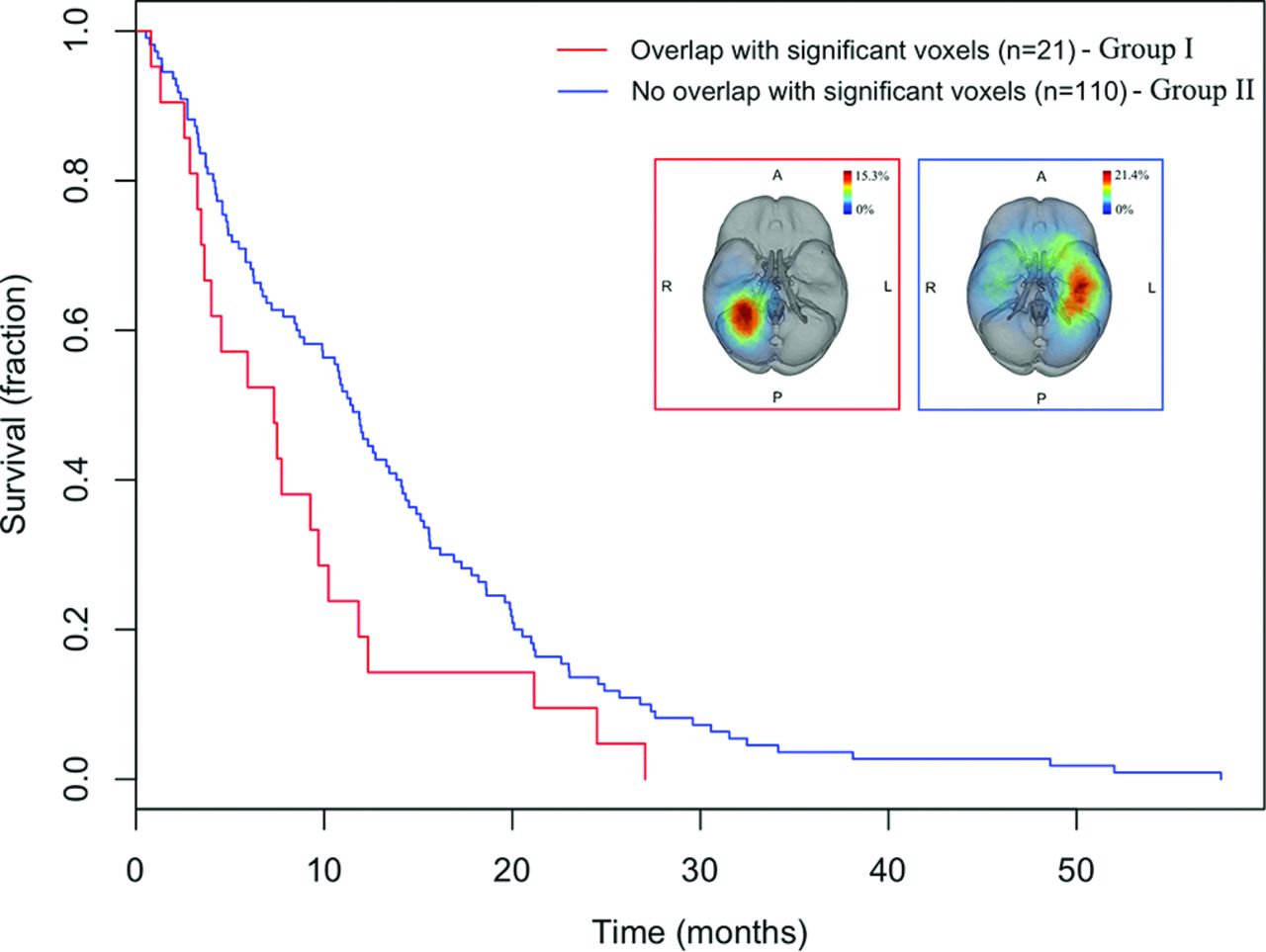
  • Fig 3.
    • Download figure
    • Open in new tab
    • Download powerpoint
    Fig 3.

    Kaplan-Meier survival curves of patients with GBMs depict decreased overall survival in TCGA patients with an overlap (group I) versus nonoverlap (group II) with the voxels significantly associated with survival identified from the training cohort in the test cohort (log-rank test, P = .034).

  • Fig 4.
    • Download figure
    • Open in new tab
    • Download powerpoint
    Fig 4.

    Axial postcontrast T1-weighted images of 4 patients from group II. A, A 69-year-old man with a right parietal GBM and an overall survival of 27 months. B, A 49-year-old man with a right temporal GBM and an overall survival of 25 months. C, A 63-year-old man with a left temporal GBM and an overall survival of 21 months. D, A 36-year-old woman with a left frontal GBM, an overall survival of 6 months, and the smallest tumor volume in the group of intermediate tumors.

Tables

  • Figures
    • View popup
    Table 1:

    Summary of clinical variables in the training cohort

    Survival Groups
    Poor (OS ≤ 11 mo)Medium (11 < OS < 17 mo)Good (OS ≥ 17 mo)
    Total No.1523863
    Median age (yr)69.057.259.5
    % Male61.260.557.1
    Median survival (mo)4.114.121.2
    Mean survival (mo)4.814.026.3
    Mean CEL volume (cm3)34.134.028.9
    STR/GTR/biopsy only (No.)53/13/8617/8/1317/36/10
    • Note:—STR indicates subtotal resection; GTR, gross total resection.

    • View popup
    Table 2:

    Univariate and multivariate Cox survival analysis of clinical variables

    Univariate CoxMultivariate Cox
    HR (95% CI)P ValueHR (95% CI)P Value
    TCGA test cohorta
        Age younger than 64 yr0.19 (0.26–0.55)4.1e–7b0.36 (0.24–0.53)3.7e–7b
        Male sex1.05 (0.73–1.50).79––
        CEL tumor volume (cm3)
            Large1.68 (1.10–2.54).015b1.80 (1.18–2.75).0064b
            Intermediate––––
            Small0.90 (0.58–1.37).621.11 (0.71–1.71).65
        Right laterality0.927 (0.65–1.32).67––
        STR2.52 (0.73–8.68).14––
        Tumor location = right periatrial location1.66 (1.03–2.67).036b2.0 (1.01–2.64).045b
    Restricted set of intermediate and large tumorsc
        Age younger than 64 yr0.19 (0.26–0.55)4.1e–70.46 (0.30–0.72).00062
        Tumor location = right periatrial location1.84 (1.10–3.08).0191.87 (1.11–3.15).018
    • Note:—HR indicates hazard ratio; STR, subtotal resection.

    • ↵a Age, CEL tumor volume, and tumor location were independently significant in multivariate analysis (overall P < 3.52e–8). Surgical resection is subtotal resection or gross total resection, available for 24 cases in the TCGA test cohort.

    • ↵b P value < .05, indicating the variable is significant.

    • ↵c Tumor location remained significant in Cox analysis performed on the restricted set of intermediate and large tumors.

PreviousNext
Back to top

In this issue

American Journal of Neuroradiology: 37 (4)
American Journal of Neuroradiology
Vol. 37, Issue 4
1 Apr 2016
  • Table of Contents
  • Index by author
  • Complete Issue (PDF)
Advertisement
Print
Download PDF
Email Article

Thank you for your interest in spreading the word on American Journal of Neuroradiology.

NOTE: We only request your email address so that the person you are recommending the page to knows that you wanted them to see it, and that it is not junk mail. We do not capture any email address.

Enter multiple addresses on separate lines or separate them with commas.
Computational Identification of Tumor Anatomic Location Associated with Survival in 2 Large Cohorts of Human Primary Glioblastomas
(Your Name) has sent you a message from American Journal of Neuroradiology
(Your Name) thought you would like to see the American Journal of Neuroradiology web site.
CAPTCHA
This question is for testing whether or not you are a human visitor and to prevent automated spam submissions.
Cite this article
T.T. Liu, A.S. Achrol, L.A. Mitchell, W.A. Du, J.J. Loya, S.A. Rodriguez, A. Feroze, E.M. Westbroek, K.W. Yeom, J.M. Stuart, S.D. Chang, G.R. Harsh, D.L. Rubin
Computational Identification of Tumor Anatomic Location Associated with Survival in 2 Large Cohorts of Human Primary Glioblastomas
American Journal of Neuroradiology Apr 2016, 37 (4) 621-628; DOI: 10.3174/ajnr.A4631

Citation Manager Formats

  • BibTeX
  • Bookends
  • EasyBib
  • EndNote (tagged)
  • EndNote 8 (xml)
  • Medlars
  • Mendeley
  • Papers
  • RefWorks Tagged
  • Ref Manager
  • RIS
  • Zotero
0 Responses
Respond to this article
Share
Bookmark this article
Computational Identification of Tumor Anatomic Location Associated with Survival in 2 Large Cohorts of Human Primary Glioblastomas
T.T. Liu, A.S. Achrol, L.A. Mitchell, W.A. Du, J.J. Loya, S.A. Rodriguez, A. Feroze, E.M. Westbroek, K.W. Yeom, J.M. Stuart, S.D. Chang, G.R. Harsh, D.L. Rubin
American Journal of Neuroradiology Apr 2016, 37 (4) 621-628; DOI: 10.3174/ajnr.A4631
del.icio.us logo Twitter logo Facebook logo Mendeley logo
  • Tweet Widget
  • Facebook Like
  • Google Plus One
Purchase

Jump to section

  • Article
    • Abstract
    • ABBREVIATIONS:
    • Materials and Methods
    • Results
    • Discussion
    • Conclusions
    • Acknowledgments
    • Footnotes
    • REFERENCES
  • Figures & Data
  • Supplemental
  • Info & Metrics
  • Responses
  • References
  • PDF

Related Articles

  • PubMed
  • Google Scholar

Cited By...

  • Overview of prognostic factors in adult glioma; a 10-year experience at a single institution
  • Interactions between ploidy and resource availability shape clonal interference at initiation and recurrence of glioblastoma
  • Study protocol: retrospectively mining multisite clinical data to presymptomatically predict seizure onset for individual patients with Sturge-Weber
  • Glioblastoma heterogeneity and the tumour microenvironment: implications for preclinical research and development of new treatments
  • Crossref
  • Google Scholar

This article has not yet been cited by articles in journals that are participating in Crossref Cited-by Linking.

More in this TOC Section

  • Diagnostic Neuroradiology of Monoclonal Antibodies
  • Clinical Outcomes After Chiari I Decompression
  • Segmentation of Brain Metastases with BLAST
Show more Adult Brain

Similar Articles

Advertisement

Indexed Content

  • Current Issue
  • Accepted Manuscripts
  • Article Preview
  • Past Issues
  • Editorials
  • Editor's Choice
  • Fellows' Journal Club
  • Letters to the Editor
  • Video Articles

Cases

  • Case Collection
  • Archive - Case of the Week
  • Archive - Case of the Month
  • Archive - Classic Case

More from AJNR

  • Trainee Corner
  • Imaging Protocols
  • MRI Safety Corner
  • Book Reviews

Multimedia

  • AJNR Podcasts
  • AJNR Scantastics

Resources

  • Turnaround Time
  • Submit a Manuscript
  • Submit a Video Article
  • Submit an eLetter to the Editor/Response
  • Manuscript Submission Guidelines
  • Statistical Tips
  • Fast Publishing of Accepted Manuscripts
  • Graphical Abstract Preparation
  • Imaging Protocol Submission
  • Evidence-Based Medicine Level Guide
  • Publishing Checklists
  • Author Policies
  • Become a Reviewer/Academy of Reviewers
  • News and Updates

About Us

  • About AJNR
  • Editorial Board
  • Editorial Board Alumni
  • Alerts
  • Permissions
  • Not an AJNR Subscriber? Join Now
  • Advertise with Us
  • Librarian Resources
  • Feedback
  • Terms and Conditions
  • AJNR Editorial Board Alumni

American Society of Neuroradiology

  • Not an ASNR Member? Join Now

© 2025 by the American Society of Neuroradiology All rights, including for text and data mining, AI training, and similar technologies, are reserved.
Print ISSN: 0195-6108 Online ISSN: 1936-959X

Powered by HighWire