Advertisement

Main menu

  • Home
  • Content
    • Current Issue
    • Accepted Manuscripts
    • Article Preview
    • Past Issue Archive
    • Video Articles
    • AJNR Case Collection
    • Case of the Week Archive
    • Case of the Month Archive
    • Classic Case Archive
  • Special Collections
    • AJNR Awards
    • Low-Field MRI
    • Alzheimer Disease
    • ASNR Foundation Special Collection
    • Photon-Counting CT
    • View All
  • Multimedia
    • AJNR Podcasts
    • AJNR SCANtastic
    • Trainee Corner
    • MRI Safety Corner
    • Imaging Protocols
  • For Authors
    • Submit a Manuscript
    • Submit a Video Article
    • Submit an eLetter to the Editor/Response
    • Manuscript Submission Guidelines
    • Statistical Tips
    • Fast Publishing of Accepted Manuscripts
    • Graphical Abstract Preparation
    • Imaging Protocol Submission
    • Author Policies
  • About Us
    • About AJNR
    • Editorial Board
    • Editorial Board Alumni
  • More
    • Become a Reviewer/Academy of Reviewers
    • Subscribers
    • Permissions
    • Alerts
    • Feedback
    • Advertisers
    • ASNR Home

User menu

  • Alerts
  • Log in

Search

  • Advanced search
American Journal of Neuroradiology

American Journal of Neuroradiology

ASHNR American Society of Functional Neuroradiology ASHNR American Society of Pediatric Neuroradiology ASSR
  • Alerts
  • Log in

Advanced Search

  • Home
  • Content
    • Current Issue
    • Accepted Manuscripts
    • Article Preview
    • Past Issue Archive
    • Video Articles
    • AJNR Case Collection
    • Case of the Week Archive
    • Case of the Month Archive
    • Classic Case Archive
  • Special Collections
    • AJNR Awards
    • Low-Field MRI
    • Alzheimer Disease
    • ASNR Foundation Special Collection
    • Photon-Counting CT
    • View All
  • Multimedia
    • AJNR Podcasts
    • AJNR SCANtastic
    • Trainee Corner
    • MRI Safety Corner
    • Imaging Protocols
  • For Authors
    • Submit a Manuscript
    • Submit a Video Article
    • Submit an eLetter to the Editor/Response
    • Manuscript Submission Guidelines
    • Statistical Tips
    • Fast Publishing of Accepted Manuscripts
    • Graphical Abstract Preparation
    • Imaging Protocol Submission
    • Author Policies
  • About Us
    • About AJNR
    • Editorial Board
    • Editorial Board Alumni
  • More
    • Become a Reviewer/Academy of Reviewers
    • Subscribers
    • Permissions
    • Alerts
    • Feedback
    • Advertisers
    • ASNR Home
  • Follow AJNR on Twitter
  • Visit AJNR on Facebook
  • Follow AJNR on Instagram
  • Join AJNR on LinkedIn
  • RSS Feeds

AJNR Awards, New Junior Editors, and more. Read the latest AJNR updates

Getting new auth cookie, if you see this message a lot, tell someone!
Research ArticlePEDIATRICS
Open Access

MRI Evaluation of Non-Necrotic T2-Hyperintense Foci in Pediatric Diffuse Intrinsic Pontine Glioma

O. Clerk-Lamalice, W.E. Reddick, X. Li, Y. Li, A. Edwards, J.O. Glass and Z. Patay
American Journal of Neuroradiology October 2016, 37 (10) 1930-1937; DOI: https://doi.org/10.3174/ajnr.A4814
O. Clerk-Lamalice
aFrom the Departments of Diagnostic Imaging (O.C.-L., W.E.R., A.E., J.O.G., Z.P.)
  • Find this author on Google Scholar
  • Find this author on PubMed
  • Search for this author on this site
  • ORCID record for O. Clerk-Lamalice
W.E. Reddick
aFrom the Departments of Diagnostic Imaging (O.C.-L., W.E.R., A.E., J.O.G., Z.P.)
  • Find this author on Google Scholar
  • Find this author on PubMed
  • Search for this author on this site
  • ORCID record for W.E. Reddick
X. Li
bBiostatistics (X.L., Y.L.), St. Jude Children's Research Hospital, Memphis, Tennessee.
  • Find this author on Google Scholar
  • Find this author on PubMed
  • Search for this author on this site
  • ORCID record for X. Li
Y. Li
bBiostatistics (X.L., Y.L.), St. Jude Children's Research Hospital, Memphis, Tennessee.
  • Find this author on Google Scholar
  • Find this author on PubMed
  • Search for this author on this site
  • ORCID record for Y. Li
A. Edwards
aFrom the Departments of Diagnostic Imaging (O.C.-L., W.E.R., A.E., J.O.G., Z.P.)
  • Find this author on Google Scholar
  • Find this author on PubMed
  • Search for this author on this site
  • ORCID record for A. Edwards
J.O. Glass
aFrom the Departments of Diagnostic Imaging (O.C.-L., W.E.R., A.E., J.O.G., Z.P.)
  • Find this author on Google Scholar
  • Find this author on PubMed
  • Search for this author on this site
  • ORCID record for J.O. Glass
Z. Patay
aFrom the Departments of Diagnostic Imaging (O.C.-L., W.E.R., A.E., J.O.G., Z.P.)
  • Find this author on Google Scholar
  • Find this author on PubMed
  • Search for this author on this site
  • ORCID record for Z. Patay
  • Article
  • Figures & Data
  • Info & Metrics
  • Responses
  • References
  • PDF
Loading

Article Figures & Data

Figures

  • Tables
  • Fig 1.
    • Download figure
    • Open in new tab
    • Download powerpoint
    Fig 1.

    Axial MR images centered on the pons and showing non-necrotic T2HrF (long arrow). T2-weighted image (A), ADC map (B), T1-weighted postcontrast subtraction image (C), and CBV map (D). These images show a well-defined, fairly voluminous T2HrF within the left hemipons (a smaller similar lesion may be present on the right side, too), which is associated with mass effect, slightly increased signal in ADC (B), lack of contrast enhancement after IV gadolinium injection (C), and moderately increased CBV (D).

  • Fig 2.
    • Download figure
    • Open in new tab
    • Download powerpoint
    Fig 2.

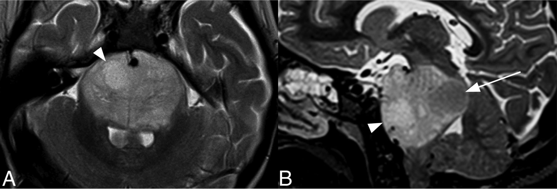
    Axial (A) and sagittal (B) T2-weighted MR images of a DIPG with both non-necrotic T2HrF (arrowhead) and T2HoF (long arrow).

  • Fig 3.
    • Download figure
    • Open in new tab
    • Download powerpoint
    Fig 3.

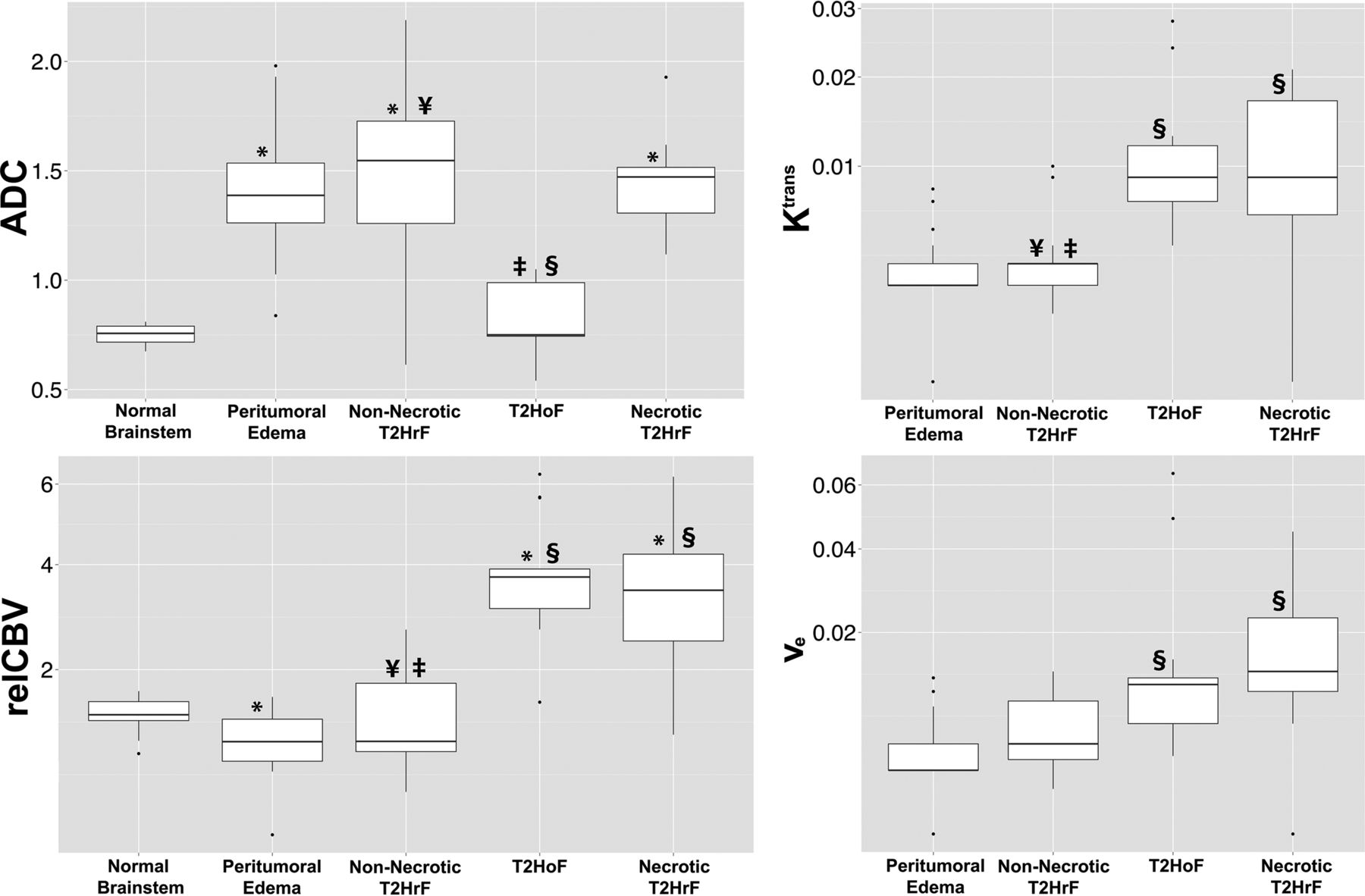
    Boxplots of ADC, rCBV, Ktrans, and ve for the different ROI types analyzed in DIPG. The y-axis of boxplots was rescaled for rCBV, Ktrans, and ve. Error bars represent SDs. Statistical differences between groups (P < .05) are signified as follows: The asterisk indicates normal brain stem, ¥, T2HoF; ‡, necrotic T2HrF; §, edema.

  • Fig 4.
    • Download figure
    • Open in new tab
    • Download powerpoint
    Fig 4.

    Feature comparison of the 4 ROIs. A, Normal brain stem. B, A non-necrotic T2HrF is a well-circumscribed intratumoral area exhibiting high T2 signal and is often associated with local mass effect on surrounding structures, shown by splaying transverse pontocerebellar fibers. C, T2HoF are characterized by low T2 signal and are locally expansile. D, Necrotic T2HrF exhibit irregular margins, central T2 hypersignal, peripheral T2 hyposignal, and postcontrast signal enhancement. On the basis of their advanced MR imaging features, we speculate that non-necrotic T2HrF, T2HoF, and necrotic T2HrF, while possibly coexisting, may indicate sequential steps in the evolution of tumor cell populations (clones).

Tables

  • Figures
  • Measurement of advanced MRI-based surrogate biomarkers in 5 regionsa

    RegionADC (×10−3 mm2/s)rCBVKtrans (min−1)ve
    Normal brain stem (n = 17)0.75 ± 0.041.36 ± 0.21NANA
    Peritumoral edema (n = 22)1.42 ± 0.27b1.04 ± 0.31b0.0028 ± 0.00200.0035 ± 0.0030
    Non-necrotic T2HrF (n = 16)1.48 ± 0.41b,c1.38 ± 0.68c,d0.0034 ± 0.0025c,d0.0057 ± 0.0042
    T2HoF (n = 13)0.82 ± 0.16d,e3.82 ± 1.32b,e0.0112 ± 0.0071e0.0163 ± 0.0184e
    Necrotic T2HrF (n = 9)1.47 ± 0.23b3.61 ± 1.63b,e0.0108 ± 0.0072e0.0171 ± 0.0132e
    • Note:—NA indicates not applicable.

    • ↵a Mean values ± SD are shown. Statistical differences between groups (P < .05) are signified as follows:

    • ↵b Normal brain stem.

    • ↵c T2HoF.

    • ↵d Necrotic T2HrF.

    • ↵e Edema.

PreviousNext
Back to top

In this issue

American Journal of Neuroradiology: 37 (10)
American Journal of Neuroradiology
Vol. 37, Issue 10
1 Oct 2016
  • Table of Contents
  • Index by author
  • Complete Issue (PDF)
Advertisement
Print
Download PDF
Email Article

Thank you for your interest in spreading the word on American Journal of Neuroradiology.

NOTE: We only request your email address so that the person you are recommending the page to knows that you wanted them to see it, and that it is not junk mail. We do not capture any email address.

Enter multiple addresses on separate lines or separate them with commas.
MRI Evaluation of Non-Necrotic T2-Hyperintense Foci in Pediatric Diffuse Intrinsic Pontine Glioma
(Your Name) has sent you a message from American Journal of Neuroradiology
(Your Name) thought you would like to see the American Journal of Neuroradiology web site.
CAPTCHA
This question is for testing whether or not you are a human visitor and to prevent automated spam submissions.
Cite this article
O. Clerk-Lamalice, W.E. Reddick, X. Li, Y. Li, A. Edwards, J.O. Glass, Z. Patay
MRI Evaluation of Non-Necrotic T2-Hyperintense Foci in Pediatric Diffuse Intrinsic Pontine Glioma
American Journal of Neuroradiology Oct 2016, 37 (10) 1930-1937; DOI: 10.3174/ajnr.A4814

Citation Manager Formats

  • BibTeX
  • Bookends
  • EasyBib
  • EndNote (tagged)
  • EndNote 8 (xml)
  • Medlars
  • Mendeley
  • Papers
  • RefWorks Tagged
  • Ref Manager
  • RIS
  • Zotero
0 Responses
Respond to this article
Share
Bookmark this article
MRI Evaluation of Non-Necrotic T2-Hyperintense Foci in Pediatric Diffuse Intrinsic Pontine Glioma
O. Clerk-Lamalice, W.E. Reddick, X. Li, Y. Li, A. Edwards, J.O. Glass, Z. Patay
American Journal of Neuroradiology Oct 2016, 37 (10) 1930-1937; DOI: 10.3174/ajnr.A4814
del.icio.us logo Twitter logo Facebook logo Mendeley logo
  • Tweet Widget
  • Facebook Like
  • Google Plus One
Purchase

Jump to section

  • Article
    • Abstract
    • ABBREVIATIONS:
    • Materials and Methods
    • Results
    • Discussion
    • Conclusions
    • Acknowledgments
    • Footnotes
    • References
  • Figures & Data
  • Info & Metrics
  • Responses
  • References
  • PDF

Related Articles

  • No related articles found.
  • PubMed
  • Google Scholar

Cited By...

  • No citing articles found.
  • Crossref
  • Google Scholar

This article has not yet been cited by articles in journals that are participating in Crossref Cited-by Linking.

More in this TOC Section

  • Early Ultrasonic Monitoring of Brain Growth and Later Neurodevelopmental Outcome in Very Preterm Infants
  • Diagnostic Value of Sylvian Fissure Hyperechogenicity in Fetal SAH
  • ACTA2-Related Dysgyria: An Under-Recognized Malformation of Cortical Development
Show more Pediatrics

Similar Articles

Advertisement

Indexed Content

  • Current Issue
  • Accepted Manuscripts
  • Article Preview
  • Past Issues
  • Editorials
  • Editor's Choice
  • Fellows' Journal Club
  • Letters to the Editor
  • Video Articles

Cases

  • Case Collection
  • Archive - Case of the Week
  • Archive - Case of the Month
  • Archive - Classic Case

More from AJNR

  • Trainee Corner
  • Imaging Protocols
  • MRI Safety Corner
  • Book Reviews

Multimedia

  • AJNR Podcasts
  • AJNR Scantastics

Resources

  • Turnaround Time
  • Submit a Manuscript
  • Submit a Video Article
  • Submit an eLetter to the Editor/Response
  • Manuscript Submission Guidelines
  • Statistical Tips
  • Fast Publishing of Accepted Manuscripts
  • Graphical Abstract Preparation
  • Imaging Protocol Submission
  • Evidence-Based Medicine Level Guide
  • Publishing Checklists
  • Author Policies
  • Become a Reviewer/Academy of Reviewers
  • News and Updates

About Us

  • About AJNR
  • Editorial Board
  • Editorial Board Alumni
  • Alerts
  • Permissions
  • Not an AJNR Subscriber? Join Now
  • Advertise with Us
  • Librarian Resources
  • Feedback
  • Terms and Conditions
  • AJNR Editorial Board Alumni

American Society of Neuroradiology

  • Not an ASNR Member? Join Now

© 2025 by the American Society of Neuroradiology All rights, including for text and data mining, AI training, and similar technologies, are reserved.
Print ISSN: 0195-6108 Online ISSN: 1936-959X

Powered by HighWire