Advertisement

Main menu

  • Home
  • Content
    • Current Issue
    • Accepted Manuscripts
    • Article Preview
    • Past Issue Archive
    • Video Articles
    • AJNR Case Collection
    • Case of the Week Archive
    • Case of the Month Archive
    • Classic Case Archive
  • Special Collections
    • AJNR Awards
    • Low-Field MRI
    • Alzheimer Disease
    • ASNR Foundation Special Collection
    • Photon-Counting CT
    • View All
  • Multimedia
    • AJNR Podcasts
    • AJNR SCANtastic
    • Trainee Corner
    • MRI Safety Corner
    • Imaging Protocols
  • For Authors
    • Submit a Manuscript
    • Submit a Video Article
    • Submit an eLetter to the Editor/Response
    • Manuscript Submission Guidelines
    • Statistical Tips
    • Fast Publishing of Accepted Manuscripts
    • Graphical Abstract Preparation
    • Imaging Protocol Submission
    • Author Policies
  • About Us
    • About AJNR
    • Editorial Board
    • Editorial Board Alumni
  • More
    • Become a Reviewer/Academy of Reviewers
    • Subscribers
    • Permissions
    • Alerts
    • Feedback
    • Advertisers
    • ASNR Home

User menu

  • Alerts
  • Log in

Search

  • Advanced search
American Journal of Neuroradiology

American Journal of Neuroradiology

ASHNR American Society of Functional Neuroradiology ASHNR American Society of Pediatric Neuroradiology ASSR
  • Alerts
  • Log in

Advanced Search

  • Home
  • Content
    • Current Issue
    • Accepted Manuscripts
    • Article Preview
    • Past Issue Archive
    • Video Articles
    • AJNR Case Collection
    • Case of the Week Archive
    • Case of the Month Archive
    • Classic Case Archive
  • Special Collections
    • AJNR Awards
    • Low-Field MRI
    • Alzheimer Disease
    • ASNR Foundation Special Collection
    • Photon-Counting CT
    • View All
  • Multimedia
    • AJNR Podcasts
    • AJNR SCANtastic
    • Trainee Corner
    • MRI Safety Corner
    • Imaging Protocols
  • For Authors
    • Submit a Manuscript
    • Submit a Video Article
    • Submit an eLetter to the Editor/Response
    • Manuscript Submission Guidelines
    • Statistical Tips
    • Fast Publishing of Accepted Manuscripts
    • Graphical Abstract Preparation
    • Imaging Protocol Submission
    • Author Policies
  • About Us
    • About AJNR
    • Editorial Board
    • Editorial Board Alumni
  • More
    • Become a Reviewer/Academy of Reviewers
    • Subscribers
    • Permissions
    • Alerts
    • Feedback
    • Advertisers
    • ASNR Home
  • Follow AJNR on Twitter
  • Visit AJNR on Facebook
  • Follow AJNR on Instagram
  • Join AJNR on LinkedIn
  • RSS Feeds

AJNR Awards, New Junior Editors, and more. Read the latest AJNR updates

Research ArticleHEAD & NECK

Evaluating Instantaneous Perfusion Responses of Parotid Glands to Gustatory Stimulation Using High-Temporal-Resolution Echo-Planar Diffusion-Weighted Imaging

T.-W. Chiu, Y.-J. Liu, H.-C. Chang, Y.-H. Lee, J.-C. Lee, K. Hsu, C.-W. Wang, J.-M. Yang, H.-H. Hsu and C.-J. Juan
American Journal of Neuroradiology October 2016, 37 (10) 1909-1915; DOI: https://doi.org/10.3174/ajnr.A4852
T.-W. Chiu
aFrom the Departments of Radiology (T.-W.C., C.-W.W., H.-H.H., C.-J.J.)
cDepartment of Medicine (T.-W.C.), Taipei Medical University, Taipei, Taiwan
  • Find this author on Google Scholar
  • Find this author on PubMed
  • Search for this author on this site
  • ORCID record for T.-W. Chiu
Y.-J. Liu
cDepartment of Medicine (T.-W.C.), Taipei Medical University, Taipei, Taiwan
  • Find this author on Google Scholar
  • Find this author on PubMed
  • Search for this author on this site
  • ORCID record for Y.-J. Liu
H.-C. Chang
eDepartment of Diagnostic Radiology (H.-C.C.), The University of Hong Kong, Hong Kong
  • Find this author on Google Scholar
  • Find this author on PubMed
  • Search for this author on this site
  • ORCID record for H.-C. Chang
Y.-H. Lee
cDepartment of Medicine (T.-W.C.), Taipei Medical University, Taipei, Taiwan
  • Find this author on Google Scholar
  • Find this author on PubMed
  • Search for this author on this site
  • ORCID record for Y.-H. Lee
J.-C. Lee
fDepartment of Otolaryngology-Head and Neck Surgery (J.-C.L.), Tri-Service General Hospital, National Defense Medical Center, Taipei, Taiwan
gDepartment of Biological Science and Technology (J.-C.L., J.-M.Y.), Institute of Bioinformatics and Systems Biology, National Chiao Tung University, Hsinchu, Taiwan
  • Find this author on Google Scholar
  • Find this author on PubMed
  • Search for this author on this site
  • ORCID record for J.-C. Lee
K. Hsu
bDentistry (K.H.), National Defense Medical Center, Taipei, Taiwan
  • Find this author on Google Scholar
  • Find this author on PubMed
  • Search for this author on this site
  • ORCID record for K. Hsu
C.-W. Wang
aFrom the Departments of Radiology (T.-W.C., C.-W.W., H.-H.H., C.-J.J.)
hDepartment of Radiology (C.-W.W., H.-H.H., C.-J.J.), Tri-Service General Hospital, Taipei, Taiwan.
  • Find this author on Google Scholar
  • Find this author on PubMed
  • Search for this author on this site
  • ORCID record for C.-W. Wang
J.-M. Yang
gDepartment of Biological Science and Technology (J.-C.L., J.-M.Y.), Institute of Bioinformatics and Systems Biology, National Chiao Tung University, Hsinchu, Taiwan
  • Find this author on Google Scholar
  • Find this author on PubMed
  • Search for this author on this site
  • ORCID record for J.-M. Yang
H.-H. Hsu
aFrom the Departments of Radiology (T.-W.C., C.-W.W., H.-H.H., C.-J.J.)
hDepartment of Radiology (C.-W.W., H.-H.H., C.-J.J.), Tri-Service General Hospital, Taipei, Taiwan.
  • Find this author on Google Scholar
  • Find this author on PubMed
  • Search for this author on this site
  • ORCID record for H.-H. Hsu
C.-J. Juan
aFrom the Departments of Radiology (T.-W.C., C.-W.W., H.-H.H., C.-J.J.)
hDepartment of Radiology (C.-W.W., H.-H.H., C.-J.J.), Tri-Service General Hospital, Taipei, Taiwan.
  • Find this author on Google Scholar
  • Find this author on PubMed
  • Search for this author on this site
  • ORCID record for C.-J. Juan
  • Article
  • Figures & Data
  • Info & Metrics
  • Responses
  • References
  • PDF
Loading

Article Figures & Data

Figures

  • Tables
  • Fig 1.
    • Download figure
    • Open in new tab
    • Download powerpoint
    Fig 1.

    Demonstration of data acquisition and arrangement of DWI. A, The first 2 images were discarded. T2-weighted images (b0) and diffusion-weighted images (b200) were arranged alternatively for 75 dynamic scans. For DWI, diffusion gradients were applied along the x, y, and z axes periodically. B, Lemon juice was introduced into the oral cavity via a connecting tube at the 11th scan and swallowed at 21st scan. Figure 1B is courtesy of Cheng-Hsuan Juan.

  • Fig 2.
    • Download figure
    • Open in new tab
    • Download powerpoint
    Fig 2.

    Signal intensity–time curves before and after fifth-order Butterworth low-pass filtering on DWI (b200). High-frequency noises (gray peaks) are apparently reduced, while the trend of time-series data in response to gustatory stimulation is preserved. a.u. indicates arbitrary unit.

  • Fig 3.
    • Download figure
    • Open in new tab
    • Download powerpoint
    Fig 3.

    Magnetic susceptibility artifacts on T2WI and DWI. Mild distortion and signal loss involving the bilateral maxillary sinuses (stars) occur on all T2WI and DWI scans. During swallowing, the parotid glands are still free from artifacts, though more extensive artifacts involve the nasal cavity, oropharynx, and bilateral masticator spaces (circles). The anterior margins of the parotid glands are indicated by arrows. S indicates magnetic susceptibility artifact score.

  • Fig 4.
    • Download figure
    • Open in new tab
    • Download powerpoint
    Fig 4.

    Magnetic susceptibility artifacts involving the bilateral masticator spaces and parotid glands (long arrows) are observed during swallowing on DWI in a volunteer with metallic dental implants. Artifacts involving the oral cavity (stars) and left masticator space (short arrows) are evident on all T2WI and DWI. S indicates magnetic susceptibility artifact score.
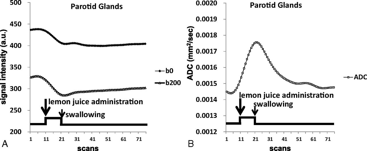
  • Fig 5.
    • Download figure
    • Open in new tab
    • Download powerpoint
    Fig 5.

    Averaged signal intensity–time curves of T2WI (b0) and DWI (b200) (A) and ADC time curves of the parotid glands (B). Lemon juice is administrated (long arrow) at the 11th scan and is swallowed (short arrow) at the 21st scan. a.u. indicates arbitrary unit.
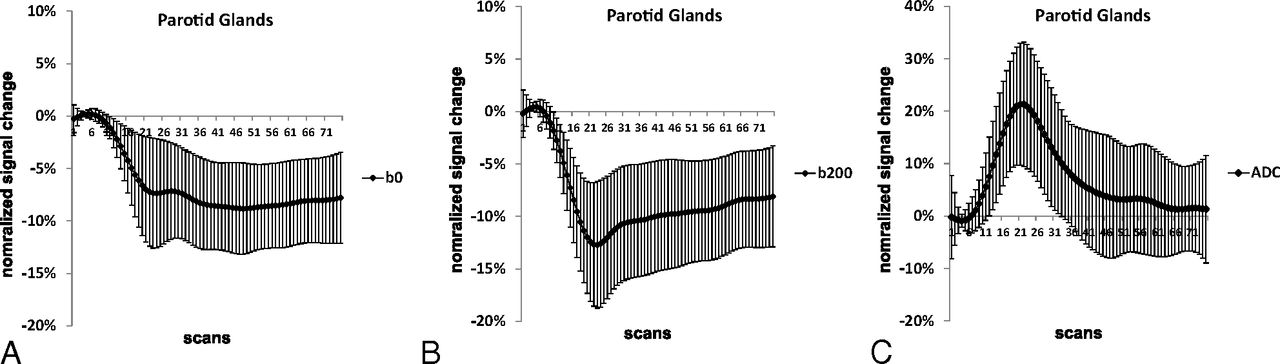
  • Fig 6.
    • Download figure
    • Open in new tab
    • Download powerpoint
    Fig 6.

    Normalized signal change–time curves (mean ± SD) of the parotid glands. During gustatory stimulation, the normalized signal change decreases rapidly on T2WI (A) and DWI (B), while it increases rapidly on ADC (C). After swallowing, the normalized signal change remains stagnant on T2WI, increases slowly on DWI, and reduces rapidly on ADC.

Tables

  • Figures
  • Magnetic susceptibility artifact scores of T2WI and DWI

    MSA ScoreAt the BaselineDuring SwallowingAt the End
    Rater 1Rater 2Rater 1Rater 2Rater 1Rater 2
    T2WI (b0)
        0000000
        1010101
        2201920192019
        3111111
    DWI (b200)
        0000000
        1003200
        2202017182020
        3111111
    • Note:—MSA indicates magnetic susceptibility artifacts.

PreviousNext
Back to top

In this issue

American Journal of Neuroradiology: 37 (10)
American Journal of Neuroradiology
Vol. 37, Issue 10
1 Oct 2016
  • Table of Contents
  • Index by author
  • Complete Issue (PDF)
Advertisement
Print
Download PDF
Email Article

Thank you for your interest in spreading the word on American Journal of Neuroradiology.

NOTE: We only request your email address so that the person you are recommending the page to knows that you wanted them to see it, and that it is not junk mail. We do not capture any email address.

Enter multiple addresses on separate lines or separate them with commas.
Evaluating Instantaneous Perfusion Responses of Parotid Glands to Gustatory Stimulation Using High-Temporal-Resolution Echo-Planar Diffusion-Weighted Imaging
(Your Name) has sent you a message from American Journal of Neuroradiology
(Your Name) thought you would like to see the American Journal of Neuroradiology web site.
CAPTCHA
This question is for testing whether or not you are a human visitor and to prevent automated spam submissions.
Cite this article
T.-W. Chiu, Y.-J. Liu, H.-C. Chang, Y.-H. Lee, J.-C. Lee, K. Hsu, C.-W. Wang, J.-M. Yang, H.-H. Hsu, C.-J. Juan
Evaluating Instantaneous Perfusion Responses of Parotid Glands to Gustatory Stimulation Using High-Temporal-Resolution Echo-Planar Diffusion-Weighted Imaging
American Journal of Neuroradiology Oct 2016, 37 (10) 1909-1915; DOI: 10.3174/ajnr.A4852

Citation Manager Formats

  • BibTeX
  • Bookends
  • EasyBib
  • EndNote (tagged)
  • EndNote 8 (xml)
  • Medlars
  • Mendeley
  • Papers
  • RefWorks Tagged
  • Ref Manager
  • RIS
  • Zotero
0 Responses
Respond to this article
Share
Bookmark this article
Evaluating Instantaneous Perfusion Responses of Parotid Glands to Gustatory Stimulation Using High-Temporal-Resolution Echo-Planar Diffusion-Weighted Imaging
T.-W. Chiu, Y.-J. Liu, H.-C. Chang, Y.-H. Lee, J.-C. Lee, K. Hsu, C.-W. Wang, J.-M. Yang, H.-H. Hsu, C.-J. Juan
American Journal of Neuroradiology Oct 2016, 37 (10) 1909-1915; DOI: 10.3174/ajnr.A4852
del.icio.us logo Twitter logo Facebook logo Mendeley logo
  • Tweet Widget
  • Facebook Like
  • Google Plus One
Purchase

Jump to section

  • Article
    • Abstract
    • ABBREVIATIONS:
    • Materials and Methods
    • Results
    • Discussion
    • Conclusions
    • Acknowledgments
    • Footnotes
    • References
  • Figures & Data
  • Info & Metrics
  • Responses
  • References
  • PDF

Related Articles

  • No related articles found.
  • PubMed
  • Google Scholar

Cited By...

  • No citing articles found.
  • Crossref
  • Google Scholar

This article has not yet been cited by articles in journals that are participating in Crossref Cited-by Linking.

More in this TOC Section

  • Revisiting CT Signs of Unilateral Vocal Fold Paralysis: A Single, Blinded Study
  • Anosmia in COVID-19 Associated with Injury to the Olfactory Bulbs Evident on MRI
  • COVID-19–Associated Bifacial Weakness with Paresthesia Subtype of Guillain-Barré Syndrome
Show more Head & Neck

Similar Articles

Advertisement

Indexed Content

  • Current Issue
  • Accepted Manuscripts
  • Article Preview
  • Past Issues
  • Editorials
  • Editor's Choice
  • Fellows' Journal Club
  • Letters to the Editor
  • Video Articles

Cases

  • Case Collection
  • Archive - Case of the Week
  • Archive - Case of the Month
  • Archive - Classic Case

More from AJNR

  • Trainee Corner
  • Imaging Protocols
  • MRI Safety Corner
  • Book Reviews

Multimedia

  • AJNR Podcasts
  • AJNR Scantastics

Resources

  • Turnaround Time
  • Submit a Manuscript
  • Submit a Video Article
  • Submit an eLetter to the Editor/Response
  • Manuscript Submission Guidelines
  • Statistical Tips
  • Fast Publishing of Accepted Manuscripts
  • Graphical Abstract Preparation
  • Imaging Protocol Submission
  • Evidence-Based Medicine Level Guide
  • Publishing Checklists
  • Author Policies
  • Become a Reviewer/Academy of Reviewers
  • News and Updates

About Us

  • About AJNR
  • Editorial Board
  • Editorial Board Alumni
  • Alerts
  • Permissions
  • Not an AJNR Subscriber? Join Now
  • Advertise with Us
  • Librarian Resources
  • Feedback
  • Terms and Conditions
  • AJNR Editorial Board Alumni

American Society of Neuroradiology

  • Not an ASNR Member? Join Now

© 2025 by the American Society of Neuroradiology All rights, including for text and data mining, AI training, and similar technologies, are reserved.
Print ISSN: 0195-6108 Online ISSN: 1936-959X

Powered by HighWire