Advertisement

Main menu

  • Home
  • Content
    • Current Issue
    • Accepted Manuscripts
    • Article Preview
    • Past Issue Archive
    • Video Articles
    • AJNR Case Collection
    • Case of the Week Archive
    • Case of the Month Archive
    • Classic Case Archive
  • Special Collections
    • AJNR Awards
    • Low-Field MRI
    • Alzheimer Disease
    • ASNR Foundation Special Collection
    • Photon-Counting CT
    • View All
  • Multimedia
    • AJNR Podcasts
    • AJNR SCANtastic
    • Trainee Corner
    • MRI Safety Corner
    • Imaging Protocols
  • For Authors
    • Submit a Manuscript
    • Submit a Video Article
    • Submit an eLetter to the Editor/Response
    • Manuscript Submission Guidelines
    • Statistical Tips
    • Fast Publishing of Accepted Manuscripts
    • Graphical Abstract Preparation
    • Imaging Protocol Submission
    • Author Policies
  • About Us
    • About AJNR
    • Editorial Board
    • Editorial Board Alumni
  • More
    • Become a Reviewer/Academy of Reviewers
    • Subscribers
    • Permissions
    • Alerts
    • Feedback
    • Advertisers
    • ASNR Home

User menu

  • Alerts
  • Log in

Search

  • Advanced search
American Journal of Neuroradiology

American Journal of Neuroradiology

ASHNR American Society of Functional Neuroradiology ASHNR American Society of Pediatric Neuroradiology ASSR
  • Alerts
  • Log in

Advanced Search

  • Home
  • Content
    • Current Issue
    • Accepted Manuscripts
    • Article Preview
    • Past Issue Archive
    • Video Articles
    • AJNR Case Collection
    • Case of the Week Archive
    • Case of the Month Archive
    • Classic Case Archive
  • Special Collections
    • AJNR Awards
    • Low-Field MRI
    • Alzheimer Disease
    • ASNR Foundation Special Collection
    • Photon-Counting CT
    • View All
  • Multimedia
    • AJNR Podcasts
    • AJNR SCANtastic
    • Trainee Corner
    • MRI Safety Corner
    • Imaging Protocols
  • For Authors
    • Submit a Manuscript
    • Submit a Video Article
    • Submit an eLetter to the Editor/Response
    • Manuscript Submission Guidelines
    • Statistical Tips
    • Fast Publishing of Accepted Manuscripts
    • Graphical Abstract Preparation
    • Imaging Protocol Submission
    • Author Policies
  • About Us
    • About AJNR
    • Editorial Board
    • Editorial Board Alumni
  • More
    • Become a Reviewer/Academy of Reviewers
    • Subscribers
    • Permissions
    • Alerts
    • Feedback
    • Advertisers
    • ASNR Home
  • Follow AJNR on Twitter
  • Visit AJNR on Facebook
  • Follow AJNR on Instagram
  • Join AJNR on LinkedIn
  • RSS Feeds

AJNR Awards, New Junior Editors, and more. Read the latest AJNR updates

Research ArticleExtracranial Vascular
Open Access

Intervention versus Aggressive Medical Therapy for Cognition in Severe Asymptomatic Carotid Stenosis

C.-J. Lin, F.-C. Chang, K.-H. Chou, P.-C. Tu, Y.-H. Lee, C.-P. Lin, P.-N. Wang and I.-H. Lee
American Journal of Neuroradiology October 2016, 37 (10) 1889-1897; DOI: https://doi.org/10.3174/ajnr.A4798
C.-J. Lin
aFrom the Departments of Neurology (C.-J.L., P.-N.W., I.-H.L.)
dInstitute of Brain Science (C.-J.L., P.-C.T., I.-H.L.)
  • Find this author on Google Scholar
  • Find this author on PubMed
  • Search for this author on this site
  • ORCID record for C.-J. Lin
F.-C. Chang
bRadiology (F.-C.C.)
  • Find this author on Google Scholar
  • Find this author on PubMed
  • Search for this author on this site
  • ORCID record for F.-C. Chang
K.-H. Chou
eInstitute of Neuroscience (K.-H.C., C.-P.L.)
  • Find this author on Google Scholar
  • Find this author on PubMed
  • Search for this author on this site
  • ORCID record for K.-H. Chou
P.-C. Tu
cMedical Education and Research (P.-C.T.), Taipei Veterans General Hospital, Taipei, Taiwan
dInstitute of Brain Science (C.-J.L., P.-C.T., I.-H.L.)
  • Find this author on Google Scholar
  • Find this author on PubMed
  • Search for this author on this site
  • ORCID record for P.-C. Tu
Y.-H. Lee
fDepartment and Institute of Physiology (Y.-H.L.)
  • Find this author on Google Scholar
  • Find this author on PubMed
  • Search for this author on this site
  • ORCID record for Y.-H. Lee
C.-P. Lin
eInstitute of Neuroscience (K.-H.C., C.-P.L.)
  • Find this author on Google Scholar
  • Find this author on PubMed
  • Search for this author on this site
  • ORCID record for C.-P. Lin
P.-N. Wang
aFrom the Departments of Neurology (C.-J.L., P.-N.W., I.-H.L.)
gSchool of Medicine (P.-N.W.), National Yang-Ming University, Taipei, Taiwan.
  • Find this author on Google Scholar
  • Find this author on PubMed
  • Search for this author on this site
  • ORCID record for P.-N. Wang
I.-H. Lee
aFrom the Departments of Neurology (C.-J.L., P.-N.W., I.-H.L.)
dInstitute of Brain Science (C.-J.L., P.-C.T., I.-H.L.)
  • Find this author on Google Scholar
  • Find this author on PubMed
  • Search for this author on this site
  • ORCID record for I.-H. Lee
  • Article
  • Figures & Data
  • Info & Metrics
  • Responses
  • References
  • PDF
Loading

Article Figures & Data

Figures

  • Tables
  • Fig 1.
    • Download figure
    • Open in new tab
    • Download powerpoint
    Fig 1.

    A, Procedure-related microemboli based on the diffusion-weighted images are overlaid on a standard Montreal Neurological Institute template from 12 of 25 patients in the stent-placement group, indicated by different colors. B, The increases (red-yellow) of fractional anisotropy (the white matter skeleton is shown in green) at 3 months after aggressive medical therapy alone (Med, upper row) or combined carotid artery stent placement (Med+CAS, middle row) and the between-group comparisons (lower row). The carotid stenotic side was set to the right in all subjects. The third column from the left represents the high-power views of the insets. Note significant FA increases at the posterior corpus callosum (arrowheads) and the posterior periventricular white matter ipsilateral to the CAS in the stent-placement group.

  • Fig 2.
    • Download figure
    • Open in new tab
    • Download powerpoint
    Fig 2.

    A and B, The functional connectivity correlation maps of both groups (Med indicates medical group; Med+CAS, stent placement group) before (pre) and 3 months after treatment (post). The carotid stenotic side was set to the right. Hollow circles indicate the predefined ROIs for individual networks at the right brain. Color bars represent T values. Q indicates the false discovery rate–corrected P value. The stent-placement group, not the medical group, showed within-group enhancement of Fc at the medial prefrontal cortex (MPF, T = 5.27, cluster size = 47, Q = .027) of the default mode network (DMN) and at the insular cortex (INS; T = 5.35, cluster size = 56, Q = .040) of the dorsal attention network (DAN) (arrowheads). C, The bar chart of the aforementioned cluster sizes with increased Fc is shown. FPN indicates frontoparietal network.
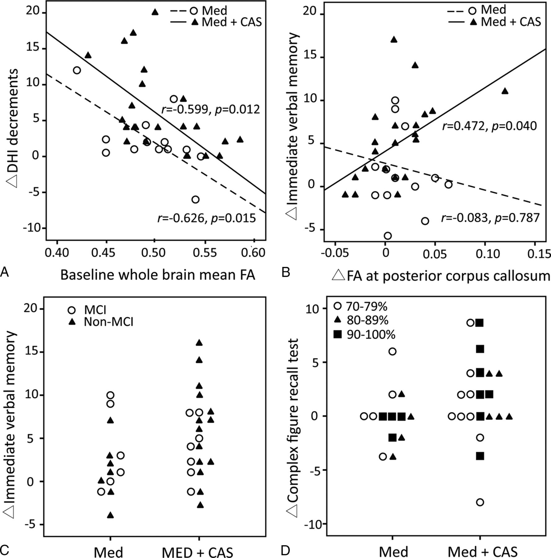
  • Fig 3.
    • Download figure
    • Open in new tab
    • Download powerpoint
    Fig 3.

    Scatterplots of the correlation analyses in the medical (Med) and the stent placement group (Med+CAS). A, The baseline whole-brain mean fractional anisotropy negatively correlates with dizziness alleviation (decreases in Dizziness Handicap Inventory [DHI]) in both groups. B, The focal FA increases in the posterior corpus callosum positively correlate with the improvement of immediate verbal memory only in the stent-placement group. C, The relationship is shown between the baseline presence of mild cognitive impairment/vascular cognitive impairment no dementia and the improvement of immediate verbal memory in the 2 groups. D, The baseline stenotic degree is not related to the changes of Complex Figure Test (Recall) scores.

Tables

  • Figures
    • View popup
    Table 1:

    Baseline characteristics

    MedMed+CASP Value
    Age (yr)68.8 ± 8.871.4 ± 7.8.348
    Male sex (%)11 (73)21 (84).687
    Education (yr)12 (7.5–12)10 (7–12).602
    Stenotic degree (%)81.3 ± 12.081.0 ± 9.8.928
    Total occlusion (No.) (%)3 (20)0 (0).543
    Hypertension (No.) (%)12 (80)20 (80).611
        Mean BP (mm Hg)95.1 ± 9.595.3 ± 10.2.887
    Diabetes mellitus (No.) (%)7 (47)9 (36).268
        HbA1c (%)6.6 ± 1.26.4 ± 0.4.254
    Hypercholesterolemia (No.) (%)9 (60)17 (68).444
        LDL (mg/dL)85.3 ± 20.189.1 ± 14.7.851
    Smoking (No.) (%)5 (33)9 (36).542
    Atrial fibrillation (No.) (%)1 (6.6)0 (0).118
    Double antiplatelets (No.) (%)8 (53)18 (72).345
    Statins (No.) (%)8 (53)15 (60).488
    Dizziness Handicap Inventory14.7 ± 19.518.3 ± 13.8.541
    Mini-Mental State Examination28.4 ± 1.228.2 ± 1.8.641
    Verbal memory tests
        Total immediate recall38.3 ± 10.546.1 ± 7.9.086
        Delayed recall7.3 ± 2.38.1 ± 2.3.571
    Attention tests
        Symbol Digit Modalities Test45.5 ± 16.341.9 ± 20.1.559
    Executive function tests
        Modified Trail-Making Test A16.4 ± 8.622.4 ± 15.1.214
        Modified Trail-Making Test B43.6 ± 28.953.1 ± 31.1.455
        Stroop Color and Word Test33. ± 13.432.9 ± 14.6.819
    Complex visuospatial perception
        Complex Figure Test (Copy)16.4 ± 1.215.5 ± 1.8.109
        Complex Figure Test (Recall)10.4 ± 4.59.7 ± 4.2.631
    MCI/VCIND (No.) (%)6 (40%)7 (28%).318
    Scheltens leukoaraiosis score5.2 ± 2.75.4 ± 3.1.889
    Hippocampal volume (mL)3.3 ± 0.33.2 ± 0.2.533
    Ipsilateral hemispheric FA0.50 ± 0.010.49 ± 0.01.471
    Contralateral hemispheric FA0.51 ± 0.010.50 ± 0.01.375
    • Note:—Med indicates medical therapy alone; Med+CAS, medical therapy combined with carotid artery stent placement; BP, blood pressure; LDL, low-density lipoprotein; HbA1c, hemoglobin A1c test.

    • View popup
    Table 2:

    Interval changes within and between groups

    ΔChangesMedMed+CASP Value
    Dizziness Handicap Inventory−2.7 ± 4.8−6.7 ± 7.1a.045a
    Neuropsychological tests
        Mini-Mental State Examination−0.2 ± 1.70.1 ± 1.0.525
        Total immediate recall2.2 ± 5.64.2 ± 5.6a.296
        Delayed recall−0.3 ± 1.20.6 ± 1.7.050
        Symbol Digit Modalities Test1.7 ± 5.22.3 ± 4.4.710
        Modified Trail-Making Test A−0.1 ± 8.1−2.6 ± 4.5.309
        Modified Trail-Making Test B1.7 ± 31.9−4.9 ± 13.1.405
        Stroop Color and Word Test1.7 ± 5.82.9 ± 6.4.545
        Complex Figure Test (Copy)0.2 ± 1.41.1 ± 1.4a.064
        Complex Figure Test (Recall)0.1 ± 3.11.7 ± 3.7.157
    Scheltens leukoaraiosis score0.1 ± 0.30.2 ± 0.5.561
    Ipsilateral hemispheric FA0.002 ± 0.040.009 ± 0.004.447
    Contralateral hemispheric FA0.002 ± 0.050.004 ± 0.005.681
    • Note:—Med indicates medical therapy alone.

    • ↵a For dizziness and imaging measures, P < .05 was defined as significant. For the 9 neuropsychological tests, P < .0056 was defined as significant with the Bonferroni correction.

PreviousNext
Back to top

In this issue

American Journal of Neuroradiology: 37 (10)
American Journal of Neuroradiology
Vol. 37, Issue 10
1 Oct 2016
  • Table of Contents
  • Index by author
  • Complete Issue (PDF)
Advertisement
Print
Download PDF
Email Article

Thank you for your interest in spreading the word on American Journal of Neuroradiology.

NOTE: We only request your email address so that the person you are recommending the page to knows that you wanted them to see it, and that it is not junk mail. We do not capture any email address.

Enter multiple addresses on separate lines or separate them with commas.
Intervention versus Aggressive Medical Therapy for Cognition in Severe Asymptomatic Carotid Stenosis
(Your Name) has sent you a message from American Journal of Neuroradiology
(Your Name) thought you would like to see the American Journal of Neuroradiology web site.
CAPTCHA
This question is for testing whether or not you are a human visitor and to prevent automated spam submissions.
Cite this article
C.-J. Lin, F.-C. Chang, K.-H. Chou, P.-C. Tu, Y.-H. Lee, C.-P. Lin, P.-N. Wang, I.-H. Lee
Intervention versus Aggressive Medical Therapy for Cognition in Severe Asymptomatic Carotid Stenosis
American Journal of Neuroradiology Oct 2016, 37 (10) 1889-1897; DOI: 10.3174/ajnr.A4798

Citation Manager Formats

  • BibTeX
  • Bookends
  • EasyBib
  • EndNote (tagged)
  • EndNote 8 (xml)
  • Medlars
  • Mendeley
  • Papers
  • RefWorks Tagged
  • Ref Manager
  • RIS
  • Zotero
0 Responses
Respond to this article
Share
Bookmark this article
Intervention versus Aggressive Medical Therapy for Cognition in Severe Asymptomatic Carotid Stenosis
C.-J. Lin, F.-C. Chang, K.-H. Chou, P.-C. Tu, Y.-H. Lee, C.-P. Lin, P.-N. Wang, I.-H. Lee
American Journal of Neuroradiology Oct 2016, 37 (10) 1889-1897; DOI: 10.3174/ajnr.A4798
del.icio.us logo Twitter logo Facebook logo Mendeley logo
  • Tweet Widget
  • Facebook Like
  • Google Plus One
Purchase

Jump to section

  • Article
    • Abstract
    • ABBREVIATIONS:
    • Materials and Methods
    • Results
    • Discussion
    • Conclusions
    • Acknowledgments
    • Footnotes
    • References
  • Figures & Data
  • Info & Metrics
  • Responses
  • References
  • PDF

Related Articles

  • No related articles found.
  • PubMed
  • Google Scholar

Cited By...

  • No citing articles found.
  • Crossref
  • Google Scholar

This article has not yet been cited by articles in journals that are participating in Crossref Cited-by Linking.

More in this TOC Section

  • Proximal Vertebral Artery Variants and Embryology
  • High-Risk Plaque Features in Carotid MRI
  • Nonstenotic Carotid Plaques and Stroke Review
Show more Extracranial Vascular

Similar Articles

Advertisement

Indexed Content

  • Current Issue
  • Accepted Manuscripts
  • Article Preview
  • Past Issues
  • Editorials
  • Editor's Choice
  • Fellows' Journal Club
  • Letters to the Editor
  • Video Articles

Cases

  • Case Collection
  • Archive - Case of the Week
  • Archive - Case of the Month
  • Archive - Classic Case

More from AJNR

  • Trainee Corner
  • Imaging Protocols
  • MRI Safety Corner
  • Book Reviews

Multimedia

  • AJNR Podcasts
  • AJNR Scantastics

Resources

  • Turnaround Time
  • Submit a Manuscript
  • Submit a Video Article
  • Submit an eLetter to the Editor/Response
  • Manuscript Submission Guidelines
  • Statistical Tips
  • Fast Publishing of Accepted Manuscripts
  • Graphical Abstract Preparation
  • Imaging Protocol Submission
  • Evidence-Based Medicine Level Guide
  • Publishing Checklists
  • Author Policies
  • Become a Reviewer/Academy of Reviewers
  • News and Updates

About Us

  • About AJNR
  • Editorial Board
  • Editorial Board Alumni
  • Alerts
  • Permissions
  • Not an AJNR Subscriber? Join Now
  • Advertise with Us
  • Librarian Resources
  • Feedback
  • Terms and Conditions
  • AJNR Editorial Board Alumni

American Society of Neuroradiology

  • Not an ASNR Member? Join Now

© 2025 by the American Society of Neuroradiology All rights, including for text and data mining, AI training, and similar technologies, are reserved.
Print ISSN: 0195-6108 Online ISSN: 1936-959X

Powered by HighWire