Advertisement

Main menu

  • Home
  • Content
    • Current Issue
    • Accepted Manuscripts
    • Article Preview
    • Past Issue Archive
    • Video Articles
    • AJNR Case Collection
    • Case of the Week Archive
    • Case of the Month Archive
    • Classic Case Archive
  • Special Collections
    • AJNR Awards
    • Low-Field MRI
    • Alzheimer Disease
    • ASNR Foundation Special Collection
    • Photon-Counting CT
    • View All
  • Multimedia
    • AJNR Podcasts
    • AJNR SCANtastic
    • Trainee Corner
    • MRI Safety Corner
    • Imaging Protocols
  • For Authors
    • Submit a Manuscript
    • Submit a Video Article
    • Submit an eLetter to the Editor/Response
    • Manuscript Submission Guidelines
    • Statistical Tips
    • Fast Publishing of Accepted Manuscripts
    • Graphical Abstract Preparation
    • Imaging Protocol Submission
    • Author Policies
  • About Us
    • About AJNR
    • Editorial Board
    • Editorial Board Alumni
  • More
    • Become a Reviewer/Academy of Reviewers
    • Subscribers
    • Permissions
    • Alerts
    • Feedback
    • Advertisers
    • ASNR Home

User menu

  • Alerts
  • Log in

Search

  • Advanced search
American Journal of Neuroradiology

American Journal of Neuroradiology

ASHNR American Society of Functional Neuroradiology ASHNR American Society of Pediatric Neuroradiology ASSR
  • Alerts
  • Log in

Advanced Search

  • Home
  • Content
    • Current Issue
    • Accepted Manuscripts
    • Article Preview
    • Past Issue Archive
    • Video Articles
    • AJNR Case Collection
    • Case of the Week Archive
    • Case of the Month Archive
    • Classic Case Archive
  • Special Collections
    • AJNR Awards
    • Low-Field MRI
    • Alzheimer Disease
    • ASNR Foundation Special Collection
    • Photon-Counting CT
    • View All
  • Multimedia
    • AJNR Podcasts
    • AJNR SCANtastic
    • Trainee Corner
    • MRI Safety Corner
    • Imaging Protocols
  • For Authors
    • Submit a Manuscript
    • Submit a Video Article
    • Submit an eLetter to the Editor/Response
    • Manuscript Submission Guidelines
    • Statistical Tips
    • Fast Publishing of Accepted Manuscripts
    • Graphical Abstract Preparation
    • Imaging Protocol Submission
    • Author Policies
  • About Us
    • About AJNR
    • Editorial Board
    • Editorial Board Alumni
  • More
    • Become a Reviewer/Academy of Reviewers
    • Subscribers
    • Permissions
    • Alerts
    • Feedback
    • Advertisers
    • ASNR Home
  • Follow AJNR on Twitter
  • Visit AJNR on Facebook
  • Follow AJNR on Instagram
  • Join AJNR on LinkedIn
  • RSS Feeds

AJNR Awards, New Junior Editors, and more. Read the latest AJNR updates

Research ArticleAdult Brain
Open Access

White Matter Hyperintensity Volume and Cerebral Perfusion in Older Individuals with Hypertension Using Arterial Spin-Labeling

J.W. van Dalen, H.J.M.M. Mutsaerts, A.J. Nederveen, H. Vrenken, M.D. Steenwijk, M.W.A. Caan, C.B.L.M. Majoie, W.A. van Gool and E. Richard
American Journal of Neuroradiology October 2016, 37 (10) 1824-1830; DOI: https://doi.org/10.3174/ajnr.A4828
J.W. van Dalen
aFrom the Departments of Neurology (J.W.v.D., W.A.v.G., E.R.)
  • Find this author on Google Scholar
  • Find this author on PubMed
  • Search for this author on this site
  • ORCID record for J.W. van Dalen
H.J.M.M. Mutsaerts
bRadiology (H.J.M.M.M., A.J.N., M.W.A.C., C.B.L.M.M.), Academic Medical Center, University of Amsterdam, Amsterdam, the Netherlands
  • Find this author on Google Scholar
  • Find this author on PubMed
  • Search for this author on this site
  • ORCID record for H.J.M.M. Mutsaerts
A.J. Nederveen
bRadiology (H.J.M.M.M., A.J.N., M.W.A.C., C.B.L.M.M.), Academic Medical Center, University of Amsterdam, Amsterdam, the Netherlands
  • Find this author on Google Scholar
  • Find this author on PubMed
  • Search for this author on this site
  • ORCID record for A.J. Nederveen
H. Vrenken
cDepartments of Radiology and Nuclear Medicine (H.V., M.D.S.)
dPhysics and Medical Technology (H.V.), Neuroscience Campus Amsterdam, VU University Medical Center Amsterdam, Amsterdam, the Netherlands
  • Find this author on Google Scholar
  • Find this author on PubMed
  • Search for this author on this site
  • ORCID record for H. Vrenken
M.D. Steenwijk
cDepartments of Radiology and Nuclear Medicine (H.V., M.D.S.)
  • Find this author on Google Scholar
  • Find this author on PubMed
  • Search for this author on this site
  • ORCID record for M.D. Steenwijk
M.W.A. Caan
bRadiology (H.J.M.M.M., A.J.N., M.W.A.C., C.B.L.M.M.), Academic Medical Center, University of Amsterdam, Amsterdam, the Netherlands
  • Find this author on Google Scholar
  • Find this author on PubMed
  • Search for this author on this site
  • ORCID record for M.W.A. Caan
C.B.L.M. Majoie
bRadiology (H.J.M.M.M., A.J.N., M.W.A.C., C.B.L.M.M.), Academic Medical Center, University of Amsterdam, Amsterdam, the Netherlands
  • Find this author on Google Scholar
  • Find this author on PubMed
  • Search for this author on this site
  • ORCID record for C.B.L.M. Majoie
W.A. van Gool
aFrom the Departments of Neurology (J.W.v.D., W.A.v.G., E.R.)
  • Find this author on Google Scholar
  • Find this author on PubMed
  • Search for this author on this site
  • ORCID record for W.A. van Gool
E. Richard
aFrom the Departments of Neurology (J.W.v.D., W.A.v.G., E.R.)
eDepartment of Neurology (E.R.), Radboud University Medical Center, Nijmegen, the Netherlands.
  • Find this author on Google Scholar
  • Find this author on PubMed
  • Search for this author on this site
  • ORCID record for E. Richard
  • Article
  • Figures & Data
  • Supplemental
  • Info & Metrics
  • Responses
  • References
  • PDF
Loading

Article Figures & Data

Figures

  • Tables
  • Fig 1.
    • Download figure
    • Open in new tab
    • Download powerpoint
    Fig 1.

    Regional cerebral blood flow. Cerebral blood in the gray matter, white matter, normal-appearing white matter unaffected by WMHs (NAWM), and white matter hyperintensities. Shown are means (SDs) and P values of paired sample t tests.
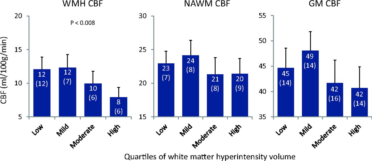
  • Fig 2.
    • Download figure
    • Open in new tab
    • Download powerpoint
    Fig 2.

    Cerebral blood flow per quartiles of WMH load. Cerebral blood flow in the gray matter, normal-appearing white matter unaffected by WMHs, and white matter hyperintensities in subgroups based on quartiles of WMH volume. Shown are means (SDs) and significant P values of 1-way analysis of variance.

  • Fig 3.
    • Download figure
    • Open in new tab
    • Download powerpoint
    Fig 3.

    Scatterplots of relations between CBF and WMH volume, adjusted for total brain volume. Lines denote the regression line with 95% CI. Log WMH volume is logarithmically transformed.

Tables

  • Figures
    • View popup
    Table 1:

    General characteristicsa

    Characteristics (n = 181)
    Age (yr)77 (2)
    Female96 (53%)
    MMSE29 (28–30)
    BMI (kg/m2)26 (24–28)
    History of stroke or TIA19 (11%)
    History of cardiovascular disease41 (23%)
    Diabetes mellitus20 (11%)
    Smoking status
        Never82 (45%)
        Former88 (49%)
        Current11 (6%)
    Antihypertensive drug use108 (60%)
    World Health Organization hypertension grade:
        Normotension47 (26%)
        Grade I hypertension73 (41%)
        Grade II hypertension38 (21%)
        Grade III hypertension20 (11%)
    Systolic blood pressure (mm Hg)148 (138–165)
    Diastolic blood pressure (mm Hg)81 (74–90)
    Brain parenchymal fraction0.61 (0.025)
    WMH volume (mL)6.5 (3.6–11.2)
    • Note:—MMSE indicates Mini-Mental State Examination; BMI, body mass index.

    • ↵a Reported are means and SDs, numbers and valid percentages, or medians with interquartile range. Cardiovascular disease comprises peripheral arterial disease, angina pectoris, and myocardial infarction. Brain parenchymal fraction = (total cerebral volume)/total intracranial volume.

    • View popup
    Table 2:

    Association between cerebral perfusion and white matter hyperintensity volumea

    PredictorModel 1Model 2
    βP ValueR2βP ValueR2
    CBF in WMH−.248.0010.06−.201.0290.06
    CBF in NAWM−.035.6430.00.175.0980.05
    CBF in GM−.065.3820.00.175.1330.05
    • ↵a R2 is the adjusted R2 representing the proportion of variation in white matter hyperintensity volume explained by all variables in the model, corrected for the number of variables—model 1: adjusted for total brain volume; model 2: adjusted for total brain volume, age, antihypertensive use, brain parenchymal fraction, and transit time.

PreviousNext
Back to top

In this issue

American Journal of Neuroradiology: 37 (10)
American Journal of Neuroradiology
Vol. 37, Issue 10
1 Oct 2016
  • Table of Contents
  • Index by author
  • Complete Issue (PDF)
Advertisement
Print
Download PDF
Email Article

Thank you for your interest in spreading the word on American Journal of Neuroradiology.

NOTE: We only request your email address so that the person you are recommending the page to knows that you wanted them to see it, and that it is not junk mail. We do not capture any email address.

Enter multiple addresses on separate lines or separate them with commas.
White Matter Hyperintensity Volume and Cerebral Perfusion in Older Individuals with Hypertension Using Arterial Spin-Labeling
(Your Name) has sent you a message from American Journal of Neuroradiology
(Your Name) thought you would like to see the American Journal of Neuroradiology web site.
CAPTCHA
This question is for testing whether or not you are a human visitor and to prevent automated spam submissions.
Cite this article
J.W. van Dalen, H.J.M.M. Mutsaerts, A.J. Nederveen, H. Vrenken, M.D. Steenwijk, M.W.A. Caan, C.B.L.M. Majoie, W.A. van Gool, E. Richard
White Matter Hyperintensity Volume and Cerebral Perfusion in Older Individuals with Hypertension Using Arterial Spin-Labeling
American Journal of Neuroradiology Oct 2016, 37 (10) 1824-1830; DOI: 10.3174/ajnr.A4828

Citation Manager Formats

  • BibTeX
  • Bookends
  • EasyBib
  • EndNote (tagged)
  • EndNote 8 (xml)
  • Medlars
  • Mendeley
  • Papers
  • RefWorks Tagged
  • Ref Manager
  • RIS
  • Zotero
0 Responses
Respond to this article
Share
Bookmark this article
White Matter Hyperintensity Volume and Cerebral Perfusion in Older Individuals with Hypertension Using Arterial Spin-Labeling
J.W. van Dalen, H.J.M.M. Mutsaerts, A.J. Nederveen, H. Vrenken, M.D. Steenwijk, M.W.A. Caan, C.B.L.M. Majoie, W.A. van Gool, E. Richard
American Journal of Neuroradiology Oct 2016, 37 (10) 1824-1830; DOI: 10.3174/ajnr.A4828
del.icio.us logo Twitter logo Facebook logo Mendeley logo
  • Tweet Widget
  • Facebook Like
  • Google Plus One
Purchase

Jump to section

  • Article
    • Abstract
    • ABBREVIATIONS:
    • Materials and Methods
    • Results
    • Discussion
    • Conclusions
    • Acknowledgments
    • Footnotes
    • References
  • Figures & Data
  • Supplemental
  • Info & Metrics
  • Responses
  • References
  • PDF

Related Articles

  • PubMed
  • Google Scholar

Cited By...

  • Mid-life association between cardiovascular risk factors and cerebral blood flow in a multi-ethnic population
  • Associations between white matter hyperintensity burden, cerebral blood flow and transit time in small vessel disease: an updated meta-analysis
  • Crossref
  • Google Scholar

This article has not yet been cited by articles in journals that are participating in Crossref Cited-by Linking.

More in this TOC Section

  • Diagnostic Neuroradiology of Monoclonal Antibodies
  • Clinical Outcomes After Chiari I Decompression
  • Segmentation of Brain Metastases with BLAST
Show more Adult Brain

Similar Articles

Advertisement

Indexed Content

  • Current Issue
  • Accepted Manuscripts
  • Article Preview
  • Past Issues
  • Editorials
  • Editor's Choice
  • Fellows' Journal Club
  • Letters to the Editor
  • Video Articles

Cases

  • Case Collection
  • Archive - Case of the Week
  • Archive - Case of the Month
  • Archive - Classic Case

More from AJNR

  • Trainee Corner
  • Imaging Protocols
  • MRI Safety Corner
  • Book Reviews

Multimedia

  • AJNR Podcasts
  • AJNR Scantastics

Resources

  • Turnaround Time
  • Submit a Manuscript
  • Submit a Video Article
  • Submit an eLetter to the Editor/Response
  • Manuscript Submission Guidelines
  • Statistical Tips
  • Fast Publishing of Accepted Manuscripts
  • Graphical Abstract Preparation
  • Imaging Protocol Submission
  • Evidence-Based Medicine Level Guide
  • Publishing Checklists
  • Author Policies
  • Become a Reviewer/Academy of Reviewers
  • News and Updates

About Us

  • About AJNR
  • Editorial Board
  • Editorial Board Alumni
  • Alerts
  • Permissions
  • Not an AJNR Subscriber? Join Now
  • Advertise with Us
  • Librarian Resources
  • Feedback
  • Terms and Conditions
  • AJNR Editorial Board Alumni

American Society of Neuroradiology

  • Not an ASNR Member? Join Now

© 2025 by the American Society of Neuroradiology All rights, including for text and data mining, AI training, and similar technologies, are reserved.
Print ISSN: 0195-6108 Online ISSN: 1936-959X

Powered by HighWire