Advertisement

Main menu

  • Home
  • Content
    • Current Issue
    • Accepted Manuscripts
    • Article Preview
    • Past Issue Archive
    • Video Articles
    • AJNR Case Collection
    • Case of the Week Archive
    • Case of the Month Archive
    • Classic Case Archive
  • Special Collections
    • AJNR Awards
    • Low-Field MRI
    • Alzheimer Disease
    • ASNR Foundation Special Collection
    • Photon-Counting CT
    • View All
  • Multimedia
    • AJNR Podcasts
    • AJNR SCANtastic
    • Trainee Corner
    • MRI Safety Corner
    • Imaging Protocols
  • For Authors
    • Submit a Manuscript
    • Submit a Video Article
    • Submit an eLetter to the Editor/Response
    • Manuscript Submission Guidelines
    • Statistical Tips
    • Fast Publishing of Accepted Manuscripts
    • Graphical Abstract Preparation
    • Imaging Protocol Submission
    • Author Policies
  • About Us
    • About AJNR
    • Editorial Board
    • Editorial Board Alumni
  • More
    • Become a Reviewer/Academy of Reviewers
    • Subscribers
    • Permissions
    • Alerts
    • Feedback
    • Advertisers
    • ASNR Home

User menu

  • Alerts
  • Log in

Search

  • Advanced search
American Journal of Neuroradiology

American Journal of Neuroradiology

ASHNR American Society of Functional Neuroradiology ASHNR American Society of Pediatric Neuroradiology ASSR
  • Alerts
  • Log in

Advanced Search

  • Home
  • Content
    • Current Issue
    • Accepted Manuscripts
    • Article Preview
    • Past Issue Archive
    • Video Articles
    • AJNR Case Collection
    • Case of the Week Archive
    • Case of the Month Archive
    • Classic Case Archive
  • Special Collections
    • AJNR Awards
    • Low-Field MRI
    • Alzheimer Disease
    • ASNR Foundation Special Collection
    • Photon-Counting CT
    • View All
  • Multimedia
    • AJNR Podcasts
    • AJNR SCANtastic
    • Trainee Corner
    • MRI Safety Corner
    • Imaging Protocols
  • For Authors
    • Submit a Manuscript
    • Submit a Video Article
    • Submit an eLetter to the Editor/Response
    • Manuscript Submission Guidelines
    • Statistical Tips
    • Fast Publishing of Accepted Manuscripts
    • Graphical Abstract Preparation
    • Imaging Protocol Submission
    • Author Policies
  • About Us
    • About AJNR
    • Editorial Board
    • Editorial Board Alumni
  • More
    • Become a Reviewer/Academy of Reviewers
    • Subscribers
    • Permissions
    • Alerts
    • Feedback
    • Advertisers
    • ASNR Home
  • Follow AJNR on Twitter
  • Visit AJNR on Facebook
  • Follow AJNR on Instagram
  • Join AJNR on LinkedIn
  • RSS Feeds

AJNR Awards, New Junior Editors, and more. Read the latest AJNR updates

Research ArticleAdult Brain

Improved Automatic Detection of New T2 Lesions in Multiple Sclerosis Using Deformation Fields

M. Cabezas, J.F. Corral, A. Oliver, Y. Díez, M. Tintoré, C. Auger, X. Montalban, X. Lladó, D. Pareto and À. Rovira
American Journal of Neuroradiology October 2016, 37 (10) 1816-1823; DOI: https://doi.org/10.3174/ajnr.A4829
M. Cabezas
aFrom the Section of Neuroradiology, Department of Radiology (M.C., J.F.C., C.A., D.P., À.R.)
cVisió per Computador i Robòtica group (M.C., A.O., Y.D., X.L.), University of Girona, Girona, Spain.
  • Find this author on Google Scholar
  • Find this author on PubMed
  • Search for this author on this site
  • ORCID record for M. Cabezas
J.F. Corral
aFrom the Section of Neuroradiology, Department of Radiology (M.C., J.F.C., C.A., D.P., À.R.)
  • Find this author on Google Scholar
  • Find this author on PubMed
  • Search for this author on this site
A. Oliver
cVisió per Computador i Robòtica group (M.C., A.O., Y.D., X.L.), University of Girona, Girona, Spain.
  • Find this author on Google Scholar
  • Find this author on PubMed
  • Search for this author on this site
  • ORCID record for A. Oliver
Y. Díez
cVisió per Computador i Robòtica group (M.C., A.O., Y.D., X.L.), University of Girona, Girona, Spain.
  • Find this author on Google Scholar
  • Find this author on PubMed
  • Search for this author on this site
  • ORCID record for Y. Díez
M. Tintoré
bCentre d'Esclerosi Múltiple de Catalunya, Department of Neurology/Neuroimmunology (M.T., X.M.), Vall d'Hebron University Hospital, Vall d'Hebron Research Institute, Autonomous University of Barcelona, Barcelona, Spain
  • Find this author on Google Scholar
  • Find this author on PubMed
  • Search for this author on this site
  • ORCID record for M. Tintoré
C. Auger
aFrom the Section of Neuroradiology, Department of Radiology (M.C., J.F.C., C.A., D.P., À.R.)
  • Find this author on Google Scholar
  • Find this author on PubMed
  • Search for this author on this site
  • ORCID record for C. Auger
X. Montalban
bCentre d'Esclerosi Múltiple de Catalunya, Department of Neurology/Neuroimmunology (M.T., X.M.), Vall d'Hebron University Hospital, Vall d'Hebron Research Institute, Autonomous University of Barcelona, Barcelona, Spain
  • Find this author on Google Scholar
  • Find this author on PubMed
  • Search for this author on this site
  • ORCID record for X. Montalban
X. Lladó
cVisió per Computador i Robòtica group (M.C., A.O., Y.D., X.L.), University of Girona, Girona, Spain.
  • Find this author on Google Scholar
  • Find this author on PubMed
  • Search for this author on this site
  • ORCID record for X. Lladó
D. Pareto
aFrom the Section of Neuroradiology, Department of Radiology (M.C., J.F.C., C.A., D.P., À.R.)
  • Find this author on Google Scholar
  • Find this author on PubMed
  • Search for this author on this site
  • ORCID record for D. Pareto
À. Rovira
aFrom the Section of Neuroradiology, Department of Radiology (M.C., J.F.C., C.A., D.P., À.R.)
  • Find this author on Google Scholar
  • Find this author on PubMed
  • Search for this author on this site
  • ORCID record for À. Rovira
  • Article
  • Figures & Data
  • Info & Metrics
  • Responses
  • References
  • PDF
Loading

Article Figures & Data

Figures

  • Tables
  • Fig 1.
    • Download figure
    • Open in new tab
    • Download powerpoint
    Fig 1.

    Example of the deformation field inside a new lesion. All arrows point to the lesion center.

  • Fig 2.
    • Download figure
    • Open in new tab
    • Download powerpoint
    Fig 2.

    Example of the deformation field for 2 sections. The first image does not contain lesions and presents large deformations with no clear sinking patterns, while in the second image with a lesion, all the arrows inside the lesion point to the center.

  • Fig 3.
    • Download figure
    • Open in new tab
    • Download powerpoint
    Fig 3.

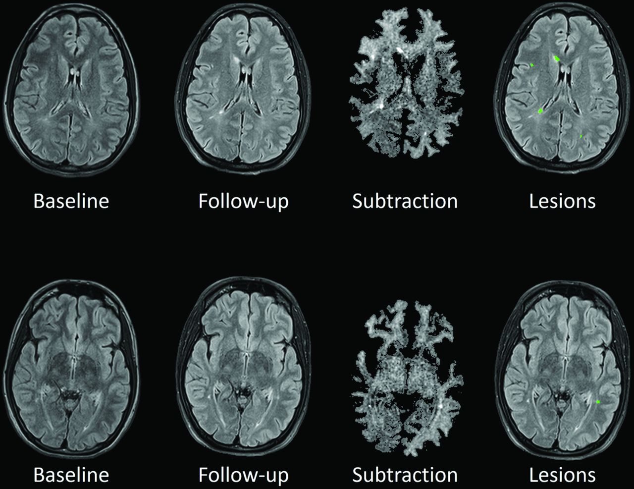
    New lesion detection. For each row, the first image is the baseline image, the second is the follow-up image, the third is the subtraction, and the fourth is the lesion analysis over the follow-up image (green = true-positive). The patient has a large number of TPs (100%), with a small number of FPs (0%).

  • Fig 4.
    • Download figure
    • Open in new tab
    • Download powerpoint
    Fig 4.

    Correlation between the number of ground truth lesions and the number of automatically detected ones (Pearson coefficient = 0.81, P = 2.2688e-09).

Tables

  • Figures
    • View popup
    Table 1:

    Lesion detections obtained for our data base using various approaches

    ImageMethodASDTPfFPfDSC (Lesions)DSC (Volume)
    PDThreshold25.8092.2893.180.110.31
    Intensity rules2021.9080.6183.010.240.35
    DF19.9173.1877.020.300.37
    T2Threshold25.2293.8995.880.070.25
    Intensity rules2022.2264.0986.350.170.25
    DF17.7681.7980.840.260.34
    T2-FLAIRThreshold27.2290.2492.790.100.26
    Intensity rules2021.1778.3480.770.250.31
    DF21.1481.2277.110.300.33
    CombinationThreshold13.0791.0585.610.220.45
    Intensity rules2030.8051.6235.870.460.37
    Proposal7.8970.9317.800.680.52
    • Note:—ASD indicates average surface distance.

    • View popup
    Table 2:

    Permutation test ranking of DSC values for the approaches applied on each image separatelya

    MethodMean P Value
    Rank 1 (<1 σ)T2-FLAIR-DF.75
    PD-DF.56
    T2-DF.53
    Rank 2 (<2 σ)T2-FLAIR20.22
    PD20.16
    Rank 3 (<3 σ)T220−.22
    PD-threshold−.56
    T2-FLAIR-threshold−.67
    T2-threshold−.78
    • ↵a Methods were ranked relative to the mean and SD of the method with the highest DSC value. Methods in the same rank have similar results, whereas methods in different ranks show significant differences.

    • View popup
    Table 3:

    Analysis of the TPf before and after postprocessing with deformation fields for different sizesa

    ImageMethod3–1011–5050+
    CombinationCombination (threshold)71.4372.3895.16
    Proposal42.8648.5777.42
    • ↵a Lesions between 3 and 10 voxels are considered small; lesions between 11 and 50 voxels, medium; and lesions with >50 voxels, large.

PreviousNext
Back to top

In this issue

American Journal of Neuroradiology: 37 (10)
American Journal of Neuroradiology
Vol. 37, Issue 10
1 Oct 2016
  • Table of Contents
  • Index by author
  • Complete Issue (PDF)
Advertisement
Print
Download PDF
Email Article

Thank you for your interest in spreading the word on American Journal of Neuroradiology.

NOTE: We only request your email address so that the person you are recommending the page to knows that you wanted them to see it, and that it is not junk mail. We do not capture any email address.

Enter multiple addresses on separate lines or separate them with commas.
Improved Automatic Detection of New T2 Lesions in Multiple Sclerosis Using Deformation Fields
(Your Name) has sent you a message from American Journal of Neuroradiology
(Your Name) thought you would like to see the American Journal of Neuroradiology web site.
CAPTCHA
This question is for testing whether or not you are a human visitor and to prevent automated spam submissions.
Cite this article
M. Cabezas, J.F. Corral, A. Oliver, Y. Díez, M. Tintoré, C. Auger, X. Montalban, X. Lladó, D. Pareto, À. Rovira
Improved Automatic Detection of New T2 Lesions in Multiple Sclerosis Using Deformation Fields
American Journal of Neuroradiology Oct 2016, 37 (10) 1816-1823; DOI: 10.3174/ajnr.A4829

Citation Manager Formats

  • BibTeX
  • Bookends
  • EasyBib
  • EndNote (tagged)
  • EndNote 8 (xml)
  • Medlars
  • Mendeley
  • Papers
  • RefWorks Tagged
  • Ref Manager
  • RIS
  • Zotero
0 Responses
Respond to this article
Share
Bookmark this article
Improved Automatic Detection of New T2 Lesions in Multiple Sclerosis Using Deformation Fields
M. Cabezas, J.F. Corral, A. Oliver, Y. Díez, M. Tintoré, C. Auger, X. Montalban, X. Lladó, D. Pareto, À. Rovira
American Journal of Neuroradiology Oct 2016, 37 (10) 1816-1823; DOI: 10.3174/ajnr.A4829
del.icio.us logo Twitter logo Facebook logo Mendeley logo
  • Tweet Widget
  • Facebook Like
  • Google Plus One
Purchase

Jump to section

  • Article
    • Abstract
    • ABBREVIATIONS:
    • Materials and Methods
    • Results
    • Discussion
    • Conclusions
    • Footnotes
    • References
  • Figures & Data
  • Info & Metrics
  • Responses
  • References
  • PDF

Related Articles

  • No related articles found.
  • PubMed
  • Google Scholar

Cited By...

  • Evaluation of the Statistical Detection of Change Algorithm for Screening Patients with MS with New Lesion Activity on Longitudinal Brain MRI
  • Evaluation of the Statistical Detection of Change Algorithm for Screening Patients with MS with New Lesion Activity on Longitudinal Brain MRI
  • Evaluation of statistical detection of change algorithm for triaging multiple sclerosis patients with new lesion activity on longitudinal brain MRI
  • Automatic brain lesion segmentation on standard magnetic resonance images: a scoping review
  • Detection of Volume-Changing Metastatic Brain Tumors on Longitudinal MRI Using a Semiautomated Algorithm Based on the Jacobian Operator Field
  • Crossref
  • Google Scholar

This article has not yet been cited by articles in journals that are participating in Crossref Cited-by Linking.

More in this TOC Section

  • Diagnostic Neuroradiology of Monoclonal Antibodies
  • NCCT vs. MRI for Brain Atrophy in Acute Stroke
  • Clinical Outcomes After Chiari I Decompression
Show more Adult Brain

Similar Articles

Advertisement

Indexed Content

  • Current Issue
  • Accepted Manuscripts
  • Article Preview
  • Past Issues
  • Editorials
  • Editor's Choice
  • Fellows' Journal Club
  • Letters to the Editor
  • Video Articles

Cases

  • Case Collection
  • Archive - Case of the Week
  • Archive - Case of the Month
  • Archive - Classic Case

More from AJNR

  • Trainee Corner
  • Imaging Protocols
  • MRI Safety Corner
  • Book Reviews

Multimedia

  • AJNR Podcasts
  • AJNR Scantastics

Resources

  • Turnaround Time
  • Submit a Manuscript
  • Submit a Video Article
  • Submit an eLetter to the Editor/Response
  • Manuscript Submission Guidelines
  • Statistical Tips
  • Fast Publishing of Accepted Manuscripts
  • Graphical Abstract Preparation
  • Imaging Protocol Submission
  • Evidence-Based Medicine Level Guide
  • Publishing Checklists
  • Author Policies
  • Become a Reviewer/Academy of Reviewers
  • News and Updates

About Us

  • About AJNR
  • Editorial Board
  • Editorial Board Alumni
  • Alerts
  • Permissions
  • Not an AJNR Subscriber? Join Now
  • Advertise with Us
  • Librarian Resources
  • Feedback
  • Terms and Conditions
  • AJNR Editorial Board Alumni

American Society of Neuroradiology

  • Not an ASNR Member? Join Now

© 2025 by the American Society of Neuroradiology All rights, including for text and data mining, AI training, and similar technologies, are reserved.
Print ISSN: 0195-6108 Online ISSN: 1936-959X

Powered by HighWire